Review
. 2022 Aug 25:16:967496.
doi: 10.3389/fncel.2022.967496. eCollection 2022.

Glia as a key factor in cell volume regulation processes of the central nervous system

Affiliations
Review

Glia as a key factor in cell volume regulation processes of the central nervous system

Lenin David Ochoa-de la Paz et al. Front Cell Neurosci. .

Abstract

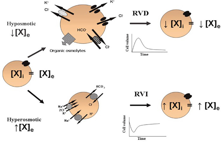
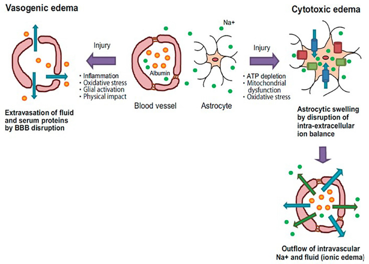
Brain edema is a pathological condition with potentially fatal consequences, related to cerebral injuries such as ischemia, chronic renal failure, uremia, and diabetes, among others. Under these pathological states, the cell volume control processes are fully compromised, because brain cells are unable to regulate the movement of water, mainly regulated by osmotic gradients. The processes involved in cell volume regulation are homeostatic mechanisms that depend on the mobilization of osmolytes (ions, organic molecules, and polyols) in the necessary direction to counteract changes in osmolyte concentration in response to water movement. The expression and coordinated function of proteins related to the cell volume regulation process, such as water channels, ion channels, and other cotransport systems in the glial cells, and considering the glial cell proportion compared to neuronal cells, leads to consider the astroglial network the main regulatory unit for water homeostasis in the central nervous system (CNS). In the last decade, several studies highlighted the pivotal role of glia in the cell volume regulation process and water homeostasis in the brain, including the retina; any malfunction of this astroglial network generates a lack of the ability to regulate the osmotic changes and water movements and consequently exacerbates the pathological condition.

Keywords: Müller cells; astrocytes; brain edema; cell volume regulation; glia.


Conflict of interest statement

The authors declare that the research was conducted in the absence of any commercial or financial relationships that could be construed as a potential conflict of interest.

Figures

Figure 1
Figure 1
Cell volume regulation. In isosmotic conditions, the extracellular (X)e and intracellular (X)i concentrations of osmolytes are in equilibrium. Acute cell volume regulation is accomplished by two mechanisms: (1) regulatory volume decrease (RVD) mediated by the net loss of intracellular osmolytes in response to cell swelling induced by a hypoosmotic environment [a reduced (X)e compared to the (X)i]; and (2) regulatory volume increase (RVI) mediated by the net accumulation of active solutes in response to cell shrinkage induced by a hyperosmotic environment [an increased (X)e compared to the (X)i]. These phenomena allow the cells to partially recover their original dimensions and in this way, prevent the deleterious effects of changes in cell volume (taken from Franco, 2003).
Figure 2
Figure 2
Pathology of vasogenic and cytotoxic edema. Vasogenic edema: after brain injury, endothelial tight junctions are disrupted by inflammatory reactions and oxidative stress. These events cause fluid and albumin extravasation, leading to the extracellular accumulation of fluid into the cerebral parenchyma. Cytotoxic edema: brain insults induce intracellular ATP depletion, resulting in mitochondrial dysfunction and oxidative stress. These events cause a disturbance of intra-extracellular ion balance. As a result, excessive inflows of extracellular fluid and Na+ into cells are induced, leading to cell swelling. Blue arrows: water flow; Green arrows: Na+ flow; Orange spheres: albumin; Green spheres: Na+; Blue columns: water channel; Green columns: ion transporter; Red columns: ion channel (taken and modified from Michinaga and Koyama, 2015).

References

    1. Abdullaev I. F., Rudkouskaya A., Schools G. P., Kimelberg H. K., Mongin A. A. (2006). Pharmacological comparison of swelling-activated excitatory amino acid release and Cl currents in cultured rat astrocytes. J. Physiol. 572, 677–689. 10.1113/jphysiol.2005.103820 - DOI - PMC - PubMed
    1. Amiry-Moghaddam M., Frydenlund D. S., Ottersen O. P. (2004). Anchoring of aquaporin-4 in brain: molecular mechanisms and implications for the physiology and pathophysiology of water transport. Neuroscience 129, 999–1010. 10.1016/j.neuroscience.2004.08.049 - DOI - PubMed
    1. Amiry-Moghaddam M., Otsuka T., Hurn P. D., Traystman R. J., Haug F. M., Froehner S. C., et al. (2003). An α-syntrophin-dependent pool of AQP4 in astroglial end-feet confers bidirectional water flow between blood and brain. Proc. Natl. Acad. Sci. U S A 100, 2106–2111. 10.1073/pnas.0437946100 - DOI - PMC - PubMed
    1. Amiry-Moghaddam M., Ottersen O. P. (2003). The molecular basis of water transport in the brain. Nat. Rev. Neurosci. 4, 991–1001. 10.1038/nrn1252 - DOI - PubMed
    1. Armstrong C. M. (2003). The Na/K pump, Cl ion and osmotic stabilization of cells. Proc. Natl. Acad. Sci. U S A 100, 6257–6262. 10.1073/pnas.0931278100 - DOI - PMC - PubMed

LinkOut - more resources