. 2022 Aug 24:9:914079.
doi: 10.3389/fnut.2022.914079. eCollection 2022.

Gypenosides ameliorate high-fat diet-induced non-alcoholic steatohepatitis via farnesoid X receptor activation

Affiliations

Gypenosides ameliorate high-fat diet-induced non-alcoholic steatohepatitis via farnesoid X receptor activation

Hongshan Li et al. Front Nutr. .

Abstract

Background: Gypenosides (Gyps), the major botanical component of Gynostemma pentaphyllum, was found to up-regulate the farnesoid X receptor (FXR) in a mouse model of non-alcoholic steatohepatitis (NASH). However, the exact role of FXR and underlying mechanisms in Gyps-mediated effects on NASH remain to be elucidated.

Purpose: This study investigated whether Gyps attenuates NASH through directly activating FXR in high-fat diet (HFD)-induced NASH, and delineated the molecular pathways involved.

Study design: A mouse model of HFD-induced NSAH was used to examine effects of Gyps on NASH with obeticholic acid (OCA) as a positive control, and the role of FXR in its mechanism of action was investigated in wild-type (WT) and FXR knockout (KO) mice.

Methods: WT or FXR KO mice were randomly assigned into four groups: normal diet (ND) group as negative control, HFD group, HFD + Gyps group, or HFD + OCA group.

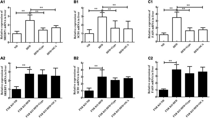
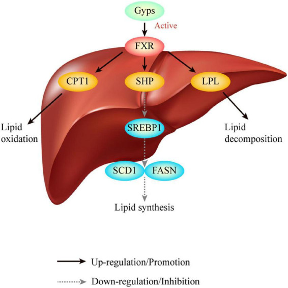
Results: Treatment with Gyps and OCA significantly improved liver histopathological abnormalities in HFD-induced NASH, reduced the non-alcoholic fatty liver disease (NAFLD) activity score (NAS), and lowered hepatic triglyceride (TG) content compared with the HFD group. In agreement with these liver tissue changes, biochemical tests of blood samples revealed that alanine aminotransferase (ALT), aspartate aminotransferase (AST), TG, total cholesterol (TC), low-density lipoprotein cholesterol (LDL-C), fasting blood glucose (FBG), and fasting insulin (FINS) levels were significantly lower in the HFD + Gyps vs. HFD group. Furthermore, Gyps and OCA treatment significantly up-regulated hepatic FXR, small heterodimer partner (SHP), carnitine palmitoyltransferase 1A (CPT1A), and lipoprotein lipase (LPL) expression, and significantly down-regulated sterol-regulatory element binding protein 1 (SREBP1), fatty acid synthetase (FASN), and stearoyl-CoA desaturase 1 (SCD1) protein levels compared with the HFD group in WT mice but not in FXR KO mice. Notably, Gyps- and OCA-mediated pharmacological effects were significantly abrogated by depletion of the FXR gene in FXR KO mice.

Conclusion: Gyps ameliorated HFD-induced NASH through the direct activation of FXR and FXR-dependent signaling pathways.

Keywords: farnesoid X receptor; gypenosides; high-fat diet; mice; non-alcoholic steatohepatitis.


Conflict of interest statement

The authors declare that the research was conducted in the absence of any commercial or financial relationships that could be construed as a potential conflict of interest.

Figures

FIGURE 1
FIGURE 1
Total-ion chromatogram and negative-ion chromatogram for gypenosides (Gyps). (A) Total-ion chromatogram for Gyps. (B) Negative-ion chromatogram for Gyps. a: gypenoside A; b: gypenoside XLIX. The total-ion chromatogram and negative-ion chromatogram for the corresponding standards were provided as we previously reported (12).
FIGURE 2
FIGURE 2
Effects of Gyps on food intake and body weight of WT and FXR KO mice in different groups. (A1,A2) Changes in food intake in WT and FXR KO mice in different groups. (B1,B2) Changes in body weight in WT and FXR KO mice at different time points (11, 12, 13, and 14 weeks) after treatment in WT and FXR KO mice; #p < 0.01, vs. ND group; &p < 0.01, vs. HFD group. (C1,C2) Changes in body weight in WT and FXR KO mice at the end of experiments in WT and FXR KO mice. WT, wide-type; KO, knockout; ND, normal diet; HFD, high-fat diet; Gyps, gypenosides. **p < 0.01.
FIGURE 3
FIGURE 3
Effects of Gyps on histopathological alterations of liver sections and hepatic TG levels in WT and FXR KO mice. (A) Representative images of liver histopathological alterations in different groups. H&E staining of liver sections of WT mice (upper panels) and FXR KO mice (lower panels) in the ND, HFD, and HFD + Gyps groups. The characteristic histological findings of hepatic steatosis were observed in the HFD group, including many fat droplets in the cytoplasm, scattered inflammatory cell infiltration, and balloon-like degeneration. (B) Comparison of activity grade between groups in WT and FXR KO mice according to NAS score of liver pathology. (C) Levels of hepatic TG in liver tissue of mice in each group. Comparison of hepatic TG levels between groups in WT and FXR KO mice. WT, wild-type C57BL/6 mice (n = 9); FXR–/–, FXR knockout (KO) C57BL/6 mice (n = 6); FXR, farnesoid X receptor; TG, triglyceride; ND, normal diet; HFD, high-fat diet; Gyps, gypenosides; NAS, non-alcoholic fatty liver disease (NAFLD) activity score **p < 0.01.
FIGURE 4
FIGURE 4
Effects of Gyps on biochemical parameters in WT and FXR KO mice. Alterations in serum levels of (A1,A2) liver transaminases (ALT, AST), (B1–B4) lipid profiles (TG, TC, LDL-C, HDL-C), and (C1a–C3b) blood sugar levels (FINS, FBG, HOMA-IR) in the ND, HFD, or HFD + Gyps groups in WT and FXR KO mice. WT, wild-type C57BL/6 mice (n = 9); KO, FXR knockout C57BL/6 mice (n = 6); TG, triglyceride; FXR, farnesoid X receptor; ALT, alanine aminotransferase; AST, aspartate aminotransferase; TC, total cholesterol; LDL-C, low-density lipoprotein cholesterol; HDL-C, high-density lipoprotein cholesterol; ND, normal diet; HFD, high-fat diet; Gyps, gypenosides; FINS, fasting insulin; FBG, fasting blood glucose; HOMA-IR, homeostasis model assessment insulin resistance *p < 0.05, **p < 0.01.
FIGURE 5
FIGURE 5
Effects of Gyps on hepatic FXR mRNA and protein expression levels in WT and FXR KO mice. (A) Quantification of hepatic FXR protein levels in different groups in WT mice. (B) WB analysis of hepatic FXR and β-actin protein expression in WT mice. (C) WB analysis of hepatic FXR and GAPDH protein expression in FXR KO mice. (D) Relative expression levels of FXR mRNA in different groups in WT mice. (E) WB analysis of hepatic FXR protein expression in the ND and FXR KO ND groups. (F) Quantification of hepatic FXR protein levels in the ND and FXR KO ND groups. FXR, farnesoid X receptor; GAPDH, glyceraldehyde 3-phosphate dehydrogenase; ND, normal diet; HFD, high-fat diet; Gyps, gypenosides; WT, wild-type; KO, knockout; WB, western blot **p < 0.01.
FIGURE 6
FIGURE 6
Effects of Gyps on hepatic SHP mRNA and protein levels in WT and FXR KO mice. (A1) Quantification of hepatic SHP protein levels in different groups of WT mice. (A2) Quantification of hepatic SHP protein levels in different groups in FXR KO mice. (B1) Relative expression levels of SHP mRNA in different groups in WT and FXR KO mice. (B2) Quantification of hepatic SHP protein levels in the ND and FXR KO ND groups. (C1) WB analysis of hepatic SHP protein expression in different groups of WT mice. (C2) WB analysis of hepatic SHP protein expression in different groups of FXR KO mice. (C3) WB analysis of hepatic SHP protein expression in ND and FXR KO ND groups. FXR, farnesoid X receptor; SHP, small heterodimer partner; ND, normal diet; HFD, high-fat diet; Gyps, gypenosides; WT, wild-type; KO, knockout; WB, western blot. *p < 0.05, **p < 0.01.
FIGURE 7
FIGURE 7
Effects of Gyps on hepatic SREBP1, SCD1, and FASN protein levels in WT and FXR KO mice. (A1,A2) Quantification of hepatic SREBP1 protein levels in the different groups of WT and FXR KO mice. (B1,B2) Quantification of hepatic SCD1 protein levels in different groups of WT and FXR KO mice. (C1,C2) Quantification of hepatic FASN protein levels in different groups of WT and FXR KO mice. (D1,D2) WB analysis of hepatic SREBP1, SCD1, and FASN protein expression in different groups of WT and FXR KO mice. SREBP1, sterol-regulatory element binding protein 1; SCD1, stearoyl-CoA desaturase 1; FASN, fatty acid synthetase; ND, normal diet; HFD, high-fat diet; Gyps, gypenosides; WT, wild-type; KO, knockout; WB, western blot **p < 0.01.
FIGURE 8
FIGURE 8
Effects of Gyps on hepatic SREBP1, SCD1, and FASN mRNA levels in WT and FXR KO mice. (A1,A2) Relative expression levels of SREBP1 mRNA in the different groups in WT and FXR KO mice. (B1,B2) Relative expression levels of SCD1 mRNA in different groups in WT and FXR KO mice. (C1,C2) Relative expression levels of FASN mRNA in different groups in WT and FXR KO mice. SREBP1, sterol-regulatory element binding protein 1; SCD1, stearoyl-CoA desaturase 1; FASN, fatty acid synthetase; ND, normal diet; HFD, high-fat diet; Gyps, gypenosides; WT, wild-type; KO, knockout; WB, western blot. **p < 0.01.
FIGURE 9
FIGURE 9
Effects of Gyps on hepatic CPT1A and LPL mRNA and protein expression in WT and FXR KO mice. (A1,A2) Quantification of hepatic CPT1A protein levels in different groups of WT and FXR KO mice. (a1,a2) Relative expression levels of CPT1 mRNA in different groups in WT and FXR KO mice. (B1,B2) Quantification of hepatic LPL protein levels in different groups of WT and FXR KO mice. (b1,b2) Relative expression levels of LPL mRNA in different groups in WT and FXR KO mice. (C1,C2) WB analysis of hepatic CPT1A and LPL protein expression in different groups of WT and FXR KO mice. CPT1A, carnitine palmitoyltransferase 1A; LPL, lipoprotein lipase; ND, normal diet; HFD, high-fat diet; Gyps, gypenosides; WT, wild-type; KO, knockout; WB, western blot. *p < 0.05, **p < 0.01.
FIGURE 10
FIGURE 10
Graphical abstract. Gyps treatment significantly increased hepatic expression of FXR and its target SHP, and led to the up-regulation of CPT1 and LPL, and down-regulation of SREBP1, FASN and SCD1 protein levels in WT mice but not FXR KO mice. Ultimately, Gyps improves lipid metabolism in a mouse model of NASH through the activation of FXR. Gyps, gypenosides; FXR, farnesoid X receptor; SHP, small heterodimer partner; SREBP1, sterol-regulatory element binding protein 1; SCD1, stearoyl-CoA desaturase 1; FASN, fatty acid synthetase; CPT1A, carnitine palmitoyltransferase 1A; LPL, lipoprotein lipase.

References

    1. Pierantonelli I, Svegliati-Baroni G. Nonalcoholic fatty liver disease: basic pathogenetic mechanisms in the progression from NAFLD to NASH. Transplantation. (2019) 103:e1–13. 10.1097/tp.0000000000002480 - DOI - PubMed
    1. Santhekadur PK, Kumar DP, Sanyal AJ. Preclinical models of non-alcoholic fatty liver disease. J Hepatol. (2018) 68:230–7. 10.1016/j.jhep.2017.10.031 - DOI - PMC - PubMed
    1. Younossi Z, Tacke F, Arrese M, Chander Sharma B, Mostafa I, Bugianesi E, et al. Global perspectives on nonalcoholic fatty liver disease and nonalcoholic steatohepatitis. Hepatology. (2019) 69:2672–82. 10.1002/hep.30251 - DOI - PubMed
    1. Hung CK, Bodenheimer HC, Jr. Current treatment of nonalcoholic fatty liver disease/nonalcoholic steatohepatitis. Clin Liver Dis. (2018) 22:175–87. 10.1016/j.cld.2017.08.012 - DOI - PubMed
    1. Li K, Ma C, Li H, Dev S, He J, Qu X. Medicinal value and potential therapeutic mechanisms of Gynostemma pentaphyllum (Thunb.) makino and its derivatives: an overview. Curr Top Med Chem. (2019) 19:2855–67. 10.2174/1568026619666191114104718 - DOI - PubMed