

G lugea sp. infecting Sardinella aurita in Algeria
- PMID: 36091289
- PMCID: PMC9458822
- DOI: 10.1007/s12639-022-01483-5
G lugea sp. infecting Sardinella aurita in Algeria
Abstract
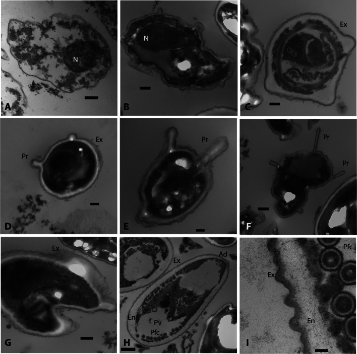
Parasitological examination of the commercially important pelagic fish Sardinella aurita Valenciennes, 1847 (Clupeidae) from the Eastern coast of Algeria revealed xenomas in the peritoneal cavity, suggesting a microsporidian infection. The prevalence of the disease was approximately 30% on average, higher in smaller individuals and showing significant seasonal variation. The xenomas contained numerous ellipsoidal spores, surrounded by a dense layer of connective tissue. Spore sizes were 6.10 ± 0.38 µm length and 3.54 ± 0.43 µm width. Ultrastructural examination by transmission electron microscopy showed various development stages of the parasite, including meronts, sporonts, sporoblasts and mature spores. The internal organization of the mature spores, with a single nucleus, prominent posterior vacuole, a lamellar polaroplast and an isofilar polar tube arranged in a single row, was typical of the genus Glugea. The DNA sequence of the small subunit ribosomal RNA gene confirmed that this parasite belongs to the genus Glugea. Genetic and morphologic comparison with G. sardinellensis, a species previously described in the same host from Tunisia shows many similarities, although some molecular and morphometric inconsistencies precluded the unambiguous assignment of our samples to G. sardinellensis. At the same time, we do not find sufficient grounds to erect a new taxon for our parasite. We discuss the implications of our findings for the current state of the systematics of Glugea.
Keywords: Algeria; Glugea; Histology; Microsporidia; SSU rDNA; Sardinella aurita; Ultrastructure.
© Indian Society for Parasitology 2022.
Conflict of interest statement
Conflict of interestAll the authors declare that they have no conflict of interest.
Figures


Similar articles
-
Ultrastructural and molecular characterization of Glugea serranus n. sp., a microsporidian infecting the blacktail comber, Serranus atricauda (Teleostei: Serranidae), in the Madeira Archipelago (Portugal).Parasitol Res. 2016 Oct;115(10):3963-72. doi: 10.1007/s00436-016-5162-7. Epub 2016 Jun 10. Parasitol Res. 2016. PMID: 27287485
-
Ultrastructural description and phylogeny of a novel microsporidian, Glugea eda n. sp. from the striated fusilier, Caesio striata, in the Red Sea off Saudi Arabia.Acta Trop. 2020 Apr;204:105331. doi: 10.1016/j.actatropica.2020.105331. Epub 2020 Jan 8. Acta Trop. 2020. PMID: 31923380
-
Mass mortality event of round sardinella Sardinella aurita Valenciennes associated with Glugea Thélohan, 1891 microsporidian infection off the southern Italian coast.J Fish Dis. 2024 Oct;47(10):e13995. doi: 10.1111/jfd.13995. Epub 2024 Jul 2. J Fish Dis. 2024. PMID: 38953156
-
Pseudokabatana alburnus n. gen. n. sp., (Microsporidia) from the liver of topmouth culter Culter alburnus (Actinopterygii, Cyprinidae) from China.Parasitol Res. 2019 Jun;118(6):1689-1699. doi: 10.1007/s00436-019-06303-z. Epub 2019 Apr 11. Parasitol Res. 2019. PMID: 30976967 Review.
-
Fish microsporidia: fine structural diversity and phylogeny.Int J Parasitol. 2003 Feb;33(2):107-27. doi: 10.1016/s0020-7519(02)00252-7. Int J Parasitol. 2003. PMID: 12633649 Review.
References
-
- Abdel-Baki AS, Dkhil MA, Al-Quraishy S. Seasonality and prevalence of microsporidium sp. infecting lizard fish, Saurida undosquamis from the Arab Gulf. J King Saud Univ (science) 2009;21:195–198. doi: 10.1016/j.jksus.2009.10.007. - DOI
-
- Abdel-Baki AS, Al-Quraishy S, Al-Qahtani H, Dkhil MA, Azevedo C. Morphological and ultrastructural description of Pleistophora dammami sp. n. infecting the intestinal wall of Saurida undosquamis from the Arabian Gulf, Saudi Arabia. Parasitol Res. 2012;111:413–418. doi: 10.1007/s00436-012-2855-4. - DOI - PubMed
-
- Abdel-Baki AS, Al-Quraishy S, Rocha S, Dkhil MA, Casal G, Azevedo C (2015b) Ultrastructure and phylogeny of Glugea nagelia sp. n. (Microsporidia: Glugeidae), infecting the intestinal wall of the yellowfin hind, Cephalopholis hemistiktos (Actinopterygii: Serranidae), from the Red Sea. Folia Parasitol 62:007 - PubMed
LinkOut - more resources
Full Text Sources