

doi: 10.1002/jgh3.12802.
eCollection 2022 Sep.
Case of pneumatosis cystoides intestinalis with intra-abdominal free air developed during treatment with voglibose
Affiliations
- PMID: 36091325
- PMCID: PMC9446400
- DOI: 10.1002/jgh3.12802
Item in Clipboard
Case of pneumatosis cystoides intestinalis with intra-abdominal free air developed during treatment with voglibose
JGH Open.
.
Abstract
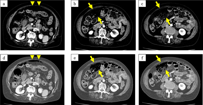
Contrast-enhanced computed tomography (CT) scan (portal phase) at the onset. Emphysema is detected in the ileal wall (b, c: Arrows) and free air is detected in the abdominal cavity (a: Arrowhead). CT scan imaging settings: (a-c) window level (WL) 60 and window width (WW) 300. (d-f) WL 0 and WW 433. By changing the imaging settings, intestinal emphysema and free air can be more easily identified.
Keywords: glucosidases; humans; pneumatosis cystoides intestinalis.
© 2022 The Authors. JGH Open published by Journal of Gastroenterology and Hepatology Foundation and John Wiley & Sons Australia, Ltd.
Figures
Contrast‐enhanced computed tomography (CT) scan (portal phase) at the onset. Emphysema is detected in the ileal wall (b, c: Arrows) and free air is detected in the abdominal cavity (a: Arrowhead). CT scan imaging settings: (a‐c) window level (WL) 60 and window width (WW) 300. (d‐f) WL 0 and WW 433. By changing the imaging settings, intestinal emphysema and free air can be more easily identified.

Computed tomography (CT) scan on the third day of onset: (a, c): Both intestinal emphysema (arrows) and free air (arrowheads) remain, but there is no increase in free air. CT scan 2 weeks after onset: (b, d): Intestinal emphysema and free air have resolved. Imaging settings: (a, b) Window level (WL) 30 and window width (WW) 300. (c, d) WL 0 and WW 433.
Similar articles
-
Pneumatosis cystoides intestinalis: case report and review of literature.Clin J Gastroenterol. 2020 Feb;13(1):31-36. doi: 10.1007/s12328-019-00999-3. Epub 2019 Jun 3. Clin J Gastroenterol. 2020. PMID: 31161540 Review.
-
Pneumatosis cystoides intestinalis: lung window setting on CT.Clin Case Rep. 2017 Sep 12;5(11):1896-1897. doi: 10.1002/ccr3.1151. eCollection 2017 Nov. Clin Case Rep. 2017. PMID: 29152295 Free PMC article.
-
Pneumatosis cystoides intestinalis presenting as pneumoperitoneum in a patient with chronic obstructive pulmonary disease: a case report.J Med Case Rep. 2017 Feb 28;11(1):55. doi: 10.1186/s13256-017-1198-2. J Med Case Rep. 2017. PMID: 28241852 Free PMC article.
-
Successful treatment with hyperbaric oxygen therapy for pneumatosis cystoides intestinalis as a complication of granulomatosis with polyangiitis: a case report.J Med Case Rep. 2017 Sep 17;11(1):263. doi: 10.1186/s13256-017-1421-1. J Med Case Rep. 2017. PMID: 28917259 Free PMC article.
-
Pneumatosis cystoides intestinalis associated with etoposide in hematological malignancies: a case report and a literature review.BMC Gastroenterol. 2022 Mar 28;22(1):150. doi: 10.1186/s12876-022-02219-8. BMC Gastroenterol. 2022. PMID: 35346061 Free PMC article. Review.
Cited by
-
Pneumatosis Intestinalis With Abdominal Wall Emphysema in Hypothermia.Cureus. 2023 Feb 13;15(2):e34909. doi: 10.7759/cureus.34909. eCollection 2023 Feb. Cureus. 2023. PMID: 36938220 Free PMC article.
References
-
- Galandiuk S, Fazio VW. Pneumatosis cystoides intestinalis. A review of the literature. Dis. Colon Rectum. 1986; 29: 358–63. - PubMed
-
- Yale CE, Balish E, Wu JP. The bacterial etiology of pneumatosis cystoides intestinalis. Arch. Surg. 1974; 109: 89–94. - PubMed
-
- Yamaguchi K, Shirai T, Shimakura K et al. Pneumatosis cystoides intestinalis and trichloroethylene exposure. Am. J. Gastroenterol. 1985; 80: 753–7. - PubMed
-
- Keyting WS, McCarver RR, Kovarik JL, Daywitt AL. Pneumatosis intestinalis: a new concept. Radiology. 1961; 76: 733–41. - PubMed
LinkOut - more resources
Full Text Sources
Miscellaneous
