. 2022 Aug 25:10:953034.
doi: 10.3389/fbioe.2022.953034. eCollection 2022.

Effects of HSD11B1 knockout and overexpression on local cortisol production and differentiation of mesenchymal stem cells

Affiliations

Effects of HSD11B1 knockout and overexpression on local cortisol production and differentiation of mesenchymal stem cells

Angelique Kragl et al. Front Bioeng Biotechnol. .

Abstract

Exogenous glucocorticoids increase the risk for osteoporosis, but the role of endogenous glucocorticoids remains elusive. Here, we describe the generation and validation of a loss- and a gain-of-function model of the cortisol producing enzyme 11β-HSD1 (HSD11B1) to modulate the endogenous glucocorticoid conversion in SCP-1 cells - a model for human mesenchymal stem cells capable of adipogenic and osteogenic differentiation. CRISPR-Cas9 was successfully used to generate a cell line carrying a single base duplication and a 5 bp deletion in exon 5, leading to missense amino acid sequences after codon 146. These inactivating genomic alterations were validated by deep sequencing and by cloning with subsequent capillary sequencing. 11β-HSD1 protein levels were reduced by 70% in the knockout cells and cortisol production was not detectable. Targeted chromosomal integration was used to stably overexpress HSD11B1. Compared to wildtype cells, HSD11B1 overexpression resulted in a 7.9-fold increase in HSD11B1 mRNA expression, a 5-fold increase in 11β-HSD1 protein expression and 3.3-fold increase in extracellular cortisol levels under adipogenic differentiation. The generated cells were used to address the effects of 11β-HSD1 expression on adipogenic and osteogenic differentiation. Compared to the wildtype, HSD11B1 overexpression led to a 3.7-fold increase in mRNA expression of lipoprotein lipase (LPL) and 2.5-fold increase in lipid production under adipogenic differentiation. Under osteogenic differentiation, HSD11B1 knockout led to enhanced alkaline phosphatase (ALP) activity and mRNA expression, and HSD11B1 overexpression resulted in a 4.6-fold and 11.7-fold increase in mRNA expression of Dickkopf-related protein 1 (DKK1) and LPL, respectively. Here we describe a HSD11B1 loss- and gain-of-function model in SCP-1 cells at genetic, molecular and functional levels. We used these models to study the effects of endogenous cortisol production on mesenchymal stem cell differentiation and demonstrate an 11β-HSD1 dependent switch from osteogenic to adipogenic differentiation. These results might help to better understand the role of endogenous cortisol production in osteoporosis on a molecular and cellular level.

Keywords: 11β-HSD1; CRISPR-Cas9; glucocorticoids; osteoporosis; targeted chromosomal integration.


Conflict of interest statement

The authors declare that the research was conducted in the absence of any commercial or financial relationships that could be construed as a potential conflict of interest.

Figures
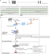
FIGURE 1
FIGURE 1
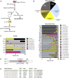
Generation of HSD11B1 knockout SCP-1 cells using CRISPR-Cas9 mediated gene editing. (A) Schematic representation of the localization of the targeted regions within HSD11B1. Two crRNAs were applied, one targeting exon 2, the other targeting exon 5. The binding sites of the crRNAs is underlined, the PAM sites are shown in bold print. (B) Workflow and components of CRISPR-Cas9 mediated gene editing applying the ribonucleoprotein approach of IDT. A crRNA is designed so as to contain a sequence complementary to the target site. It is mixed with a tracrRNA to form the gRNA. This gRNA forms a ribonucleoprotein with the Cas9 endonuclease. After transfection of target cells, Cas9 will be directed to the target site and will create a double strand break (DSB) upstream of the PAM site.
FIGURE 2
FIGURE 2
Genetic validation of HSD11B1 knockout SCP-1 cells generated using the crRNA targeting exon 5. (A) Sequencing strategy to identify sequence modifications in the CRISPR-Cas9 treated cells. (B) Summarized allele frequencies of sequence modifications detected by NGS. Others represents additional modifications that had a frequency of less than 2% (top). Read frequencies of sequence modifications across all clones determined by NGS. Others represents the clusters that were not analyzed in detail due to a read number <50 (bottom). Asterisks indicate clones with potential origin from more than one cell. (C) Read frequencies of sequence modifications determined by cloning and subsequent capillary sequencing. (D) Schematic representation of sequence modifications and consequences in clone 1C4. Positions given are based on the reference sequence NG_012081.1. (E) Potential off-target sites of the applied HSD11B1 exon 5 crRNA predicted by IDT and CCTop. Mismatches in the target sequence are marked red, hyphens indicate gaps. Sequences of the respective sites were controlled by capillary sequencing.
FIGURE 3
FIGURE 3
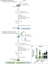
Generation and genetic validation of stably HSD11B1 overexpressing SCP-1 cells using the Flp-In™ system. (A) The host cell line is created by transfection with the pFRT/lacZeo plasmid. The FRT site is randomly integrated. The expression cell line is generated by transfection with the pcDNA5/FRT expression vector carrying HSD11B1 and the pOG44 plasmid expressing Flp recombinase. (B) HSD11B1 mRNA expression in HSD11B1 overexpressing HEK293 and SCP-1 cells compared to expression in human adipose tissue and liver. As controls untransfected SCP-1 cells and HEK293 cells transfected with the empty pcDNA5 vector were used. Shown are the HEK293-HSD clone I/6 and the SCP-1 clone I/1. Shown are means ± SEM of 2–5 biological replicates.
FIGURE 4
FIGURE 4
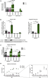
Functional validation of the HSD11B1 knockout and overexpression in SCP-1 cells after adipogenic differentiation. Functional validation has been performed by analyzing (A) mRNA expression, (B) protein expression, 11β-HSD1 activity measuring (C) extracellular cortisol levels and (D) extracellular cortisol levels over time. (A) HSD11B1 mRNA expression was determined by RT-qPCR. Shown are means ± SEM of 2–3 biological replicates. (B) Protein expression after 14 days of adipogenic differentiation as determined by Western Blot analysis (left) and Targeted Proteomics (right). A representative Western Blot is given. In both approaches, 11β-HSD1 protein levels were normalized to Na+/K+-ATPase. Cells were differentiated for 14 days. Shown are means ± SEM of 3 Western Blot quantifications from two independent differentiation experiments and 3 Targeted Proteomics measurements from a single differentiation experiment. (C) 11β-HSD1 activity was determined by cortisol measurements after a 48-h stimulation with cortisone. Shown are means ± SEM of 3–4 biological replicates. (D) 11β-HSD1 activity was determined by cortisol measurements in samples taken with every medium replacement. Absolute cortisol levels in days are depicted. Shown are means ± SEM of 3–4 biological replicates. Cells were differentiated with IBMX, indomethacin, insulin, and dexamethasone (A–C) or cortisone (D). *p ≤ 0.05, **p ≤ 0.01, ***p ≤ 0.001; $ p ≤ 0.05, §p ≤ 0.01, #p ≤ 0.001 compared to respective undifferentiated control, determined by one-way ANOVA with post-hoc Tukey’s Test.
FIGURE 5
FIGURE 5
Analyses of the effects of HSD11B1 knockout and overexpression on adipogenic differentiation. (A) Lipid droplets were visualized by Nile Red staining, nuclei were stained with DAPI. Representative images for each cell line are given. Scale bar: 100 µm. Zoom into overexpressing cells differentiated with dexamethasone (zoom factor: 5.1x, scale bar: 20 µm). (B) Nile Red was quantified directly after staining by fluorescence measurement and normalized on DAPI fluorescence. Shown are means ± SEM of 6 wells of undifferentiated and 9 wells of differentiated cells from 3 independent differentiation experiments. (C) mRNA expression of the adipogenic marker LPL. Shown are means ± SEM of 5 (undifferentiated) and 4 (differentiated) independent differentiation experiments. Cells were differentiated with IBMX, indomethacin, insulin, and cortisone or dexamethasone. *p ≤ 0.05, **p ≤ 0.01; §p ≤ 0.01, #p ≤ 0.001 compared to the respective undifferentiated cells, determined by one-way ANOVA with post-hoc Tukey’s Test.
FIGURE 6
FIGURE 6
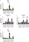
Analyses of the effects of HSD11B1 knockout and overexpression on osteogenic differentiation. mRNA expression of (A) the Wnt signaling marker DKK1, (B) the osteogenic marker ALPL, and (D) the adipogenic marker LPL. Shown are means ± SEM of 2 independent differentiation experiments (DKK1, ALPL, LPL) and 2 to 5 independent biological replicates of the undifferentiated cells (DKK1 3 replicates, ALPL 2 replicates, LPL 5 replicates). (C) ALP activity. Shown are means ± SEM of 6 wells of undifferentiated and 9 wells of differentiated cells from two independent differentiation experiments. Cells were differentiated with ascorbate, β-glycerol phosphate, 1α,25-dihydroxyvitamin D3 with or without cortisone. Mean. *p ≤ 0.05, **p ≤ 0.01, ***p ≤ 0.001; $ p ≤ 0.05, §p ≤ 0.01, #p ≤ 0.001 compared to the respective undifferentiated cells, determined by one-way ANOVA with post-hoc Tukey’s Test.

Similar articles

Cited by

References

    1. Abrahamsen B., Osmond C., Cooper C. (2015). Life expectancy in patients treated for osteoporosis: Observational cohort study using national Danish prescription data. J. Bone Min. Res. 30 (9), 1553–1559. 10.1002/jbmr.2478 - DOI - PubMed
    1. Agarwal A. K., Mune T., Monder C., White P. C. (1994). NAD(+)-dependent isoform of 11 beta-hydroxysteroid dehydrogenase. Cloning and characterization of cDNA from sheep kidney. J. Biol. Chem. 269 (42), 25959–25962. 10.1016/S0021-9258(18)47142-6 - DOI - PubMed
    1. Albiston A. L., Obeyesekere V. R., Smith R. E., Krozowski Z. S. (1994). Cloning and tissue distribution of the human 1 lβ-hydroxysteroid dehydrogenase type 2 enzyme. Mol. Cell. Endocrinol. 105 (2), R11–R17. 10.1016/0303-7207(94)90176-7 - DOI - PubMed
    1. Andrzejewska A., Catar R., Schoon J., Qazi T. H., Sass F. A., Jacobi D., et al. (2019). Multi-parameter analysis of biobanked human bone marrow stromal cells shows little influence for donor age and mild comorbidities on phenotypic and functional properties. Front. Immunol. 10, 2474. 10.3389/fimmu.2019.02474 - DOI - PMC - PubMed
    1. Best R., Nelson S. M., Walker B. R. (1997). Dexamethasone and 11-dehydrodexamethasone as tools to investigate the isozymes of 11β-hydroxysteroid dehydrogenase in vitro and in vivo . J. Endocrinol. 153 (1), 41–48. 10.1677/joe.0.1530041 - DOI - PubMed