

Paraparesis in adult aortic coarctation: Reversal by stent supported angioplasty
- PMID: 36091605
- PMCID: PMC9449749
- DOI: 10.1016/j.jccase.2022.04.009
Paraparesis in adult aortic coarctation: Reversal by stent supported angioplasty
Abstract
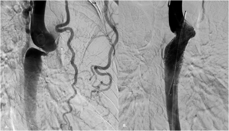
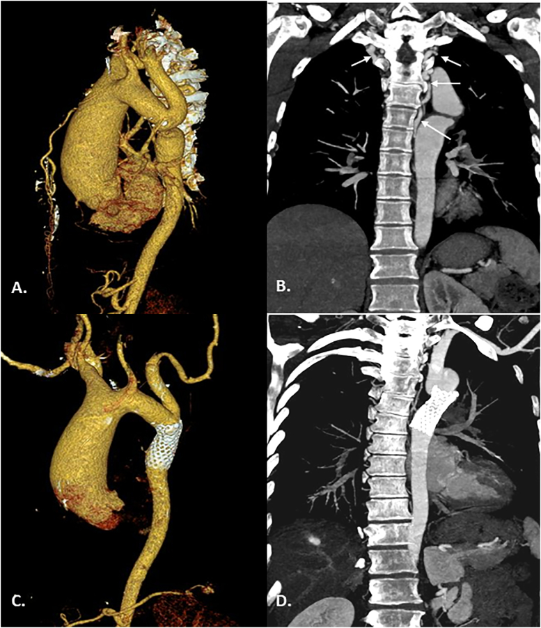
Aortic coarctation presenting with neurological complications as compressive myelopathy is rare. We report a case of a 43-year-old, hypertensive, female who presented with gradually progressive paraparesis over 4 years. She was diagnosed to be having coarctation of the aorta with intra-spinal collaterals causing compressive myelopathy. She underwent successful percutaneous endovascular implantation of a balloon-expandable aortic stent to relieve her aortic coarctation. This led to regression of her intra-spinal collaterals relieving her cord compression. This nonsurgical modality treatment proved to be safe and effective in relieving her hypertension and neurological complication of paraparesis.
Learning objectives: •To recognize that paraparesis can be a rare manifestation of coarctation of the aorta.•To highlight the importance of treating the primary pathology of coarctation of the aorta in such critically ill therapeutically challenging patients.
Keywords: Aortoplasty; Coarctation of aorta; Paraparesis.
© 2022 Japanese College of Cardiology. Published by Elsevier Ltd. All rights reserved. All rights reserved.
Conflict of interest statement
The authors declare that there is nothing to disclose.
Figures

References
-
- Trenk L., Lammers A.E., Radke R., Baumgartner H., Wort S.J., Gatzoulis M.A., et al. Neurological complications in aortic coarctation: results of a nationwide analysis based on 11,907 patients. Int J Cardiol. 2021;322:114–120. - PubMed
-
- Tan K.P., Ng F.C., Ong P.L. Paraparesis due to dilated spinal collaterals. Singapore Med J. 1979;20:454–456. - PubMed
-
- Harrer J., Dominik J., Varvarovský I., Zizka J., Harrerová L. Paraplegia as an unusual manifestation of aortic coarctation. Thorac Cardiovasc Surg. 2001;49:186–187. - PubMed
-
- Moorthy N., Ananthakrishna R., Nanjappa M.C. Percutaneous stenting of interrupted aortic arch to treat compressive myelopathy. Catheter Cardiovasc Interv. 2014;84:815–819. - PubMed
Publication types
LinkOut - more resources
Full Text Sources