Review
. 2022 Aug 15:87:e448-e461.
doi: 10.5114/pjr.2022.119032. eCollection 2022.

Magnetic resonance imaging of endometriosis: a common but often hidden, missed, and misdiagnosed entity

Affiliations
Review

Magnetic resonance imaging of endometriosis: a common but often hidden, missed, and misdiagnosed entity

Mohammad Zuber et al. Pol J Radiol. .

Abstract

Endometriosis is a common benign and chronic inflammatory gynaecological disease due to functional endometrial glands and stroma in an ectopic location outside the uterine cavity. It affects 5-10% of reproductive age group women in the peak age of 24-29 years. However, women with infertility and chronic pelvic pain have an even greater prevalence, accounting for 30-50% and 90% of cases, respectively. Although it is a common entity, patients often get a delayed diagnosis because it is often subtle (hidden), missed, or confused with mimics, leading to misdiagnosis, which significantly affects patients' quality of life because they live in constant pain from undiagnosed endometriosis. Laparoscopy followed by histopathological confirmation is the gold standard for diagnosis, but it is an invasive procedure. MRI is an excellent non-invasive modality that helps in non-invasive diagnosis, with excellent delineation of the disease extent, and thus provides a presurgical mapping of the disease, which is helpful for the operating surgeon. Radiologists should be aware of all possible spectrum and diagnose this early and provide a detailed structured report mapping the entire extent of the disease process, which helps in effective treatment planning and successful outcomes in improving patients' quality of life.

Keywords: deep infiltrative endometriosis; endometrioma; endometriosis; malignant transformation; scar endometriosis.


Conflict of interest statement

The authors report no conflict of interest.

Figures

Figure 1
Figure 1
Ultrasonographic appearance of endometrioma. Transvaginal scan shows a large well-defined unilocular cystic lesion in left adnexa adjacent to the uterus (Ut) with homogenous low-level internal echoes within giving “ground glass appearance”. The wall of the lesion is smooth with no evidence of septations or solid components within
Figure 2
Figure 2
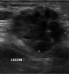
Sonographic appearance of scar endometriosis. Ultrasound image shows an ill-defined heterogeneously hypoechoic lesion with irregular margins in the infraumbilical region in the anterior midline
Figure 3
Figure 3
Diaphragmatic endometriosis. A) Contrast-enhanced computed tomography (CECT) axial image in venous phase demonstrate small cystic lesion with enhancing wall along right hemidiaphragm (white arrow). B) CECT coronal image of the same patient depicts a large well defined abdominopelvic cystic lesion (white arrow), which was operated and confirmed to be endometrioma on histopathological examination
Figure 4
Figure 4
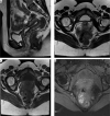
Recommended magnetic resonance imaging (MRI) sequences for endometriosis. According to the European Society of Urogenital Radiology (ESUR) guidelines, MRI protocol should include at least two 2D T2-weighted sequences at least sagittal (A) and axial (B) and a T1-weighted sequence without (C) and with fat suppression (D)
Figure 5
Figure 5
Anatomy of the female pelvis on sagittal section. A) Sketch diagram of the sagittal section of female magnetic resonance pelvis showing normal anatomy. Figure B is the corresponding sagittal T2W image demonstrating the anatomic structures that are commonly affected in endometriosis namely prevesical space (orange), peritoneal refection (green), vesicovaginal septum (light blue), vesicouterine space (pink), rectouterine space (dark blue), rectovaginal septum (violet), and presacral space (red). Figure C depicts the 3 anatomic compartments, namely, anterior (bounded by yellow line), middle (bounded by pink line), and posterior (bounded by orange line), used in reporting deep infiltrative endometriosis
Figure 6
Figure 6
Anatomy of female pelvis on axial sections: Sketch diagrams (A, B, C) and corresponding T2W axial images (D, E, F) demonstrate axial anatomy of the pelvis at the inferior level of cervix (A, D), uterocervical junction in the middle (level of torus uterinus and uterosacral ligaments in B and E) and superiorly at the level of fallopian tubes and ovaries (C, F), respectively. Round ligaments are also seen at this superior level. UB – urinary bladder, Cx – cervix, R – rectum, USL – uterosacral ligament, Ut – uterus, FT – fallopian tube, RL – round ligament, and ovaries (white circles)
Figure 7
Figure 7
Endometrioma: T1W axial image (A) of the pelvis showing well-defined, uniformly hyperintense cyst (white arrows) with no suppression of signal on T1 fat suppressed sequence (B), rather becoming more hyperintense giving a “light bulb appearance”. The lesion shows loss of the signal on T2W images (C) suggesting “T2 shading” and peripheral hypointense wall
Figure 8
Figure 8
Deep infiltrative endometriosis in vesicouterine space with contiguous bladder and uterine involvement. Coronal T2W (A) and sagittal T1 with fat suppression images (B) show obliteration of vesicouterine space with ill-defined stellate-shaped hypointense lesion extending anteriorly into the dome and posterior wall of the bladder and posteriorly involving the anterior wall of the uterus, which appears mildly anteverted. There is also presence of hyperintense foci on T1W images suggestive of haemorrhage within the glandular components
Figure 9
Figure 9
Deep infiltrative endometriosis (DIE) in rectouterine pouch. Axial T1W (A), T1W fat suppressed (B), and T2W axial image (C) depict obliteration of rectouterine pouch with ill-defined stellate shaped lesion (white arrows) appearing hypointense on both T1W and T2W images with associated retroversion of the uterus. There is also presence of multiple foci seen within the lesion, appearing hyperintense on T1W and hypointense on T2W images, suggestive of haemorrhagic foci within the ectopic glands
Figure 10
Figure 10
Scar endometrioma in a patient of post laparotomy scar. T1W (A) shows hypointense spiculated ill-defined lesion (white arrows) in the anterior abdominal wall along the subcutaneous plane with hyperintense ectopic endometrial glands on T1 fat suppressed image (B) and post contrast enhancement (C)
Figure 11
Figure 11
Umbilical endometriosis. T2W image (A) depicting umbilical scar endometriosis (white arrows) with T1W FS sequence (B, C) demonstrating ectopic endometrial glands
Figure 12
Figure 12
Pelvic floor endometriosis at episiotomy site: Axial T1W (A) and T2W (B) images show ill-defined stellate hypointense lesion in the pelvic floor at site of episiotomy scar with T1W and T2W hyperintense foci suggesting ectopic endometrial glands within the lesion

References

    1. Chamié LP, Blasbalg R, Pereira RM, et al. . Findings of pelvic endometriosis at transvaginal US, MR imaging, and laparoscopy. Radiographics 2011; 31: 77-100. - PubMed
    1. Woodward PJ, Sohaey R, Mezzetti TP Jr. Endometriosis: radiologic-pathologic correlation. Radiographics 2001; 21: 193-216. - PubMed
    1. Javiera AF, Cristian MS, Daniel GD, et al. . Endometriosis MRI: pictographic review. Rev Chil Radiol 2012; 18: 149-156.
    1. Chatman DL, Ward AB. Endometriosis in adolescents. J Reprod Health Med 1982; 27: 156-160. - PubMed
    1. Sangi-Haghpeykar H, Poindexter AN III. Epidemiology of endometriosis among parous women. Obstet Gynecol 1995; 85: 983-992. - PubMed