. 2022 Aug 25:9:976604.
doi: 10.3389/fmed.2022.976604. eCollection 2022.

COL17A1 editing via homology-directed repair in junctional epidermolysis bullosa

Affiliations

COL17A1 editing via homology-directed repair in junctional epidermolysis bullosa

Igor Petković et al. Front Med (Lausanne). .

Abstract

Background: Epidermolysis bullosa (EB), a severe genetic disorder characterized by blister formation in skin, is caused by mutations in genes encoding dermal-epidermal junction proteins that function to hold the skin layers together. CRISPR/Cas9-induced homology-directed repair (HDR) represents a promising tool for editing causal mutations in COL17A1 in the treatment of junctional epidermolysis bullosa (JEB).

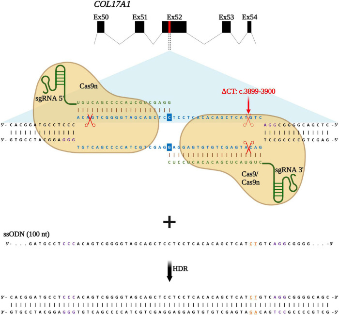
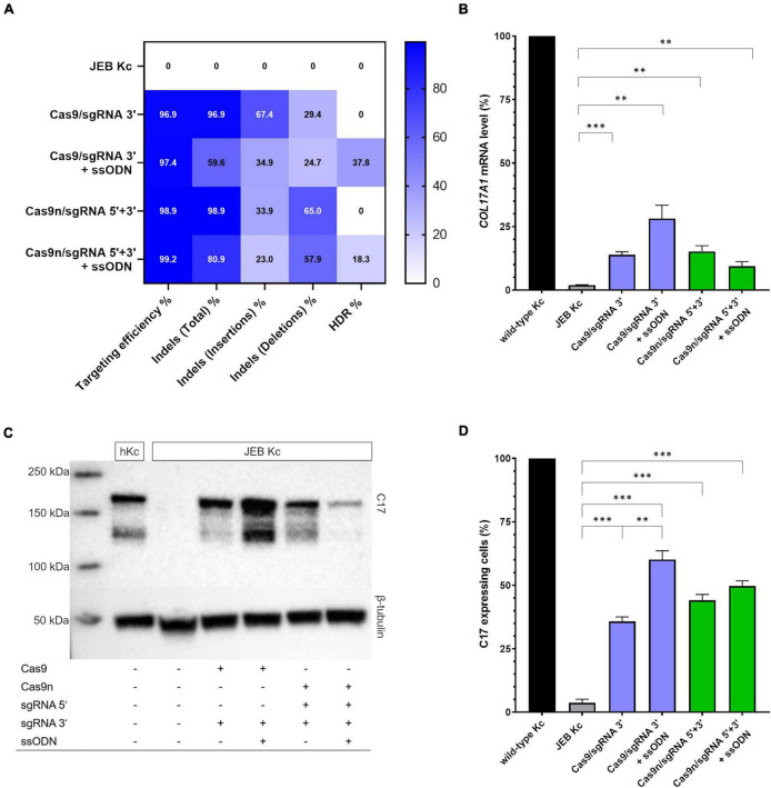
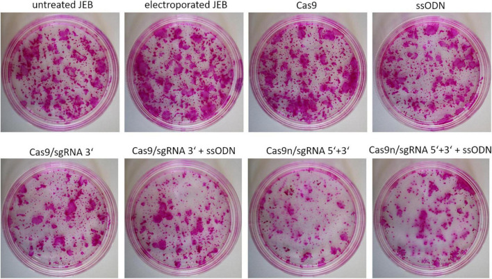
Methods: In this study, we treated primary type XVII collagen (C17)-deficient JEB keratinocytes with either Cas9 nuclease or nickase (Cas9n) ribonucleoproteins (RNP) and a single-stranded oligonucleotide (ssODN) HDR template in order to correct a causal pathogenic frameshift mutation within the COL17A1 gene.

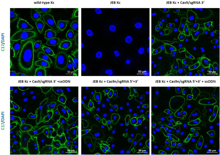
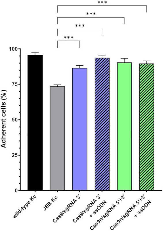
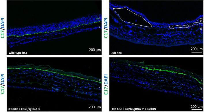
Results: As analyzed by next-generation sequencing of RNP-nucleofected keratinocytes, we observed an HDR efficiency of ∼38% when cells were treated with the high-fidelity Cas9 nuclease, a mutation-specific sgRNA, and an ssODN template. The combined induction of end-joining repair and HDR-mediated pathways resulted in a C17 restoration efficiency of up to 60% as assessed by flow cytometry. Furthermore, corrected JEB keratinocytes showed a significantly increased adhesive strength to laminin-332 and an accurate deposition of C17 along the basement membrane zone (BMZ) upon differentiation into skin equivalents.

Conclusion: Here we present a gene editing approach capable of reducing end joining-generated repair products while increasing the level of seamless HDR-mediated gene repair outcomes, thereby providing a promising CRISPR/Cas9-based gene editing approach for JEB.

Keywords: COL17A1; CRISPR/Cas9; gene editing; gene therapy; homology-directed repair (HDR); junctional epidermolysis bullosa (JEB).


Conflict of interest statement

The authors declare that the research was conducted in the absence of any commercial or financial relationships that could be construed as a potential conflict of interest.

Figures

FIGURE 1
FIGURE 1
HDR-based strategy for COL17A1 repair. sgRNA 3′ is specific for the c.3899_3900delCT mutation within exon 52 while sgRNA 5′ guides Cas9n (for paired nicking) to cut proximal to the mutation. sgRNAs (green) guide Cas9 or Cas9n (for paired nicking) to cut upstream of the PAM sequence (violet). Single-stranded oligonucleotides (ssODN) serve as repair templates carrying the wild-type COL17A1 sequence including the nucleotides C and T (orange letters) that are absent in JEB patient cells. Created with BioRender.com.
FIGURE 2
FIGURE 2
COL17A1 editing outcome in RNP and ssODN-treated primary JEB keratinocytes. (A) NGS analysis of the COL17A1 on-target locus presented as a heat map showing indel and HDR pattern of the performed editing strategies. n = 2. Mean value (B) sqRT-PCR analysis of COL17A1 mRNA expression in JEB keratinocytes upon RNP/ssODN treatment. Primary wild-type and untreated JEB cells served as controls. n = 3; Mean ± SEM; Student’s two-tailed t-test. (C) Western blot analysis of cell lysates revealed the presence of restored C17 (180 kDa). β-tubulin (50 kDa) served as loading control. (D) Flow cytometric analysis of C17-stained JEB keratinocytes revealed a high level of C17 restoration. n = 3; Mean ± SEM; Student’s two-tailed t-test. P-values (Significances): > 0.05 (not significant), * ≤ 0.05, ** ≤ 0.01, and * ≤ 0.001.
FIGURE 3
FIGURE 3
Immunofluorescence staining of RNP/ssODN-treated primary JEB keratinocytes. Immunofluorescence staining of C17 (green) revealed the accurate deposition of the restored protein in RNP and ± ssODN-treated primary JEB keratinocytes. Nuclei were stained with DAPI (blue).
FIGURE 4
FIGURE 4
Adhesive properties of RNP-treated primary JEB keratinocytes. Adhesion analysis revealed higher adhesive strength of RNP and ± ssODN-treated primary JEB keratinocytes to laminin-332 compared to untreated JEB control. n = 3, Mean ± SEM; Student’s two-tailed t-test. P-values (Significances): > 0.05 (not significant), * ≤ 0.05, ** ≤ 0.01, and *** ≤ 0.001.
FIGURE 5
FIGURE 5
Colony-forming capacity of treated JEB keratinocytes. Rhodamine stainings of JEB clones electroporated with and without Cas9 nuclease (without sgRNAs) showed no impact on cell viability and colony forming capacity. However, using either Cas9 or Cas9n with sgRNAs had a significant impact on clonal capacity of the treated cells.
FIGURE 6
FIGURE 6
C17 immunofluorescence staining of 3D skin equivalents. 3D skin equivalents from wild-type and JEB keratinocytes served as positive and negative control, respectively. The dotted line in white in the negative control indicates blisters within the BMZ. Epidermis and dermis are marked with “e” and “d.” Images show 3 × 3 tile scans of the 10x objective. C17 (green), DAPI (blue).

References

    1. Has C, Bauer JW, Bodemer C, Bolling MC, Bruckner-Tuderman L, Diem A, et al. Consensus reclassification of inherited epidermolysis bullosa and other disorders with skin fragility. Br J Dermatol. (2020) 183:614–27. 10.1111/bjd.18921 - DOI - PubMed
    1. Gurevich I, Agarwal P, Zhang P, Dolorito JA, Oliver S, Liu H, et al. In vivo topical gene therapy for recessive dystrophic epidermolysis bullosa: A phase 1 and 2 trial. Nat Med. (2022) 28:780–8. 10.1038/s41591-022-01737-y - DOI - PMC - PubMed
    1. Mavilio F, Pellegrini G, Ferrari S, Di Nunzio F, Di Iorio E, Recchia A, et al. Correction of junctional epidermolysis bullosa by transplantation of genetically modified epidermal stem cells. Nat Med. (2006) 12:1397–402. 10.1038/nm1504 - DOI - PubMed
    1. Hirsch T, Rothoeft T, Teig N, Bauer JW, Pellegrini G, De Rosa L, et al. Regeneration of the entire human epidermis using transgenic stem cells. Nature. (2017) 551:327–32. 10.1038/nature24487 - DOI - PMC - PubMed
    1. Bauer JW, Koller J, Murauer EM, De Rosa L, Enzo E, Carulli S, et al. Closure of a large chronic wound through transplantation of gene-corrected epidermal stem cells. J Invest Dermatol. (2017) 137:778–81. 10.1016/j.jid.2016.10.038 - DOI - PubMed