. 2022 Aug 24:13:964076.
doi: 10.3389/fphar.2022.964076. eCollection 2022.

A novel form of docetaxel polymeric micelles demonstrates anti-tumor and ascites-inhibitory activities in animal models as monotherapy or in combination with anti-angiogenic agents

Affiliations

A novel form of docetaxel polymeric micelles demonstrates anti-tumor and ascites-inhibitory activities in animal models as monotherapy or in combination with anti-angiogenic agents

Leilei Guo et al. Front Pharmacol. .

Abstract

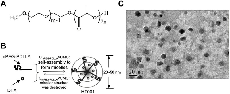
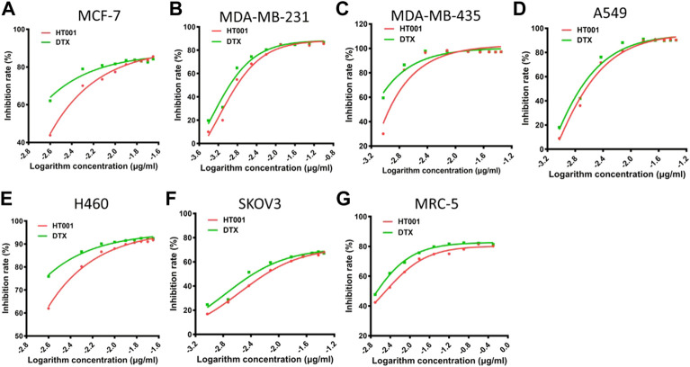
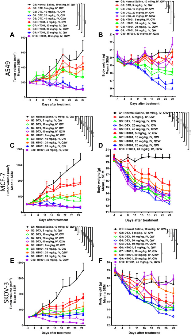
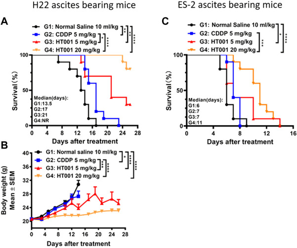
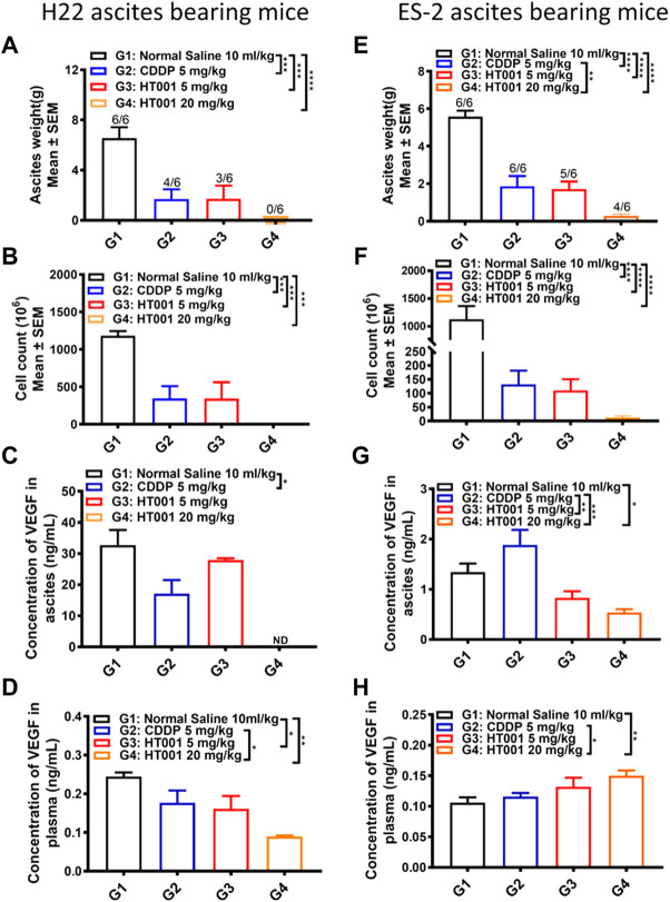
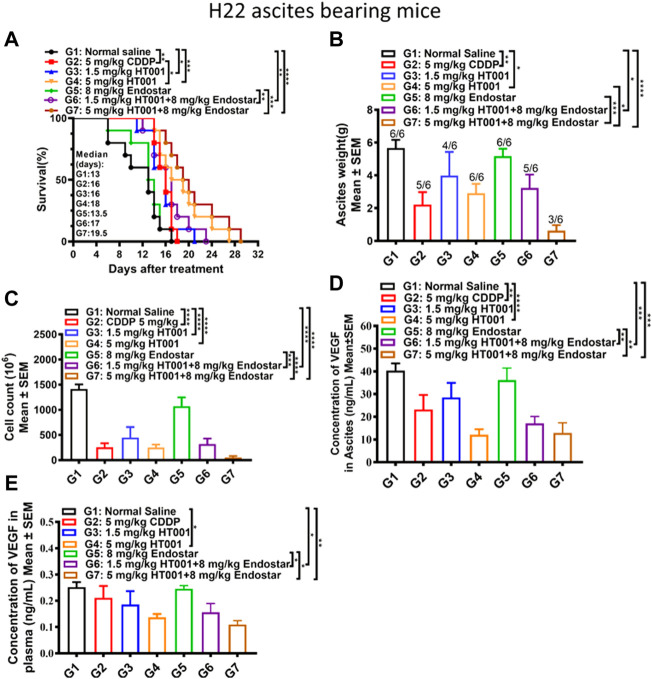
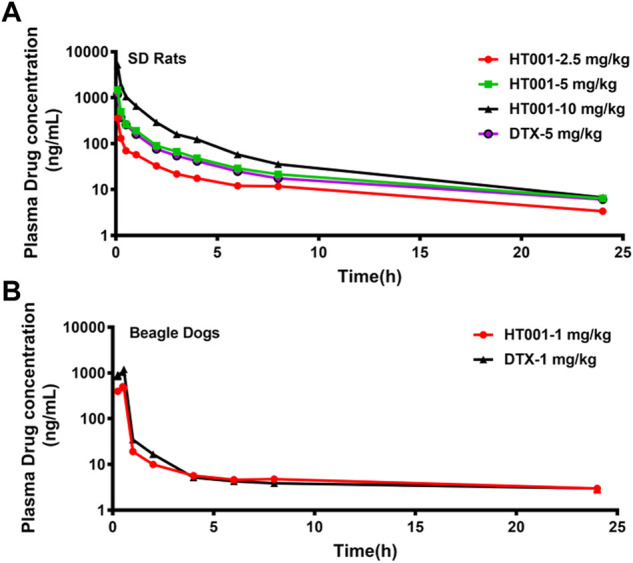
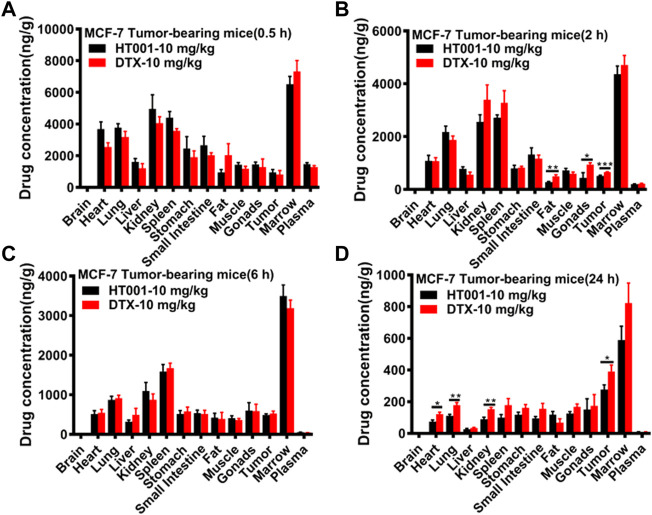
Malignant ascites (MA) is caused by intraperitoneal spread of solid tumor cells and results in a poor quality of life. Chemotherapy is a common first-line treatment for patients with MA. Taxotere ® (DTX) is widely used in solid tumor therapies. However, the low water solubility and side effects caused by additives in the formulation restrict the clinical application of docetaxel. HT001 is a clinical stage docetaxel micelle developed to overcome the solubility issue with improved safety profiles. To support clinical development and expand clinical application of HT001, this study used in vitro and in vivo approaches to investigate the anti-tumor effects of HT001 when applied as monotherapy or in combination with anti-angiogenic agents. HT001 demonstrated comparable anti-proliferative activities as docetaxel in a broad range of cancer cell lines in vitro. Furthermore, HT001 suppressed tumor growth in a dose-dependent manner in A549, MCF-7, and SKOV-3 xenograft tumor mouse models in vivo. In a hepatocellular carcinoma H22 malignant ascites-bearing mouse model, HT001 presented a dose-dependent inhibition of ascites production, prolonged animal survival, and reduced VEGF levels. When dosed at 20 mg/kg, the HT001-treated group exhibited curative results, with no ascites formation in 80% of mice at the end of the study while all the mice in the vehicle control group succumbed. Similar results were obtained in HT001 treatment of mice bearing malignant ascites produced by human ovarian cancer ES-2 cells. Notably, the combination of HT001 with Endostar not only significantly reduced ascites production but also prolonged survival of H22 ascites-bearing mice. HT001 showed similar PK and tissue distribution profiles as DTX in non-rodent hosts. Collectively, these results demonstrate potent anti-tumor activity of HT001 in multiple solid tumor models or malignant ascites models, and reveal synergistic effects with anti-angiogenic agents, supporting the clinical development and clinical expansion plans for HT001.

Keywords: anti-angiogenic agents; anti-tumor effects; ascites-inhibitory activities; combination therapy; docetaxel polymeric micelles.


Conflict of interest statement

Authors LG, XQ, LX, SZ, GY, RT, and WY were employed by the company Jiangsu Simcere Pharmaceutical Co., Ltd. The remaining authors declare that the research was conducted in the absence of any commercial or financial relationships that could be construed as a potential conflict of interest.

Figures

FIGURE 1
FIGURE 1
Synthesis and characteristics of mPEG-PDLLA and HT001. (A) The structure of polyethylene glycol monomethyl ether polylactic acid block polymer (mPEG-PDLLA). (B) The schematic process of HT001 synthesis. (C) Transmission electron microscopy image of HT001. The morphology of micelles was examined by transmission electron microscope.
FIGURE 2
FIGURE 2
Cytotoxicity of HT001 and DTX in vitro. A cell suspension of 5000 cells/well was seeded in 96-well flat-bottom tissue culture plates. Cell viability was evaluated by CCK-8 assay after 72 h treatment with HT001 or DTX. (A–G) Cell viability inhibition in different cell lines.
FIGURE 3
FIGURE 3
Anti-tumor effects of HT001 and DTX in A549, MCF-7, and SKOV-3 xenograft mouse models. A549, MCF-7, and SKOV-3 tumor-bearing mice were intravenously administrated with normal saline, DTX, or HT001 once a week or once every 2 weeks (n = 8/group). (A,C,E) Tumor growth curves of A549, MCF-7, or SKOV-3 tumor bearing mice after treatment (n = 8/group). (B,D,F) Bodyweight graphs of A549, MCF-7, or SKOV-3 tumor bearing mice after treatment. The error bars represent SEM. IV, intravenous; QW, once weekly; Q2W, once every 2 weeks; *, p < 0.05; **, p < 0.01; ***, p < 0.001; ****, p < 0.0001.
FIGURE 4
FIGURE 4
Effects of HT001 and CDDP on H22 and ES-2 ascites bearing mice. H22 or ES-2 ascites-bearing mice were intraperitoneally administrated with normal saline, 5 mg/kg CDDP, 5 mg/kg HT001, or 20 mg/kg HT001 (n = 16/group) once a week. After 7 or 8 days of administration, 6 mice from each group were sacrificed for analysis and the remaining mice were used for monitoring survival. (A,C) Kaplan-Meier survival analysis of H22 or ES-2 ascites-bearing mice. NR: not reached, more than 50% of mice survived at the end of the study and the medium survival time cannot be reached. (B) Bodyweight of H22 ascites -bearing mice. The bodyweight data points are not shown when there are less than 3 animals in a group. The error bars represent SEM. *, p < 0.05; **, p < 0.01; ***, p < 0.001; ****, p < 0.0001.
FIGURE 5
FIGURE 5
Ex vivo analysis of plasma and ascites samples from the H22 and ES-2 ascites-bearing mice. The ascites and plasma from H22 or ES-2 ascites bearing mice sacrificed 7 or 8 days after treatment as in Figure 4 were analyzed. (A,E) Ascites weight of H22 and ES-2 ascites-bearing mice. The numbers on the column represents number of mice with ascites formed/all analyzed mice. (B,F) Cell counts in ascites from H22 and ES-2 ascites bearing mice. (C,G) VEGF levels in ascites from H22 and ES-2 ascites-bearing mice. (D,H) VEGF levels in plasma from H22 and ES-2 ascites-bearing mice. Error bars represent SEM. *, p < 0.05; **, p < 0.01; ***, p < 0.001; ****, p < 0.0001.
FIGURE 6
FIGURE 6
Effects of HT001 and Endostar combination on H22 ascites-bearing mice. The H22 bearing ascites mice were intraperitoneally administrated with normal saline, 5 mg/kg CDDP, 1.5 mg/kg HT001, 5 mg/kg HT001, 8 mg/kg Endostar, 1.5 mg/kg HT001+8 mg/kg Endostar, or 5 mg/kg HT001+8 mg/kg Endostar (n = 16/group). HT001 and CDDP were administrated once a week and Endostar was administrated daily for 3 weeks. After 8 days of administration, 6 mice from each group were sacrificed for analysis and the remaining mice were used for monitoring survival. (A) Kaplan-Meier survival analysis of H22 ascites-bearing mice. (B) Ascites weight of H22 ascites mice. The numbers on the column represent number of mice with ascites formed/all analyzed mice. (C) Cell counts in ascites of H22 ascites-bearing mice. (D) VEGF levels in ascites from H22 ascites-bearing mice. (E) VEGF levels in plasma from H22 bearing ascites mice. Error bars represent SEM. *, p < 0.05; **, p < 0.01; ***, p < 0.001; ****, p < 0.0001.
FIGURE 7
FIGURE 7
Plasma concentration-time curves of docetaxel in SD Rats and Beagle dogs. (A) SD rats were intravenously administered with a single dose of 2.5, 5, 10 mg/kg HT001 or 5 mg/kg DTX (six rats per group, male: female = 1:1) and plasma drug concentrations were analyzed by LC-MS/MS. (B) Beagle dogs were intravenously administrated with a single dose of 1 mg/kg of DTX or HT001 (six dogs per group, male: female = 1:1) and plasma drug concentrations were analyzed by LC-MS/MS.
FIGURE 8
FIGURE 8
Tissue distributions of docetaxel in MCF-7 tumor-bearing mice. MCF-7 tumor-bearing Balb/c nude mice were intravenously administered with 10 mg/kg HT001 or DTX (n = 24, male: female = 1:1). (A–D) Drug concentration in plasma and different organs at each time point (0.5, 2, 6, and 24 h). Six mice from each group at each time point were sacrificed, and different organs were harvested to analyze the drug concentration by LC-MS/MS. *, p < 0.05; **, p < 0.01; ***, p < 0.001.

Similar articles

Cited by

References

    1. Angelucci A., Monache S. D., Cortellini A., Padova M. D., Ficorella C. (2018). Vessels in the storm": Searching for prognostic and predictive angiogenic factors in colorectal cancer. Int. J. Mol. Sci. 19 (1), 299. 10.3390/ijms19010299 - DOI - PMC - PubMed
    1. Bhaskari J., Krishnamoorthy L. (2015). Impact of ascites and plasma levels of VEGF-A in epithelial ovarian cancers. J. Clin. Cell. Immunol. 6, 4. 10.4172/2155-9899.1000353 - DOI
    1. Cavazzoni E., Bugiantella W., Graziosi L., Franceschini M. S., Donini A. (2013). Malignant ascites: Pathophysiology and treatment. Int. J. Clin. Oncol. 18 (1), 1–9. 10.1007/s10147-012-0396-6 - DOI - PubMed
    1. Chen X., Liu Y., Yin Y., Jin S., Ping G., Røe O. D., et al. (2012). Recombinant human endostatin (endostar) decreased recurrent ascites, pleural fluid and ascitic VEGF in a case of advanced mesothelioma. J. Chemother. 24 (4), 231–236. 10.1179/1973947812Y.0000000023 - DOI - PubMed
    1. Chilimoniuk Z., Rocka A., Stefaniak M., Tomczyk Ż., Jasielska F., Madras D., et al. (2022). Molecular methods for increasing the effectiveness of ovarian cancer treatment: A systematic review. Future Oncol. 18 (13), 1627–1650. 10.2217/fon-2021-0565 - DOI - PubMed

LinkOut - more resources