. 2022 Aug 26:13:940574.
doi: 10.3389/fphar.2022.940574. eCollection 2022.

Rhein ameliorates transverse aortic constriction-induced cardiac hypertrophy via regulating STAT3 and p38 MAPK signaling pathways

Affiliations

Rhein ameliorates transverse aortic constriction-induced cardiac hypertrophy via regulating STAT3 and p38 MAPK signaling pathways

Run-Jing Li et al. Front Pharmacol. .

Abstract

The progression from compensatory hypertrophy to heart failure is difficult to reverse, in part due to extracellular matrix fibrosis and continuous activation of abnormal signaling pathways. Although the anthraquinone rhein has been examined for its many biological properties, it is not clear whether it has therapeutic value in the treatment of cardiac hypertrophy and heart failure. In this study, we report for the first time that rhein can ameliorate transverse aortic constriction (TAC)-induced cardiac hypertrophy and other cardiac damage in vivo and in vitro. In addition, rhein can reduce cardiac hypertrophy by attenuating atrial natriuretic peptide, brain natriuretic peptide, and β-MHC expression; cardiac fibrosis; and ERK phosphorylation and transport into the nucleus. Furthermore, the inhibitory effect of rhein on myocardial hypertrophy was similar to that of specific inhibitors of STAT3 and ERK signaling. In addition, rhein at therapeutic doses had no significant adverse effects or toxicity on liver and kidney function. We conclude that rhein reduces TAC-induced cardiac hypertrophy via targeted inhibition of the molecular function of ERK and downregulates STAT3 and p38 MAPK signaling. Therefore, rhein might be a novel and effective agent for treating cardiac hypertrophy and other cardiovascular diseases.

Keywords: ERK phosphorylation; STAT; cardiac fibrosis; cardiac hypertrophy; p38 MAPK; rhein; transverse aortic constriction.


Conflict of interest statement

The authors declare that the research was conducted in the absence of any commercial or financial relationships that could be construed as a potential conflict of interest.

Figures

FIGURE 1
FIGURE 1
Rhein attenuates cardiomyocyte hypertrophy induced by AngII in vitro. (A) Chemical structure of rhein. (B,C) H9C2 and AC16 cells were stimulated with rhein (0–1,000 μM) for 24 h. Cell viabilities were measured by the MTT assay. (D) Relative expression of ANP, BNP, and β-MHC proteins in H9C2 cells treated with AngII, rhein, and telmisartan. (E–G) Quantitative PCR (qPCR) showing the mRNA levels of AngII-induced hypertrophic markers, ANP, BNP, and β-MHC, using RT-PCR analysis. Data are mean ± SD (n ≥ 3). *p < 0.05, **p < 0.01, ***p < 0.001 vs Ctrl group, #p < 0.05, ##p < 0.01, ###p < 0.001 vs AngII group (analyzed using one-way ANOVA). Telmisartan (Tel) is the positive control group.
FIGURE 2
FIGURE 2
Rhein inhibits phosphorylation and nucleation of ERK to alleviate hypertrophy. (A) H9C2 cells stained with F-actin (red), DAPI (blue), and P-ERK (green) to detect the size of cells and relative intensity of P-ERK, and immunofluorescence images were captured by fluorescence microscopy. Scale bar = 50 µm. (B) Statistical quantitative analysis of the area of H9C2 cells by Image-Pro Plus software. (C) Nucleo-cytoplasmic separation assay was used to detect the effect of rhein on AngII-stimulated P-ERK nuclear translocation in H9C2 cells. (D) Statistical quantitative analysis of the relative fluorescence intensity of P-ERK in H9C2 cells by Image-Pro Plus software. (E,F) Relative strength of P-ERK in the nucleus and cytoplasm, respectively. Data are mean ± SD (n ≥ 3). *p < 0.05, **p < 0.01, ***p < 0.001 vs Ctrl group, #p < 0.05, ##p < 0.01, ###p < 0.001 vs AngII group (analyzed using one-way ANOVA).
FIGURE 3
FIGURE 3
Rhein targets inhibition of AngII-stimulated activation of STAT3 and p38/MAPK signaling pathways in vitro. (A) Expression levels of signaling pathway proteins (total and phosphorylation of STAT3, p38, JNK, and ERK) were determined by Western blot in H9C2 cells. (B–E) Quantification of the relative changes in phosphorylation of STAT3, p38, JNK, and ERK. (F) The inhibitory effect of rhein on AngII-induced cardiac hypertrophy was compared with that of p-STAT3- and P-ERK-specific inhibitors. (G,H) Quantification of the relative changes in protein expression of ANP and BNP treated with specific inhibitors and rhein in cardiomyocytes. GAPDH was used for normalization. Data are mean ± SD (n ≥ 3). *p < 0.05, **p < 0.01, ***p < 0.001 vs Ctrl group, #p < 0.05, ##p < 0.01, ###p < 0.001 vs AngII group (analyzed using one-way ANOVA).
FIGURE 4
FIGURE 4
Rhein alleviates cardiac hypertrophy and cardiac fibrosis induced by TAC in WT mice. (A) Representative peak hemodynamics pictures. (B) Representative echocardiography pictures. (C) Morphological images of mouse hearts were exhibited from the sham, TAC, TAC + rhein (50 mg/kg), TAC + rhein (150 mg/kg), TAC + rhein (200 mg/kg), and TAC + Tel (10 mg/kg) groups. (D) Ratio of heart weight (HW) to body weight (BW). (E) Ratio of HW to tibia length in each group. (F) Paraffin sections of cardiac tissues were stained with H&E. Representative images are shown. Scale bar: 50 μm. (G) Myocardial cytoskeleton shows green fluorescence by WGA staining. Scale bar: 50 μm. (H) Masson’s trichrome staining of the hypertrophic heart. Collagen fibers are stained blue, and the myocardium is stained red. Scale bar: 50 μm. (I) Cell surface areas were quantified in the hearts of mice using Image-Pro Plus software. (J)Left ventricle fibrosis areas (of control) were quantified by Image-Pro Plus software. Data are mean ± SD (n = 6). *p < 0.05, **p < 0.01, ***p < 0.001 vs sham group, #p < 0.05, ##p < 0.01, ###p < 0.001 vs TAC group (analyzed using one-way ANOVA).
FIGURE 5
FIGURE 5
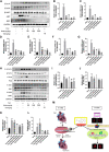
Rhein inhibits TAC operation-stimulated activation of STAT3 and p38/MAPK signaling pathways in vivo. (A) Relative expression of ANP, BNP, β-MHC, COL1A, and COL3A proteins in the left ventricle treated with TAC operation and rhein, and in the telmisartan positive control group. (B–G) mRNA expression of COL1A1, COL3A, ANP, BNP, β-MHC, and IL-1β was examined by RT-PCR. (H) The expression levels of signaling pathway proteins (total and phosphorylation of STAT3, JNK, ERK, and p38) were determined by Western blot. (I–L) Quantification of the relative changes in the phosphorylation of STAT3, p38, JNK, and ERK. (M) Schematic diagram with roles of rhein in TAC operation- and AngII-induced cardiac hypertrophy. Data are mean ± SD (n = 6). *p < 0.05, **p < 0.01, ***p < 0.001 vs sham group, #p < 0.05, ##p < 0.01, ###p < 0.001 vs TAC group.
FIGURE 6
FIGURE 6
Rhein has no obvious toxicity or side effects on normal organs. (A–E) The levels of LDH, AST, ALT, CREA-S, and UREA were tested from the serum of various mice groups. (F–G) Liver and kidney histopathology was done by H&E staining. Scale bar = 100 µm. Data are expressed as mean ± SD (n = 6). *p < 0.05 vs sham group, #p < 0.05, ##p < 0.01 vs TAC group (analyzed using one-way ANOVA).

Similar articles

Cited by

References

    1. Aikawa R., Nagai T., Kudoh S., Zou Y., Tanaka M., Tamura M., et al. (2002). Integrins play a critical role in mechanical stress-induced p38 MAPK activation. Hypertension 39 (2), 233–238. 10.1161/hy0202.102699 - DOI - PubMed
    1. Barbosa D. M., Fahlbusch P., Herzfeld de Wiza D., Jacob S., Kettel U., Al-Hasani H., et al. (2020). Rhein, a novel Histone Deacetylase (HDAC) inhibitor with antifibrotic potency in human myocardial fibrosis. Sci. Rep. 10 (1), 4888. 10.1038/s41598-020-61886-3 - DOI - PMC - PubMed
    1. Brancaccio M., Hirsch E., Notte A., Selvetella G., Lembo G., Tarone G., et al. (2006). Integrin signalling: The tug-of-war in heart hypertrophy. Cardiovasc. Res. 70 (3), 422–433. 10.1016/j.cardiores.2005.12.015 - DOI - PubMed
    1. Burchfield J. S., Xie M., Hill J. A. (2013). Pathological ventricular remodeling: Mechanisms: Part 1 of 2. Circulation 128 (4), 388–400. 10.1161/CIRCULATIONAHA.113.001878 - DOI - PMC - PubMed
    1. Chen Y., Mu L., Xing L., Li S., Fu S. (2019). Rhein alleviates renal interstitial fibrosis by inhibiting tubular cell apoptosis in rats. Biol. Res. 52 (1), 50. 10.1186/s40659-019-0257-0 - DOI - PMC - PubMed