. 2022 Aug;1(8):761-774.
doi: 10.1038/s44161-022-00116-7. Epub 2022 Aug 12.

Antigen presentation by cardiac fibroblasts promotes cardiac dysfunction

Affiliations

Antigen presentation by cardiac fibroblasts promotes cardiac dysfunction

Njabulo Ngwenyama et al. Nat Cardiovasc Res. 2022 Aug.

Erratum in

Abstract

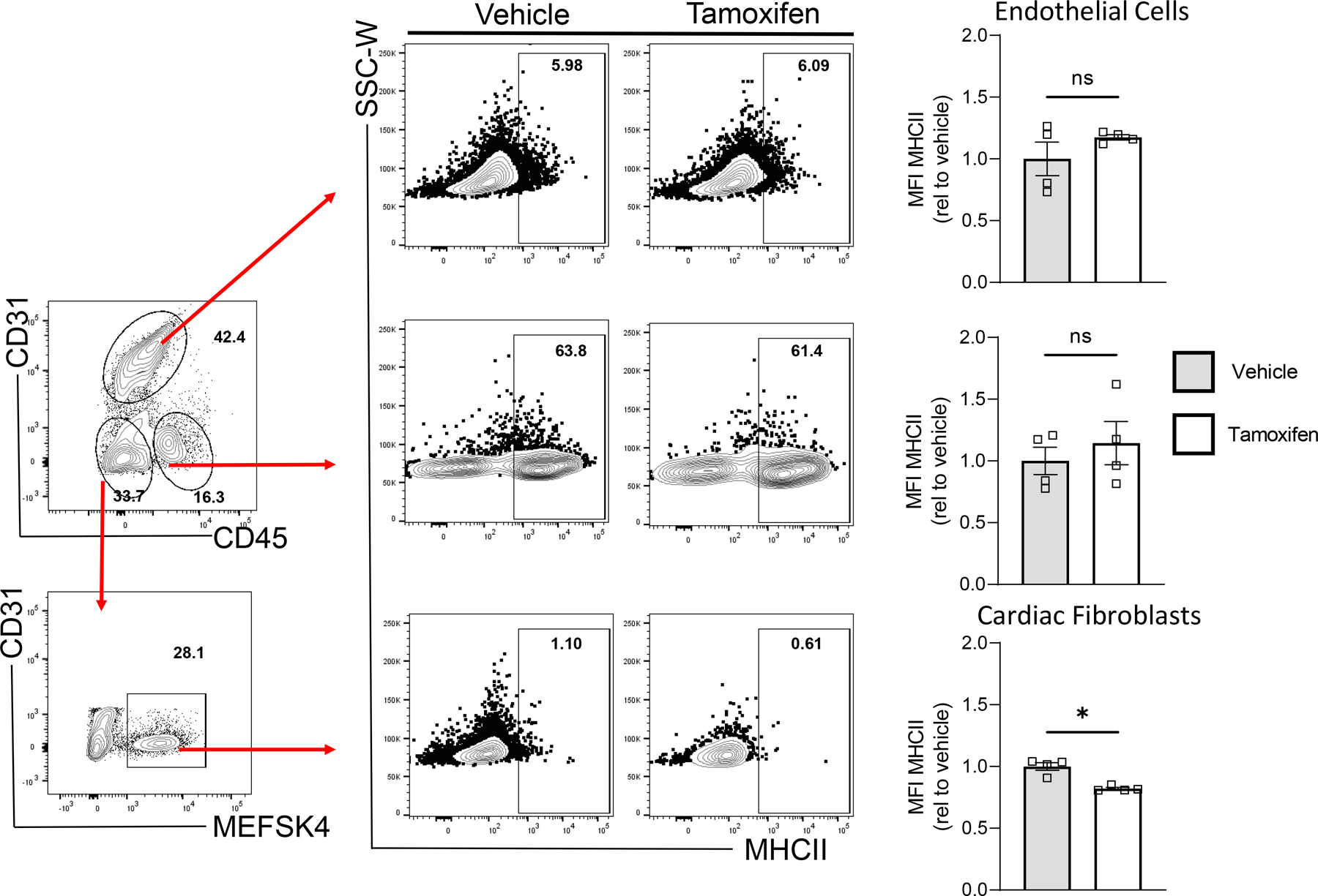
Heart failure (HF) is a leading cause of morbidity and mortality. Studies in animal models and patients with HF revealed a prominent role for CD4+ T cell immune responses in the pathogenesis of HF and highlighted an active crosstalk between cardiac fibroblasts and IFNγ producing CD4+ T cells that results in profibrotic myofibroblast transformation. Whether cardiac fibroblasts concomitantly modulate pathogenic cardiac CD4+ T cell immune responses is unknown. Here we show report that murine cardiac fibroblasts express major histocompatibility complex type II (MHCII) in two different experimental models of cardiac inflammation. We demonstrate that cardiac fibroblasts take up and process antigens for presentation to CD4+ T cells via MHCII induced by IFNγ. Conditional deletion of MhcII in cardiac fibroblasts ameliorates cardiac remodelling and dysfunction induced by cardiac pressure overload. Collectively, we demonstrate that cardiac fibroblasts function as antigen presenting cells (APCs) and contribute to cardiac fibrosis and dysfunction through IFNγ induced MHCII.


Conflict of interest statement

Competing Interests The authors declare no competing financial interests.

Figures

Extended Data Fig. 1
Extended Data Fig. 1. Cardiac fibroblasts express MHCII in vivo in response to acute T. cruzi infection.
(A) Wt mice were inoculated with 20,000 T. cruzi parasites by intraperitoneal injection and the hearts were harvested at 19 days post-infection. (B-E) Ventricular CD31-CD45-MEFSK4+ cardiac fibroblasts were analysed by flow cytometry (≥ 5,000 target cells acquired) to determine surface expression of (B-C) MHCII and (D-E) CD80. n= 4 (mock) and n=3 (Chagas) mice. Error bars represent mean ± SD. (* p≤0.05; *** p<0.001; Mann-Whitney test, two tailed).
Extended Data Fig. 2
Extended Data Fig. 2. MhcII recombination in cardiac fibroblasts occurred in a cell-specific manner.
Detection of Intact allele (310bp), recombined alleles (475 bp) and GAPDH( 156 bp) in sorted CD31-CD45-MEFSK4+ Fibroblasts and leukocytes from Tcf21iCre/+MhcIIfl/fl mice treated with vehicle or Tamoxifen. GAPDH was used as loading control.
Extended Data Fig. 3
Extended Data Fig. 3. Tamoxifen treatment of Tcf21iCre/+/ MhcIIflox/flox mice reduces MHCII surface protein expression specifically in cardiac fibroblasts
Transverse aortic constriction (TAC) surgery was performed on vehicle and Tamoxifen treated Tcf21iCre/+MhcIIfl/fl mice and the left ventricle was harvested after 4 weeks, digested, stained and analysed by flow cytometry. (A) Representative FACS plot showing gating of CD31+CD45-MESFK4- endothelial cells, CD31-CD45+MESFK4- leukocytes and CD31-CD45-MESFK4+ cardiac fibroblasts. (B-C) Representative FACS plots (B) and quantification of mean fluorescence intensity (MFI) (C) of MHC-II expression in endothelial cells (top), leukocytes (middle) and cardiac fibroblasts (bottom) in the hearts of vehicle and tamoxifen treated mice. n=4 mice/group. Error bars represent mean ± SE. (* p<0.05; Mann-Whitney test, two tailed. C: p value=0.0286).
Extended Data Fig. 4
Extended Data Fig. 4. Cardiac fibroblast MHCII expression does not affect CD4+ T cell activation in the mLNs in response to TAC
Extended Data Fig. 5
Extended Data Fig. 5. Cardiac fibroblast MHCII expression contributes to cardiomyocyte hypertrophy and to cardiac fibroblast abundance in response to pressure overload
Transverse aortic constriction (TAC) was performed on vehicle and Tamoxifen treated Tcf21iCre/+MhcIIfl/fl mice and harvested after 4 weeks. Conditional deletion of MhcII on cardiac fibroblasts was induced by administering Tamoxifen via intraperitoneal injections prior to TAC surgery, followed by Tamoxifen citrate chow for 4 weeks. (A) Wheat germ agglutinin (WGA) staining of frozen LV tissue sections was performed and used to calculate (B) mean cardiomyocyte area. Representative images of n=3 hearts (Sham), n=4 (TAC vehicle) and n=7 (TAC TMX). (C) Gross LV mass was acquired and is shown normalized to tibia length. (D-E) LV tissue was digested, stained for CD31 and MESFK4 and analysed by flow cytometry. Representative FACS plot (D) and quantification (E) of vehicle and tamoxifen treated mice hearts (n=4 mice/group). Error bars represent mean ± SE. (* p<0.01; Mann-Whitney test, two tailed: E: p=0286).
Extended Data Fig. 6
Extended Data Fig. 6. Schematic Diagram showing bidirectionality of cardiac fibroblast and Th1 cell interactions
Cardiac fibroblasts express the costimulatory molecule CD80 and efficiently capture and process extracellular antigens into small peptides that are uploaded into MHC-II molecules. In response to IFNγ stimulation, cardiac fibroblasts express MHCII and present peptide antigens that induce IFNγ + Th1 cell immune responses and promote cardiac remodelling and dysfunction.
Figure 1
Figure 1. Cardiac fibroblasts express MHCII in response to IFN𝛾 stimulation in vitro.
(A-H) CD31-CD45-MEFSK4+ primary adult murine cardiac fibroblasts were sorted from heart digests and cultured in the presence of 100U/mL recombinant murine IFN𝛾 for up to 5 days. (A) CD31-CD45-MEFSK4+ cardiac fibroblasts were analysed by flow cytometry (≥ 20,000 target cells acquired) to determine surface expression of (B-C) MHCII as well as (D-E) CD80 and (F-G) CD86 costimulatory molecules. n= 5 (Control and D3), and n=3 (D5) independent experiments and cellular preparations. (H) Immunofluorescence staining for MHCII as well as Vimentin was performed after 3 days of IFN𝛾 stimulation. n= 3 independent experiments and cardiac fibroblasts preparations. (I-L) Sorted GFP+ cardiac fibroblasts from Tcf21iCre/+; R26eGFP lineage tracing reporter mice were cultured in the presence of 100U/mL recombinant murine IFN𝛾 for 72hrs. (I) GFP+ cardiac fibroblasts were analysed by flow cytometry (≥ 5,000 target cells acquired) to determine surface expression of MHCII and CD80 and (J-K) quantified. n=3 (MCHII), and n=5 (CD 80), independent experiments and cellular preparations. (L) Immunofluorescence staining for MHCII on GFP+ cardiac fibroblasts was performed. n=3 independent experiments and cardiac fibroblasts preparations. Scale bars: 100μm. Error bars represent mean ± SD. * p<0.05, *** p<0.001; One-way ANOVA (1C, E and G) and two tailed unpaired T test (1J and K).
Figure 2
Figure 2. Cardiac fibroblasts express MHCII in response to IFN𝛾 stimulation in vivo.
(A) CD31-CD45-MEFSK4+ cardiac fibroblasts were directly analysed by flow cytometry from the LV of (B) wt mice treated with daily intraperitoneal injections of IFN𝛾 (25,000U) or PBS vehicle and harvested on day 5. Surface expression of (C-D) MHCII, (E-F) CD80 and (G-H) CD86 was determined (≥ 5,000 target cells acquired). n= 4 vehicle and n= 5 IFNγ treated mice. Error bars represent mean ± SD. * p=0.0159; two tailed Mann-Whitney test.
Figure 3
Figure 3. Cardiac fibroblasts induce antigen-specific CD4+ T cell proliferation in vitro in the presence of IFNγ.
(A) Naïve CD62LhiCD44lo CD4+ T cells were sorted by FACS from OTII bulk splenocytes and labelled with CFSE (2μM). (B-D) Primary adult murine cardiac fibroblasts and BMDCs derived from wt or MhcII−/− mice were cultured in the presence or absence of IFN𝛾 (100U/mL) for 24hrs and loaded with OVA peptide (4μg/mL) where indicated, 2hrs prior to coculture with OTII naïve CD4+ T cells. After 72hrs CFSE dilution was evaluated by flow cytometry (≥ 10,000 target cells acquired) and (D) quantified. n=3 (CFB OVA), n=5 (CFB+IFNγ+OVA; BMDC+OVA), independent experiments and cell preparations. (E) Sorted GFP+ murine cardiac fibroblasts from Tcf21iCre/+; R26eGFP lineage tracing reporter mice were cultured in the presence or absence of IFN𝛾 (100U/mL) for 24hrs and loaded with OVA peptide (4µg/mL) where indicated, 2hrs prior to coculture with OTII naïve CD4+ T cells. (F) After 72hrs CFSE dilution was evaluated by flow cytometry (≥ 5,000 target cells acquired) and (G) quantified. n=3, independent experiments and cell preparations. Error bars represent mean ± SD. ** p<0.01, *** p<0.001; one-way ANOVA.
Figure 4
Figure 4. IFNγ produced by Th1 cells induce MHCII expression by cardiac fibroblasts and promotes antigen-specific CD4+ T cell activation.
(A) OTII Th1 cells were differentiated in vitro from bulk CD4+ splenocytes, and cocultured with adult cardiac fibroblasts and OVA peptide (4μg/mL). (B-C) After 24hrs of coculture Th1 IFN𝛾 production was determined by intracellular cytokine staining (≥ 15,000 target cells acquired). n=3 control and n=6 OVA, independent experiments and cell preparations. (D-E) After 72hrs of coculture CD31-CD45-MEFSK4+ cardiac fibroblasts were analysed by flow cytometry (≥ 5,000 target cells acquired) for surface expression of MHCII, (n=3, independent experiments and cell preparations) and (F-G) antigen-specific Th1 cell proliferation via CFSE dilution (≥ 8,000 target cells acquired). n=3 independent experiments and cell preparations. Error bars represent mean ± SD. * p<0.05, ** p<0.01, two tailed student unpaired t test (4C and E ) and One-way ANOVA (4G)
Figure 5
Figure 5. Cardiac fibroblasts take up, process, and present antigens to CD4+ T cells in vitro.
(A) CD31-CD45-MEFSK4+ primary adult murine cardiac fibroblasts were cultured in the presence of DQ OVA (100μg/mL) and IFN𝛾 (100U/mL) overnight to evaluate uptake and intracellular processing of DQ OVA by flow cytometry (≥ 10,000 target cells acquired), and (B) immunofluorescence microscopy in cardiac fibroblasts and BMDCs. (n=3 independent experiments). (C-D) Wt cardiac fibroblasts and BMDCs were treated with purified OVA protein (100μg/mL) overnight and cocultured with CFSE-labelled naïve OTII CD4+ T cells in the presence of IFN𝛾 (100U/mL) for 72 hrs to evaluate CD4+ T cell proliferation by flow cytometry (≥ 5,000 target cells acquired). n=7 and n=5 (CFB untreated and OVA+IFNγ treated) and n=6 and n=7 (BMDC untreated and OVA treated), independent experiments and cell preparations. (E) Wt cardiac fibroblasts were cultured in the presence of pHrodo Green E. coli bioparticles overnight to evaluate phagocytosis by flow cytometry (≥ 7,000 target cells acquired). (F) Intracellular accumulation of bioparticles in acidified phagolysosomes was further evaluated by immunofluorescence microscopy in cardiac fibroblasts and BMDCs. n=3 independent experiments. (G-H) Wt cardiac fibroblasts and BMDCs were cultured overnight in the presence of transformed E. coli particles expressing either chicken ovalbumin or empty vector (EV), and cocultured with CFSE-labelled naïve OTII CD4+ T cells for 72 hrs to evaluate CD4+ T cell proliferation by flow cytometry (≥ 10,000 target cells acquired). n=11 BMDC EV, and n=8 independent experiments for all other conditions. Scale bars: 100μm.. Error bars represent mean ± SD. ** p<0.01, *** p<0.001; one-way ANOVA test.
Figure 6
Figure 6. Cardiac fibroblasts express MHCII in response to cardiac pressure overload.
(A-E) Transverse aortic constriction (TAC) surgery was performed on mice and harvested after 4 weeks. (A) CD31-CD45-MEFSK4+ cardiac fibroblasts from the LV of wt mice were directly analysed by flow cytometry (≥ 5,000 target cells acquired) to determine surface expression of (B-C) MHCII and (D-E) CD80. n= 3 Sham and TAC treated mice. Immunofluorescence staining of LV tissue sections was performed to detect (F) MHCII on aSMA+ cells in wt mice (white arrows) and (G) MHCII on GFP+ cells in Tcf21iCre/+; R26eGFP lineage tracing reporter mice (indicated with a white arrow) (Scale bars: 100μm). (H) Immunofluorescence staining of wt LV tissue sections was performed to evaluate colocalization of CD4+ cells and aSMA+ cells (Scale bars: 25μm). Images are representative of n=3 mice (F and H) and n=5 mice (G). Error bars represent mean ± SD. (* p<0.05, ** p<0.01; two tailed unpaired T test).
Figure 7
Figure 7. Cardiac fibroblast MHCII expression is required for cardiac dysfunction to develop in response to pressure overload.
(A) Conditional deletion of MHCII in cardiac fibroblasts was induced by administering Tamoxifen to Tcf21iCre/+MhcIIfl/fl mice. (B) CD31-CD45-MEFSK4+ cardiac fibroblasts and CD31-CD45+ leukocytes were FACS sorted from the LV of vehicle and Tamoxifen treated Tcf21iCre/+MhcIIfl/fl mice and genomic DNA was probed for the intact (P1 and P2 primers) and recombined (P1 and P3 primers) MhcII alleles by PCR using specific primers (blue arrows) that detect the recombination (MhcII excision) or the intact-non recombined- in the different conditions. Note that excision is only observed in cardiac fibroblasts from tamoxifen treated mice with the P1-P3 primers. GAPDH is shown as a loading control. Gel represents DNA from cells sorted from n=3 mice (pooled hearts). (C-D) CD31-CD45-MEFSK4+ cardiac fibroblasts from Tcf21iCre/+MhcIIfl/fl mice were cultured overnight in the presence of 4-Hydroxytamoxifen (1μM), then stimulated with IFN𝛾 (100U/mL) for 72 hrs to determine surface expression of MHCII by flow cytometry (≥ 2,000 target cells acquired. n= 4 vehicle and n= 3 for 4HO-TMX, independent experiments and cell preparations. (E-F) CD45+CD11b+CD11c+ BMDCs from Tcf21iCre/+MhcIIfl/fl mice were cultured overnight in the presence of 4-Hydroxytamoxifen (1μM), then stimulated with LPS (10ng/mL) for 72 hrs to determine surface expression of MHCII by flow cytometry (≥ 12,000 target cells acquired) n= 5, independent experiments and cell preparations. (G) Tcf21iCre/+MhcIIfl/fl mice were treated with Tamoxifen (75mg/Kg) by 5 daily I.P injections prior to TAC surgery and maintained on Tamoxifen citrate chow for 4 weeks after surgery to decrease MHCII expression in cardiac fibroblasts, or with vehicle (mice with MHCII sufficient cardiac fibroblasts). (H) MHCII expression on CD11b+ myeloid cells in the mediastinal lymph nodes (mLNs) was determined by flow cytometry. (I-J) IHC of frozen LV sections was used to determine LV CD4+ T cell infiltration, scanning the entire LV section area. Representative images of n=3 hearts (vehicle and TMX Sham), n=4 (TAC vehicle) and n=7 (TAC TMX). (K-L) Picrosirius red staining of fixed LV tissue sections was used to determine perivascular fibrosis. Representative images of n=3 hearts (vehicle and TMX Sham), n=4 (TAC vehicle) and n=7 (TAC TMX). (M-N) Transthoracic echocardiography was used to measure LV fractional shortening. Scale bars: 100μm. Each data points represents individual animal in Fig 7H, 7J, 7L and 7N. Error bars represent mean ± SD. * p<0.05; two tailed unpaired T test (7D) and One-way ANOVA (7H, J, L and N).

References

    1. Pinto AR et al. Revisiting Cardiac Cellular Composition. Circ Res 118, 400–409, doi:10.1161/CIRCRESAHA.115.307778 (2016). - DOI - PMC - PubMed
    1. Prabhu SD & Frangogiannis NG The Biological Basis for Cardiac Repair After Myocardial Infarction: From Inflammation to Fibrosis. Circ Res 119, 91–112, doi:10.1161/CIRCRESAHA.116.303577 (2016). - DOI - PMC - PubMed
    1. Kaur H et al. Targeted Ablation of Periostin-Expressing Activated Fibroblasts Prevents Adverse Cardiac Remodeling in Mice. Circ Res 118, 1906–1917, doi:10.1161/CIRCRESAHA.116.308643 (2016). - DOI - PubMed
    1. Aghajanian H et al. Targeting cardiac fibrosis with engineered T cells. Nature 573, 430–433, doi:10.1038/s41586-019-1546-z (2019). - DOI - PMC - PubMed
    1. Nevers T et al. Left Ventricular T-Cell Recruitment Contributes to the Pathogenesis of Heart Failure. Circ Heart Fail 8, 776–787, doi:10.1161/CIRCHEARTFAILURE.115.002225 (2015). - DOI - PMC - PubMed