Review
. 2022 Aug 24;7(35):30657-30672.
doi: 10.1021/acsomega.2c02675. eCollection 2022 Sep 6.

Emerging ROS-Modulating Technologies for Augmentation of the Wound Healing Process

Affiliations
Review

Emerging ROS-Modulating Technologies for Augmentation of the Wound Healing Process

Suryanarayana Polaka et al. ACS Omega. .

Abstract

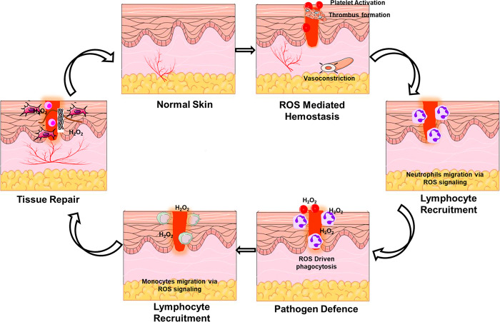
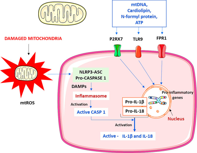
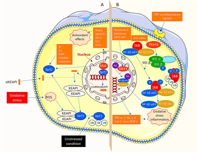
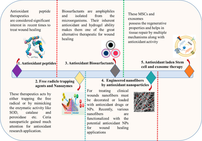
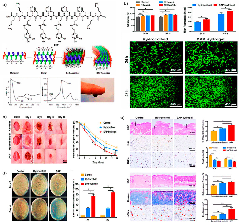
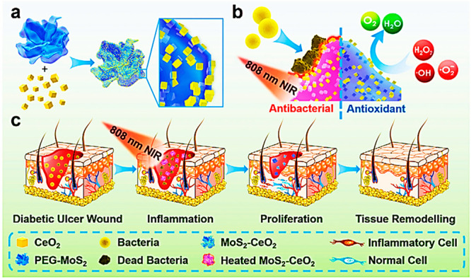
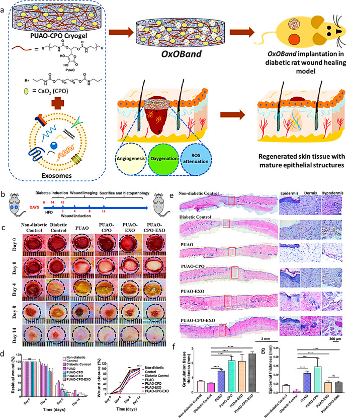
Reactive oxygen species (ROS) is considered a double-edged sword. The slightly elevated level of ROS helps in wound healing by inhibiting microbial infection. In contrast, excessive ROS levels in the wound site show deleterious effects on wound healing by extending the inflammation phase. Understanding the ROS-mediated molecular and biomolecular mechanisms and their effect on cellular homeostasis and inflammation thus substantially improves the possibility of exogenously augmenting and manipulating wound healing with the emerging antioxidant therapeutics. This review comprehensively delves into the relationship between ROS and critical phases of wound healing and the processes underpinning antioxidant therapies. The manuscript also discusses cutting-edge antioxidant therapeutics that act via ROS scavenging to enhance chronic wound healing.


Conflict of interest statement

The authors declare no competing financial interest.

Figures

Figure 1
Figure 1
ROS and their role in the various process of wound healing.
Figure 2
Figure 2
Mitochondria dysfunction mediated activation of the inflammasome. Damaged mitochondria secrete molecular patterns identified by the cytosolic and membrane receptors such as toll-like receptors (TLR) 9. NLRP3 inflammasomes get activated by interacting with various responses, including DAMPs. Further, the NLRP3 forms caspase activation and recruitment domain (CARD) with ASC and CASP1 called inflammasomes. Caspase-1 stimulates the inflammasome complex for activation, and the activated inflammasomes help convert the pro-IL-1β and pro-IL-18 into matured forms. Mitochondrial DNA (mtDNA) formyl proteins, ATP, and mitochondria ROS also help to start the NLRP3 inflammasomes directly or indirectly by receptor-mediated (FPR1, P2RX7). TLR9 specifically interacts with the mitochondrial DNA motifs and activates the signaling cascades resulting in a pro-inflammatory cytokine response. TLR-9, toll-like receptor-9; CARD, caspase activation, and recruitment domain; CASP1, caspase-1; DAMPs, danger-associated molecular patterns; IL, interleukin; mtDNA, mitochondrial DNA; mtROS, mitochondrial reactive oxygen species; FPR1, formyl peptide receptor 1; P2RX7, P2X purinoceptor.
Figure 3
Figure 3
Two major pathways in antioxidant therapy “Nrf2 pathway and the NFκB pathway”. (A) Under unstressed events, KEAP1 interrelates with Nrf2 and actin cytoskeleton to retain Nrf2 in dormant form and thus encourage both ubiquitination and deprivation of Nrf2. In addition, oxidative stress not only separates Nrf2 from KEAP1 but also translocates it to the nucleus. In the nucleus, Nrf2 heterodimerizes with Maf. It produces Nrf2-Maf heterodimer, which further attaches to ARE to form metabolic genes. For instance, NQO1, heme oxygenase-1 (HO-1), GSTs, GCL, and manganese superoxide dismutase (MnSOD) produce antioxidant effects. Additionally, oxidative stress is controlled via the activation of the Nrf2 pathway. In addition, the levels of KEAP1 are lowered by the siKEAP1 after loading them into RISC and by eliminating complementary mRNA of the KEAP1. Furthermore, Nrf2 activators, for instance, SF, CA, DMF, RTA408, and genistein, also induce the Nrf2 pathway and improve oxidative stress. (B) In resting conditions, NFκB dimers develop a complex with IkB protein in the cytoplasm. TNF-α, an inflammatory signal, encourages phosphorylation of IkB protein due to the involvement of IKK, which leads to ubiquitination and ultimately degradation of IkB. In addition, after moving active NFκB into the nucleus, it stimulates target genes such as TNF-α, NADPH oxidase (NOX)-2, cyclooxygenase-2 (COX-2), inducible nitric oxide synthase (iNOS), IL-6, and IL-1b causing both oxidative stresses as well as inflammation. Moreover, oxidative stress is controlled by hindering the NFκB pathway. Furthermore, MiR-146a shows aptitude toward targeting, inhibiting TRAF6, impeding the stimulation of IKK, and the NFκB pathway. Finally, SIRT1 activators, such as SRT1720, resveratrol, and berberine, overcome attaching of NFκB to inflammation-initiating gene promoters and their transcriptional events via stimulating SIRT1. RISC, RNA-induced silencing complex; TRAF6, tumor necrosis factor receptor-associated factor 6; ARE, antioxidant response element.
Figure 4
Figure 4
Emerging novel antioxidant therapeutic approaches for rapid wound healing.
Figure 5
Figure 5
Antioxidant and wound healing activity of DAP: (a) Chemical structure and self-assembled structural alignment of DAP; (b) percentage cell viability, mean fluorescence intensity, and live/dead assay for biocompatibility estimation of DAP; (c) photographs of wound images and their respective wound closure rates; (d) bacterial burden isolated from the infected wound in different groups at days 2 and 4 and quantitative analysis of the bacterial inhibition by various treatment at days 2 and 4; (e) immunohistological evaluation of different treatment groups H&E, IL-6, and TNF-α staining on day 6 and H&E, Masson’s trichrome, and α-smooth muscle actin (α-SMA) staining on day 14, wound re-epithelialization, number of a blood vessel per area, and fraction of collagen volume in wounded tissue. Adapted with the permissions from ref (82). Copyright 2021 American Chemical Society.
Figure 6
Figure 6
Illustration of MoS2–CeO2 nanocomposite for wound healing: (a) synthetic scheme of MoS2–CeO2 nanocomposite; (b) MoS2–CeO2 nanocomposite antioxidant and antibacterial mechanism; (c) MoS2–CeO2 nanocomposite actions on various phases of wound healing for promoting wound healing. Adapted with permission from ref (103). Copyright 2021 John Wiley and Sons.
Figure 7
Figure 7
(a) Schematic illustration of OxOBand and their properties; (b) study design for diabetic wound production and subsequent healing; (c) representative images of the wound healing on different days; (d) residual wound and % wound closure; (e) H&E staining of the wounds (left) and higher magnification images (right) on day 14 after treatment; (f) granulation tissue quantification results on day 14 after treatment; (g) quantitative epidermal thickness at the center of the wound. Adapted with permission from ref (121). Copyright 2020 Elsevier.

Similar articles

Cited by

References

    1. Martin P. Wound healing--aiming for perfect skin regeneration. Science 1997, 276 (5309), 75–81. 10.1126/science.276.5309.75. - DOI - PubMed
    2. Gurtner G. C.; Werner S.; Barrandon Y.; Longaker M. T. Wound repair and regeneration. Nature 2008, 453 (7193), 314–321. 10.1038/nature07039. - DOI - PubMed
    1. Clark R. A.Wound repair. In The molecular and cellular biology of wound repair; Springer, 1988; pp 3–50.
    1. Schafer M.; Werner S. Oxidative stress in normal and impaired wound repair. Pharmacol. Res. 2008, 58 (2), 165–171. 10.1016/j.phrs.2008.06.004. - DOI - PubMed
    1. Homayouni-Tabrizi M.; Asoodeh A.; Abbaszadegan M. R.; Shahrokhabadi K.; Nakhaie Moghaddam M. An identified antioxidant peptide obtained from ostrich (Struthio camelus) egg white protein hydrolysate shows wound healing properties. Pharm. Biol. 2015, 53 (8), 1155–1162. 10.3109/13880209.2014.962061. - DOI - PubMed
    1. Trachootham D.; Lu W.; Ogasawara M. A.; Rivera-Del Valle N.; Huang P. Redox regulation of cell survival. Antioxid. Redox Signal. 2008, 10 (8), 1343–1374. 10.1089/ars.2007.1957. - DOI - PMC - PubMed
    2. Yin X.-M.; Dong Z.. Essentials of apoptosis-A guide for basic and clinical research; Humana Press, 2009.