. 2022 Aug 24:10:920303.
doi: 10.3389/fcell.2022.920303. eCollection 2022.

Ligand-bound integrin αvβ6 internalisation and trafficking

Affiliations

Ligand-bound integrin αvβ6 internalisation and trafficking

Amelia Meecham et al. Front Cell Dev Biol. .

Abstract

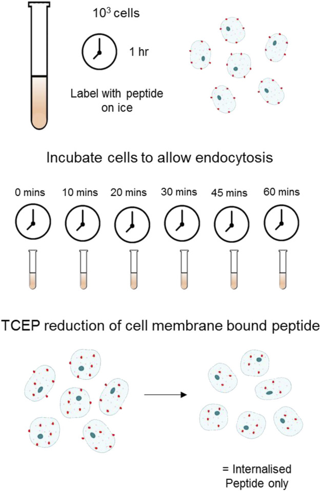
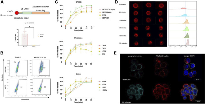
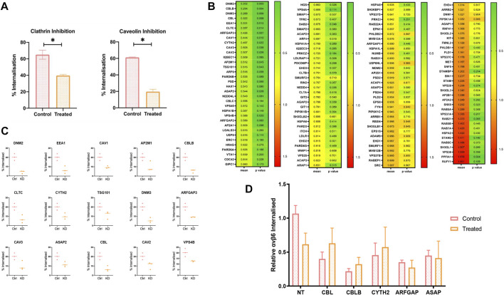
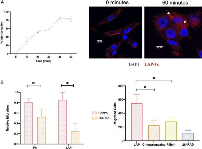
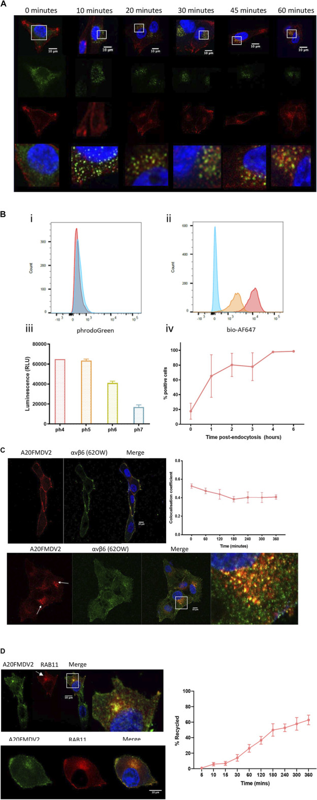
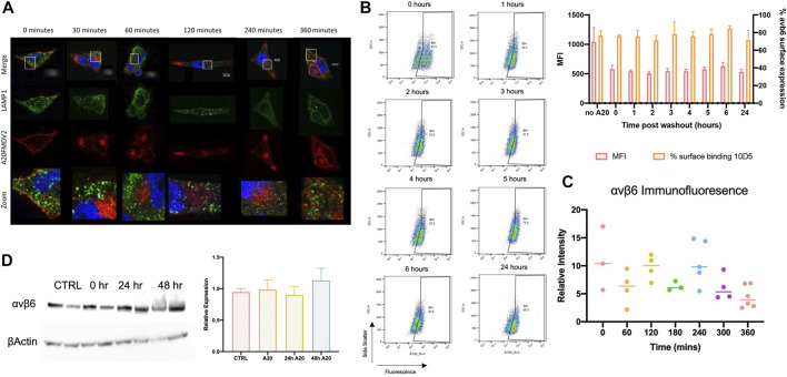
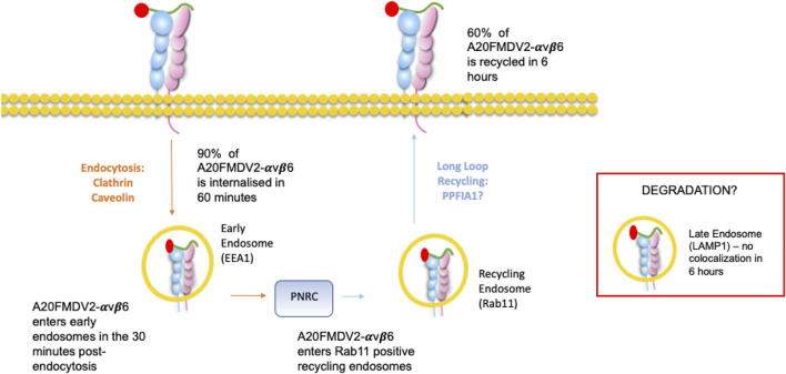
The integrin αvβ6 is expressed at low levels in most normal healthy tissue but is very often upregulated in a disease context including cancer and fibrosis. Integrins use endocytosis and trafficking as a means of regulating their surface expression and thus their functions, however little is known of how this process is regulated in the context of αvβ6. As αvβ6 is a major target for the development of therapeutics in cancer and fibrosis, understanding these dynamics is critical in the development of αvβ6-targeted therapies. Following development of a flow cytometry-based assay to measure ligand (A20FMDV2 or LAP)-bound αvβ6 endocytosis, an siRNA screen was performed to identify which genes were responsible for internalising αvβ6. These data identified 15 genes (DNM2, CBLB, DNM3, CBL, EEA1, CLTC, ARFGAP3, CAV1, CYTH2, CAV3, CAV2, IQSEC1, AP2M1, TSG101) which significantly decreased endocytosis, predominantly within dynamin-dependent pathways. Inhibition of these dynamin-dependent pathways significantly reduced αvβ6-dependent migration (αvβ6-specific migration was 547 ± 128 under control conditions, reduced to 225 ± 73 with clathrin inhibition, and 280 ± 51 with caveolin inhibition). Colocalization studies of αvβ6 with endosome markers revealed that up to 6 h post-internalisation of ligand, αvβ6 remains in Rab11-positive endosomes in a perinuclear location, with no evidence of αvβ6 degradation up to 48 h post exposure to A20FMDV2. Additionally, 60% of ligand-bound αvβ6 was recycled back to the surface by 6 h. With studies ongoing using conjugated A20FMDV2 to therapeutically target αvβ6 in cancer and fibrosis, these data have important implications. Binding of A20FMDV2 seemingly removes much of the αvβ6 from the cell membrane, and upon its recycling, a large fraction appears to still be in the ligand-bound state. While these results are observed with A20FMDV2, these data will be of value in the design of αvβ6-specific therapeutics and potentially the types of therapeutic load.

Keywords: A20FMDV2; endocytosis; internalisation; trafficking; αvβ6.


Conflict of interest statement

The authors declare that the research was conducted in the absence of any commercial or financial relationships that could be construed as a potential conflict of interest.

Figures

FIGURE 1
FIGURE 1
Schematic of Flow Cytometry Internalisation Assay using TCEP Reduction Cells labelled with a fluorescently tagged peptide were incubated at 37°C in appropriate cell culture medium + FBS for the indicated time allowing for internalisation. Following endocytosis and washing, 100 mM of TCEP was added to each sample to cleave the fluorochrome selectively from the surface bound pool of peptide.
FIGURE 2
FIGURE 2
αvβ6 bound A20FMDV2 is internalised into cells over 60 min. (A, Top) Design of the Cy3/5-SS-(GS)5-bioA20FMDV2 featuring a disulphide bond between the fluorochrome and GS linker, to allow for cleavage of the fluorochrome only. (Bottom) Pre-treating A375PB6 cells with αvβ6 specific antibody 53A2 significantly reduced binding of Cy3-SS-(GS)5-bioA20FMDV2 (74.1 ± 26.4% to 0.44 ± 0.17%, p < 0.05), confirming it retained specificity. Data represents live single cells, mean ± SD, n = 3, paired (B) A375P and A375PB6 cell lines were exposed to 100 nM of Cy3-SS-(GS)5-bioA20FMDV2 peptide for 1 hour, and binding quantified by flow cytometry, using Cy3-bioA20ran to determine the non-specific binding and gate the positive fraction. (C) Internalisation of Cy5-SS-(GS)5-bioA20FMDV2 was determined in a range of cell types of breast, pancreas and lung origin. Each panel consisted of both normal and cancer cell lines. No Significant differences were observed in the rate of internalisation between cell types. In each cell line, at least 50% of peptide was internalised in 30 min. (D) A20FMDV2 internalisation quantification using ImageStream flow cytometer. (Left) representative images of cells at each time point revealing that Cy5-(GS)5-bioA20FMDV2 is internalised from the cell membrane from 10 min of incubation at 37°C. (Right) Quantification of the relative internalisation shows that the proportion of Cy5-(GS)5-bioA20FMDV2 inside the cells at each time point is 0 min = 11.5%; 10 min = 27.8%; 20 min = 74.1%; 30 min = 76.7%; 40 min = 84.4%; 50 min = 94.6%. Data represents single live cells only. (E) Immunofluorescent staining of Cy5-(GS)5-bioA20FMDV2- αvβ6 endocytosis. αvβ6 bound A20FMDV2 is exclusively localised at the cell membrane at 0 min, while after 60 min, αvβ6 can be seen inside the cell. Images were acquired using LSM880 confocal microscope using a ×63 objective. Cy5-(GS)5-bioA20FMDV2 (Cyan), DAPI (blue) and Phalloidin (Red).
FIGURE 3
FIGURE 3
An siRNA screen to determine molecular regulators of A20FMDV2-αvβ6 endocytosis. (A) C76 cells were pre-treated with either 2 ug/ml of Chlorpromazine (clathrin inhibitor) or 10 ug/ml of Filipin (Caveolin inhibitor) before performing A20FMDV2 internalisation assay as described. Both Clathrin and Caveolin inhibition resulted in a significant decrease (p < 0.05) in the rate of internalization (65 ± 9.5% to 39 ± 2.5% and 61 ± 0.8% to 19.7 ± 3.9%, respectively) (n = 3). (B) Cherry-picked siRNA library featuring 140 gene pools was transfected in to C76 cells. 72 h post transfection an internalisation assay by flow cytometry was performed as previously described. The first column of the heat map represents the relative internalisation compared to the non-targeting, with green representing a decrease and red indicating an increase. The second column represents the p-value, with green representing a lower p value, and red a higher p value. Data represents mean values (n = 4). (C) Fifteen genes are shown from the flow cytometry screen which significantly reduced the rate of αvβ6-A02FMDV2 endocytosis in to C76 cells (p < 0.05). Data represents mean ± SD. p < 0.05, as determined by student’s t-test. (D) C76 cells were transfected with 50 mM of the indicated siRNA smartpool, 72 h later the cells were either treated with Clathrin inhibitor or vehicle control, and an internalisation assay performed as previously described using A20FMDV2. Again, compared with the non-targeting controls KD of each gene resulted in significant reduction in internalisation (NT vs. CBL: p = 0.01; CBLB: p = 0.002; CYTH2: p = 0.04; ARFGAP: p = 0.019; ASAP: p = 0.0125). However, there was no significant change in internalisation with the addition of Clathrin inhibitor, compared to the siRNA knockdown alone [NT 24 ± 4.7% vs. 14 ± 6.5% (p = 0.09), CBL 9 ± 4% vs. 14 ± 8%, CBLB 5 ± 1.6% vs. 7.4 ± 3.9%, CYTH2 10 ± 7% vs. 13 ± 11.7%, ARFGAP 8 ± 1% vs. 6.2 ± 3.5%, ASAP 10.3 ± 3.1% vs. 9.4 ± 9.9%] (n = 3). Data suggest that these five genes operate in the clathrin-mediated pathway.
FIGURE 4
FIGURE 4
Inhibition of ligand-bound αvβ6 endocytosis reduces the rate of cell migration. (A) Internalisation of LAP in C76 cells. (Left) Using flow cytometry, C76 internalised 83 ± 10% of LAP-Fc in 60 min Data represents mean ± SD (n = 3). (Right) LAP-Fc internalisation was also assessed using immunofluorescent microscopy, again revealing internalisation of LAP-Fc (red) inside the cell after 60 min. (B) αvβ6 mediated migration. (Left) Transwells were coated with either fibronectin (Fn) or LAP, and 50,000 C76 cells seeded in top of the transwell, either treated with 264RAD or an IGG control. Treatment with 264RAD caused a significant decrease in migration towards LAP but not Fn. Data represents mean ± SD (n = 3). (Right) Transwells were coated with 0.5 μg/ml of LAP and added to the top of the transwell in the presence of chlorpromazine, filipin, 264RAD or vehicle control. Significant reductions in the number of cells migrating through the transwells were observed with chlorpromazine, filipin and 264RAD.
FIGURE 5
FIGURE 5
(Continued).
FIGURE 6
FIGURE 6
αvβ6 degradation post-endocytosis. (A) Representative images show the distribution of A20FMDV2 (red) and LAMP1 (green) in A375β6 cells following Cy5-(GS)5-bioA20FMDV2 internalisation. LAMP1 staining is consistent in each time point, with no indication of changes over time (up to 360 min). (B) αvβ6 surface expression was monitored using 10D5 by flow cytometry (example dot-plots and summary histogram shown) post-exposure to A20FMDV2 up to 24 h after wash-out, with no significant differences observed in expression over this time course in the percentage of cells bound to αvβ6. There was a significant reduction in MFI of 10D5 binding with and without A20FMDV2 pretreatment (n = 3). (C) Quantification of 62OW staining intensity from images acquired in Figure 5, revealing no significance in the intensity up to 6 h post A20FMDV2 exposure. (D) αvβ6 expression was also quantified by western blotting for the β6 subunit, with no changes in expression up to 48 h post-exposure to the ligand. Data represents mean ± SD, n = 3. Blot features two technical repeats per condition.
FIGURE 7
FIGURE 7
Ligand-bound endocytosis and trafficking of αvβ6 Summary figure for this study. A20FMDV2-αvβ6 internalises by clathrin and caveolin mediated endocytosis into early endosomes, where it colocalises with EEA1 in the first 30 min post endocytosis. From the early endosome, A20FMDV2-αvβ6 accumulates in perinuclear clusters, which are positive for Rab11 (1–6 h post endocytosis). Within this 6-h time frame, 60% of internalised A20FMDV2-αvβ6 is recycled back to the cell surface.

References

    1. Ahmed N., Niu J., Dorahy D. J., Gu X., Andrews S., Meldrum C. J., et al. (2002). Direct integrin alphavbeta6-ERK binding: Implications for tumour growth. Oncogene 21 (9), 1370–1380. 10.1038/sj.onc.1205286 - DOI - PubMed
    1. Arjonen A., Alanko J., Veltel S., Ivaska J. (2012). Distinct recycling of active and inactive β1 integrins. Traffic 13 (4), 610–625. 10.1111/j.1600-0854.2012.01327.x - DOI - PMC - PubMed
    1. Asperti C., Astro V., Totaro A., Paris S., de Curtis I. (2009). Liprin-alpha1 promotes cell spreading on the extracellular matrix by affecting the distribution of activated integrins. J. Cell Sci. 122 (18), 3225–3232. 10.1242/jcs.054155 - DOI - PubMed
    1. Astro V., Asperti C., Cangi M. G., Doglioni C., de Curtis I., de CurtIs I. (2011). Liprin-α1 regulates breast cancer cell invasion by affecting cell motility, invadopodia and extracellular matrix degradation. Oncogene 30 (15), 1841–1849. 10.1038/onc.2010.562 - DOI - PubMed
    1. Balderhaar H. J. k., Ungermann C. (2013). CORVET and HOPS tethering complexes – coordinators of endosome and lysosome fusion. J. Cell Sci. 126 (6), 1307–1316. 10.1242/jcs.107805 - DOI - PubMed