. 2022 Aug 25:13:938680.
doi: 10.3389/fendo.2022.938680. eCollection 2022.

N-acetyl-L-cysteine treatment reduces beta-cell oxidative stress and pancreatic stellate cell activity in a high fat diet-induced diabetic mouse model

Affiliations

N-acetyl-L-cysteine treatment reduces beta-cell oxidative stress and pancreatic stellate cell activity in a high fat diet-induced diabetic mouse model

Meg Schuurman et al. Front Endocrinol (Lausanne). .

Abstract

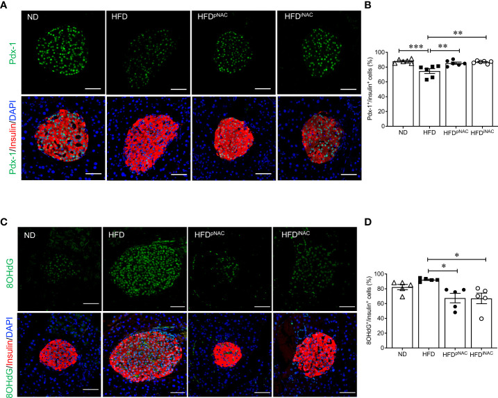
Obesity plays a major role in type II diabetes (T2DM) progression because it applies metabolic and oxidative stress resulting in dysfunctional beta-cells and activation of intra-islet pancreatic stellate cells (PaSCs) which cause islet fibrosis. Administration of antioxidant N-acetyl-L-cysteine (NAC) in vivo improves metabolic outcomes in diet-induced obese diabetic mice, and in vitro inhibits PaSCs activation. However, the effects of NAC on diabetic islets in vivo are unknown. This study examined if dosage and length of NAC treatment in HFD-induced diabetic mice effect metabolic outcomes associated with maintaining healthy beta-cells and quiescent PaSCs, in vivo. Male C57BL/6N mice were fed normal chow (ND) or high-fat (HFD) diet up to 30 weeks. NAC was administered in drinking water to HFD mice in preventative treatment (HFDpNAC) for 23 weeks or intervention treatment for 10 (HFDiNAC) or 18 (HFDiNAC+) weeks, respectively. HFDpNAC and HFDiNAC+, but not HFDiNAC, mice showed significantly improved glucose tolerance and insulin sensitivity. Hyperinsulinemia led by beta-cell overcompensation in HFD mice was significantly rescued in NAC treated mice. A reduction of beta-cell nuclear Pdx-1 localization in HFD mice was significantly improved in NAC treated islets along with significantly reduced beta-cell oxidative stress. HFD-induced intra-islet PaSCs activation, labeled by αSMA, was significantly diminished in NAC treated mice along with lesser intra-islet collagen deposition. This study determined that efficiency of NAC treatment is beneficial at maintaining healthy beta-cells and quiescent intra-islet PaSCs in HFD-induced obese T2DM mouse model. These findings highlight an adjuvant therapeutic potential in NAC for controlling T2DM progression in humans.

Keywords: HFD-induced diabetes; N-acetyl-L-cysteine (NAC); beta-cell oxidative stress; collagen fiber; pancreatic stellate cells (PaSCs).


Conflict of interest statement

The authors declare that the research was conducted in the absence of any commercial or financial relationships that could be construed as a potential conflict of interest.

Figures

Figure 1
Figure 1
Preventive NAC treatment improves glucose metabolism in HFD-induced diabetic mice. (A) Recording of bi-weekly body weight for the duration of 22 weeks (n=8-17 mice/group). Measurement of (B) body weights, (C) overnight (~16 hours) fasting blood glucose and (D) fed plasma insulin levels (n=6-17 mice/group). (E) IPGTT and area under curve (AUC, n=10-14 mice/group), (F) IPITT and AUC (n=6-12 mice/group) and (G) in vivo GSIS (n=3-5 mice/group) of ND, HFD, HFDpNAC and HFDiNAC mice at 22 weeks. Control diets (ND): open triangle; HFD: closed square; HFDpNAC: closed circle; HFDiNAC: open circle. Data are expressed as means ± SEM. *p<0.05, **p<0.01, ***p<0.001, ****p<0.0001; analyzed using one-way ANOVA followed by Tukey’s Post-Hoc test.
Figure 2
Figure 2
Histological analysis of NAC treated HFD pancreata demonstrated preserved beta-cell mass with no change in cell proliferation. (A) Representative double immunofluorescence images for islet morphology, detected by insulin (red) and glucagon (green) staining, and DAPI labeled nuclei (blue). Scale bars: 50µm. (B) Beta cell mass (C) alpha cell mass (D) beta cell size and (E) number (F) islet density in ND, HFD, HFDpNAC and HFDiNAC mouse pancrea at 22 weeks (n=5-6 pancreata/group). (G) Proliferation of beta cells quantified using Ki67 co-localization with insulin+ cells (n=6 pancreata/group). ND: open triangle; HFD: closed square. HFDpNAC: closed circle; HFDiNAC: open circle. Data are expressed as means ± SEM. *p<0.05, **p<0.01, analyzed using one-way ANOVA followed by Tukey’s Post-Hoc test.
Figure 3
Figure 3
NAC treatment preserves beta-cell identity with reduction of beta-cell oxidative stress in HFD-induced diabetic mice. Representative double immunofluorescence images of (A) Pdx-1 (green) (C) 8OHdG (green) co-stained with insulin (red), nuclei are stained with DAPI (blue). Scale bars: 50µm. Quantification of nuclear (B) Pdx-1 (D) 8OHdG in insulin+ cells of ND, HFD, HFDpNAC and HFDiNAC mouse pancreas at 22 weeks (n=5-6 pancreata/group). ND: open triangle; HFD: closed square; HFDpNAC: closed circle; HFDiNAC: open circle. Data are shown as means ± SEM. *p<0.05, **p<0.01, ***p<0.001, analyzed using one-way ANOVA followed by Tukey’s Post-Hoc test.
Figure 4
Figure 4
Preventive NAC treatment reduces intra-islet PaSCs activation and collagen deposition in HFD-induced diabetic mice. Representative double immunofluorescence images of (A) α-SMA (green) or (C) desmin (green) co-stained with insulin (red), nuclei are stained with DAPI (blue). Scale bars: 50µm. Quantification of intra-islet α-SMA (B) or desmin (D) area in ND, HFD, HFDpNAC and HFDiNAC mouse islets at 22 weeks (n=5-6 pancreata/group). (E) Representative trichrome staining images, scale bar: 50μm; and (F) quantification of intra-islet collagen deposition (n=5 pancreata/group). ND: open triangle; HFD: closed square; HFDpNAC: closed circle; HFDiNAC: open circle. Data are shown as means ± SEM. *p<0.05, **p<0.01, ***p<0.001, analyzed using one-way ANOVA followed by Tukey’s Post-Hoc test.
Figure 5
Figure 5
Prolonged intervention NAC treatment improves glucose metabolism in HFD-induced diabetic mice. (A) Recording of biweekly body weight for the duration of 30 weeks (n=8-12 mice/group). Measurement of (B) body weights, (C) overnight (~16 hours) fasting blood glucose, (D) fed plasma insulin, (E) triglycerides and (F) cholesterol levels (n=7-18 mice/group). (G) IPGTT and (H) AUC (n=5-6 mice/group), (I) IPITT and (J) AUC (n=7 mice/group) of ND, HFD and HFDiNAC+ mice at 30 weeks. ND: open triangle; HFD: closed square; HFDiNAC+: open circle. Data are expressed as means ± SEM. *p<0.05, **p<0.01, ***p<0.001, ****p<0.0001, analyzed using one-way ANOVA followed by Tukey’s Post-Hoc test.
Figure 6
Figure 6
Prolonged intervention NAC treatment preserves beta-cell mass and nuclear Pdx-1 with lower 8OHdG in HFD-induced diabetic mice. (A) Representative double immunofluorescence images for islet morphology, detected by insulin (red) and glucagon (green) staining and DAPI labeled nuclei (blue). Scale bars: 50µm. Morphometric quantification of islet density (B) beta cell mass (C) and alpha cell mass (D) (n=5-6 pancreata/group). Representative double immunofluorescence images for (E) Pdx-1 (green) or (G) 8OHdG (green) and insulin (red), nuclei are stained with DAPI (blue). Scale bars: 50µm. Quantification of nuclear Pdx-1 (F) or 8OHdG (H) in insulin+ cells of ND, HFD and HFDiNAC+ mouse pancreas at 30 weeks (n=5-6 pancreata/group). ND: open triangle; HFD: closed square). HFDiNAC+: open circle. Data are shown as means ± SEM. *p<0.05, **p<0.01, analyzed using one-way ANOVA followed by Tukey’s Post-Hoc test.
Figure 7
Figure 7
Prolonged intervention NAC treatment reduces intra-islet PaSCs activation and collagen deposition in HFD-induced diabetic mice. (A) Representative double immunofluorescence images of α-SMA (green) co-stained with insulin (red), nuclei are stained with DAPI (blue). Scale bars, 50µm; (B) quantification of intra-islet α-SMA area in ND, HFD and HFDiNAC+ mouse islets at 30 weeks (n=3 pancreata/group). (C) Representative trichrome staining images of intra-islet collagen deposition. Scale bar: 50μm. ND: open triangle; HFD: closed square; HFDiNAC+: open circle) Data are shown as means ± SEM. *p<0.05, ***p<0.001, analyzed using one-way ANOVA followed by Tukey’s Post-Hoc test.
Figure 8
Figure 8
Proposed model of NAC maintains healthy beta-cells and quiescent PaSCs in HFD-induced diabetic mouse islet. In a healthy condition under normal chow diet, intra-islet PaSCs are quiescent; they produce suitable cytokines and ECM for maintaining islet structure and facilitating healthy beta-cell function. Under high-fat diet, increased oxidative stress promoted intra-islet PaSC activation resulting in increased cytokine production and ECM deposition which diminished beta-cell identity and function. Given NAC treatment, HFD islets showed significantly reduced oxidative stress and intra-islet PaSC activation with enhanced beta-cell identity and function.

Similar articles

Cited by

References

    1. Prentki M, Nolan CJ. Islet β cell failure in type 2 diabetes. J Clin Invest (2006) 116:1802–12. doi: 10.1172/JCI29103 - DOI - PMC - PubMed
    1. Keane KN, Cruzat VF, Carlessi R, de Bittencourt PIH, Newsholme P. Molecular events linking oxidative stress and inflammation to insulin resistance and β-cell dysfunction. Oxid Med Cell Longev (2015) 2015:181643. doi: 10.1155/2015/181643 - DOI - PMC - PubMed
    1. McAdam-Marx C, Mukherjee J, Bellows BK, Unni S, Ye X, Iloeje U, et al. . Evaluation of the relationship between weight change and glycemic control after initiation of antidiabetic therapy in patients with type 2 diabetes using electronic medical record data. Diabetes Res Clin Pract (2014) 103:402–11. doi: 10.1016/j.diabres.2013.12.038 - DOI - PubMed
    1. Temelkova-Kurktschiev T, Siegert G, Bergmann S, Henkel E, Koehler C, Jaroß W, et al. . Subclinical inflammation is strongly related to insulin resistance but not to impaired insulin secretion in a high risk population for diabetes. Metabolism (2002) 51:743–9. doi: 10.1053/meta.2002.32804 - DOI - PubMed
    1. Butler AE, Janson J, Bonner-Weir S, Ritzel R, Rizza RA, Butler PC. β-cell deficit and increased β-cell apoptosis in humans with type 2 diabetes. Diabetes (2003) 52:102–10. doi: 10.2337/diabetes.52.1.102 - DOI - PubMed

Publication types