[Preprint]. 2022 Aug 31:2022.08.30.22279344.
doi: 10.1101/2022.08.30.22279344.

Recent SARS-CoV-2 infection abrogates antibody and B-cell responses to booster vaccination

Affiliations

Recent SARS-CoV-2 infection abrogates antibody and B-cell responses to booster vaccination

Clarisa M Buckner et al. medRxiv. .

Update in

Abstract

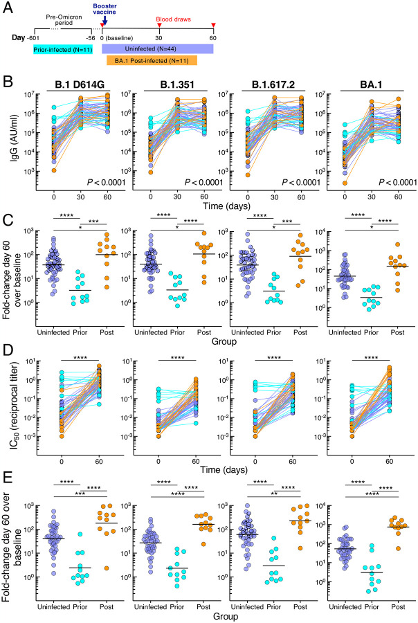
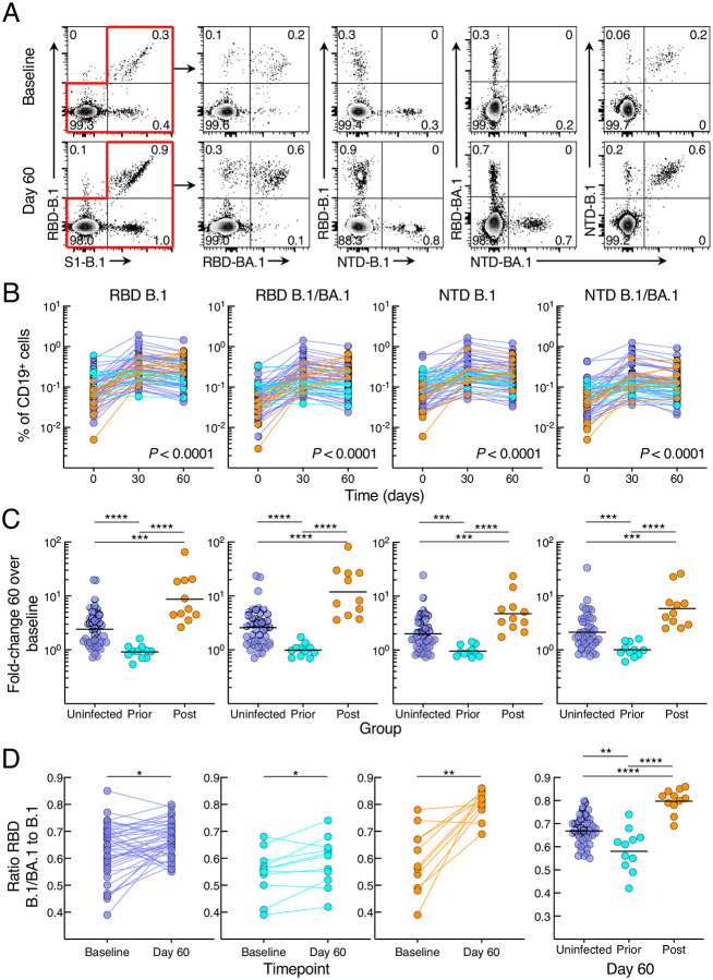
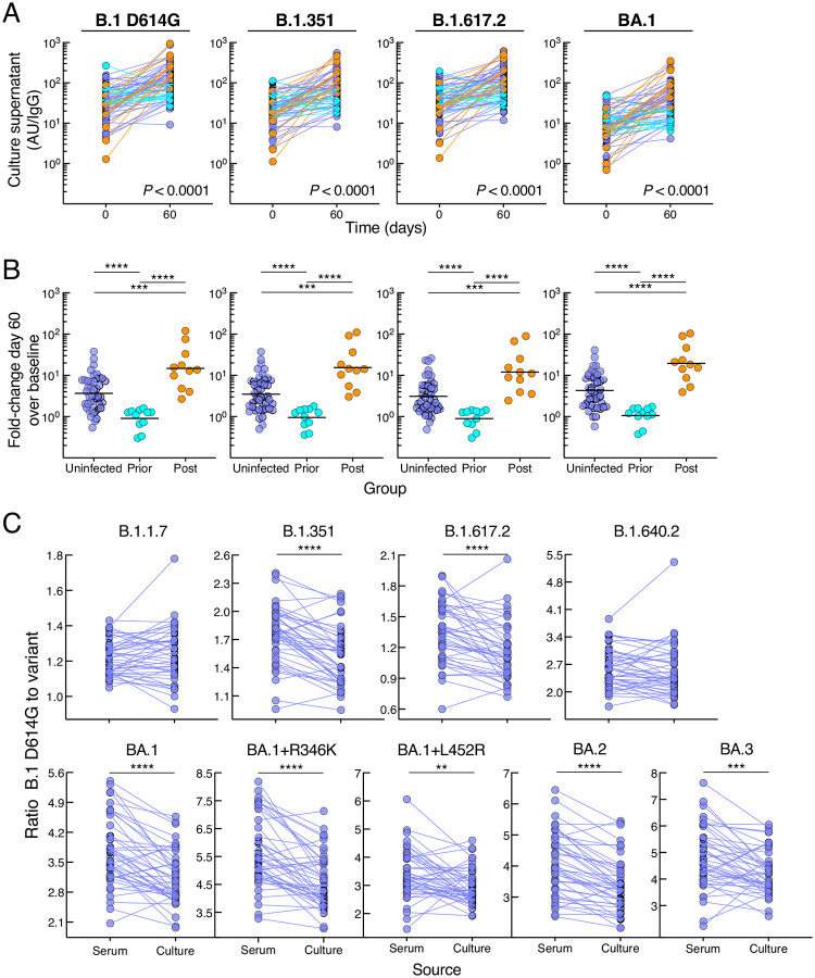
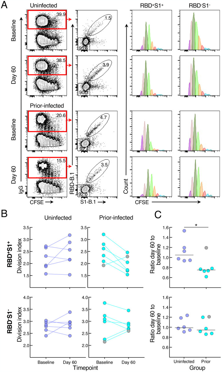
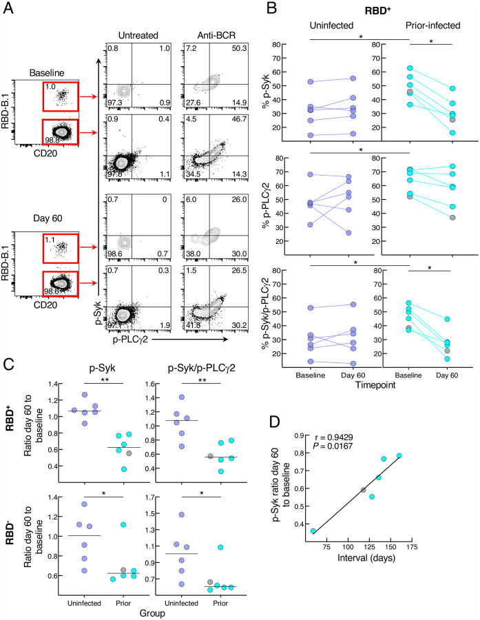
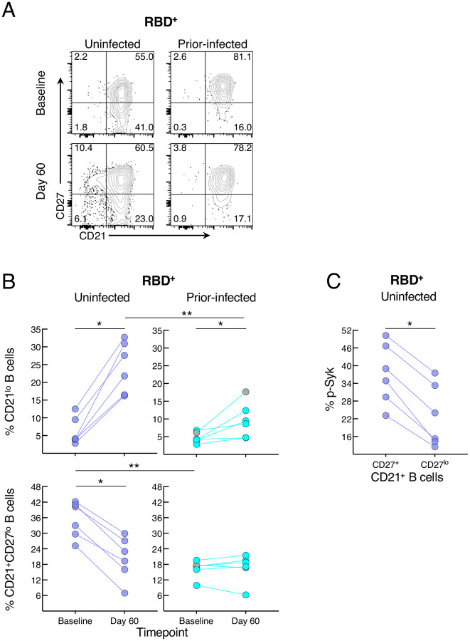
SARS-CoV-2 mRNA booster vaccines provide protection from severe disease, eliciting strong immunity that is further boosted by previous infection. However, it is unclear whether these immune responses are affected by the interval between infection and vaccination. Over a two-month period, we evaluated antibody and B-cell responses to a third dose mRNA vaccine in 66 individuals with different infection histories. Uninfected and post-boost but not previously infected individuals mounted robust ancestral and variant spike-binding and neutralizing antibodies, and memory B cells. Spike-specific B-cell responses from recent infection were elevated at pre-boost but comparatively less so at 60 days post-boost compared to uninfected individuals, and these differences were linked to baseline frequencies of CD27 lo B cells. Day 60 to baseline ratio of BCR signaling measured by phosphorylation of Syk was inversely correlated to days between infection and vaccination. Thus, B-cell responses to booster vaccines are impeded by recent infection.


Conflict of interest statement

DECLARATION OF INTERESTS

The authors declare no competing interests.

Figures

Figure 1.
Figure 1.. SARS-CoV-2 infection status alters antibody responses to booster vaccination
(A) Graphical depiction of cohort and vaccination/blood draw schedule. (B) IgG spike-binding titers expressed as AU/ml serum. (C) Fold-change day 60 over baseline in serum IgG spike-binding titers. (D) Serum neutralizing titers against SARS-CoV-2 lentiviral pseudovirus expressed as reciprocal IC50. N=41 for uninfected group. (E) Fold-change day 60 over baseline in serum neutralizing titers against SARS-CoV-2 lentiviral pseudovirus. Numerical p-values represent mixed-effect model testing of group by time interactions; see Table S2 for additional p-values. Horizontal lines represent geometrical means and coloring scheme matches groups described in Figure 1A.
Figure 2.
Figure 2.. SARS-CoV-2 infection status alters B-cell responses to booster vaccination
(A) Representative binding of B.1 and BA.1 RBD and NTD tetramers to non-naïve B cells at baseline and day 60 post booster vaccination. (B) Frequencies of B.1 and BA.1 RBD and NTD tetramer-binding B cells. (C) Fold-change day 60 over baseline of B.1 and BA.1 RBD and NTD tetramer-binding B-cell frequencies. (D) Ratio of dual B.1/BA.1 to single B.1 RBD binding to B cells. Binding frequencies represent percentage of total CD19+ B cells; see also Figure S2A for gating strategy. Numerical p-values represent mixed-effect model testing; see Table S3 for additional p-values. Horizontal lines represent geometrical means and coloring scheme matches groups described in Figure 1A.
Figure 3.
Figure 3.. SARS-CoV-2 infection status alters secreted antibody responses to booster vaccination
(A) IgG spike-binding titers in culture supernatant expressed as AU/IgG. (B) Fold-change day 60 over baseline of IgG spike-binding titers in culture supernatant. (C) Ratio of B.1 D614G to variant IgG spike-binding titers in serum and culture supernatant. Culture supernatant titers represent spike-binding IgG per total IgG. Numerical p-values represent mixed-effect model testing; see Table S5 for additional p-values. Horizontal lines represent geometrical means and coloring scheme matches groups described in Figure 1A.
Figure 4.
Figure 4.. SARS-CoV-2 recent infection decreases proliferative response of spike-specific B cells after booster vaccination
(A) Representative baseline and day 60 CFSE-dilution plots of RBD+S1+ and RBDS1 IgG-expressing B cells among stimulated PBMCs. Numbers represent percentages. (B) Division indices of RBD+S1+ and RBDS1 IgG-expressing B cells. (C) Ratio of day 60 to baseline division indices of RBD+S1+ and RBDS1 IgG-expressing B cells. CFSE-labeled PBMCs were stimulated for four days then stained with anti-IgG, B.1 S1 and RBD tetramers and other markers shown with gating strategy in Figure S4B. Division indices were measured for RBD+S1+ and RBDS1 IgG-gated B cells. Horizontal lines represent medians and coloring scheme matches groups described in Figure 1A. Gray circles refer to subject shown in Figure S4A.
Figure 5.
Figure 5.. SARS-CoV-2 recent infection decreases BCR signaling response of spike-specific B cells after booster vaccination
(A) Representative baseline and day 60 phosphorylation of Syk and PLCγ2 with and without anti-BCR treatment shown for RBD+ and RBD non-naïve B cells of a recent prior-infected subject. Numbers represent percentages. (B) Anti-BCR induced phosphorylation of Syk and PLCγ2 among RBD+ B cells, showing untreated subtracted frequencies. (C) Ratio of day 60 to baseline of anti-BCR induced phosphorylation of Syk and Syk/PLCγ2 among RBD+ and RBD B cells. (D) Correlation of p-Syk ratio day 60 to baseline and time interval between infection and vaccination. PBMCs were stained with B.1 RBD tetramer, IgD and CD27 for gating on non-naïve B cells and other markers (see Figure S2B) and incubated at 37°C for two minutes with or without anti-BCR, then stained for phosphorylated markers shown. Horizontal lines represent medians and coloring scheme matches groups described in Figure 1A. Gray circles refer to subject shown Figure S4A.
Figure 6.
Figure 6.. SARS-CoV-2 recent infection alters the phenotype of spike-specific B cells
(A) Representative expression of CD21 and CD27 among RBD+ B cells at baseline and day 60 post-boost. Numbers represent percentages. (B) Frequencies of CD21lo and CD21+CD27lo RBD+ B cells at baseline and day 60 post-boost. (C) Baseline uninfected anti-BCR induced phosphorylation of Syk among CD21+CD27lo and CD21+CD27+ RBD+ B cells. Populations shown in (A) and frequencies measured in (B) were from the CD19+ gate shown in Figure S2A and those in (C) further gated on CD21+ B cells. Horizontal lines represent medians and coloring scheme matches groups described in Figure 1A. Gray circles refer to subject shown in Figure S4A.

References

    1. Altarawneh H.N., Chemaitelly H., Ayoub H.H., Tang P., Hasan M.R., Yassine H.M., Al-Khatib H.A., Smatti M.K., Coyle P., Al-Kanaani Z., et al. (2022). Effects of Previous Infection and Vaccination on Symptomatic Omicron Infections. N Engl J Med 387, 21–34. 10.1056/NEJMoa2203965. - DOI - PMC - PubMed
    1. Andreano E., Paciello I., Piccini G., Manganaro N., Pileri P., Hyseni I., Leonardi M., Pantano E., Abbiento V., Benincasa L., et al. (2021). Hybrid immunity improves B cells and antibodies against SARS-CoV-2 variants. Nature 600, 530–535. 10.1038/s41586-021-04117-7. - DOI - PMC - PubMed
    1. Andrews S.F., Chambers M.J., Schramm C.A., Plyler J., Raab J.E., Kanekiyo M., Gillespie R.A., Ransier A., Darko S., Hu J., et al. (2019). Activation Dynamics and Immunoglobulin Evolution of Pre-existing and Newly Generated Human Memory B cell Responses to Influenza Hemagglutinin. Immunity 51, 398–410 e395. 10.1016/j.immuni.2019.06.024. - DOI - PubMed
    1. Baden L.R., El Sahly H.M., Essink B., Kotloff K., Frey S., Novak R., Diemert D., Spector S.A., Rouphael N., Creech C.B., et al. (2021). Efficacy and Safety of the mRNA-1273 SARS-CoV-2 Vaccine. N Engl J Med 384, 403–416. 10.1056/NEJMoa2035389. - DOI - PMC - PubMed
    1. Baumgarth N. (2021). The Shaping of a B Cell Pool Maximally Responsive to Infections. Annu Rev Immunol 39, 103–129. 10.1146/annurev-immunol-042718-041238. - DOI - PMC - PubMed

Publication types