

Detection of circulating tumor cells and evaluation of epithelial-mesenchymal transition patterns of circulating tumor cells in ovarian cancer
- PMID: 36093536
- PMCID: PMC9459537
- DOI: 10.21037/tcr-22-529
Detection of circulating tumor cells and evaluation of epithelial-mesenchymal transition patterns of circulating tumor cells in ovarian cancer
Abstract
Background: Circulating tumor cells (CTCs) have considered to be promising liquid biopsy in cancer due to the intact information of whole cells and the potential to reflect micrometastasis. However, CTCs research are extremely limited in ovarian cancer, probably due to their rarity. The predictive value of CTCs and circulating tumor microemboli (CTM) in metastasis remains to be elucidated in ovarian cancer. This study tried to identify CTCs/CTM in ovarian cancer with considerably positive rate. To preliminarily identify the invasive capacity of CTCs/CTM, the epithelial-mesenchymal transition (EMT) patterns of CTCs/CTM was evaluated. Moreover, for comprehensive understanding of invasiveness of disseminated cells in ovarian cancer, EMT pattern of exfoliated tumor cells in ascites were also confirmed in this study.
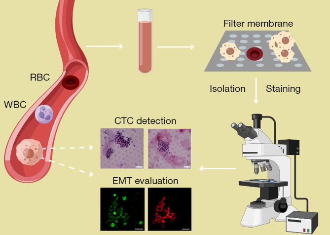
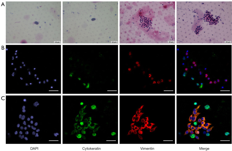
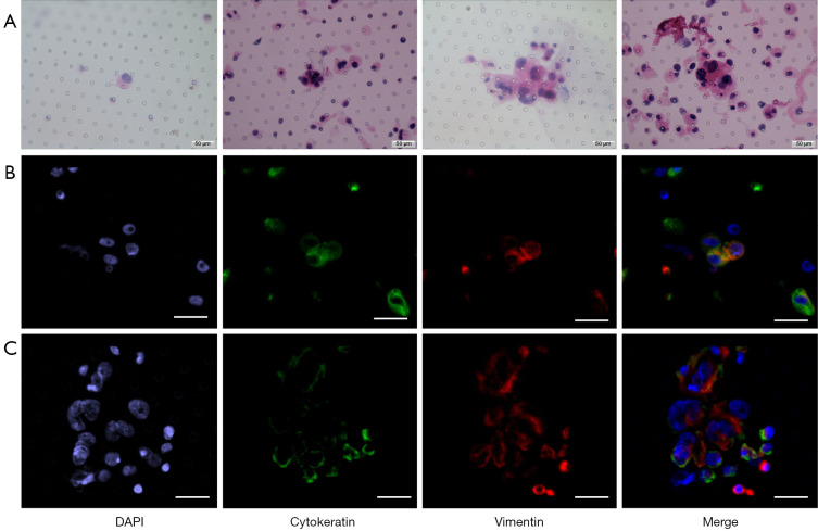
Methods: Peripheral blood samples and ascites samples were collected from 22 ovarian cancer patients. The Microfiltration combined with morphological analysis was used to detect CTC single cells or cell clusters. Microfiltration combined with morphological analysis was applied in the CTC isolation and identification. EMT was evaluated by immunofluorescence via markers including vimentin and cytokeratin.
Results: Microfiltration combined with morphological analysis was introduced to detect CTCs/CTM with a positivity rate of 40.9% in ovarian cancer patients. The number of CTC varied from 1 to 8, with CTM number from 4 to 30. CTCs/CTM of all samples have experienced EMT process. Vimentin was expressed in all CTC samples and all tumor cells in ascites, while cytokeratin was expressed in 44.4% (4/9) of CTC samples. There were no significant differences of the clinical parameters between the CTC-positive and CTC-negative patients.
Conclusions: This study showed that both CTCs/CTM and detached tumor cells in ascites might have undergone complete or partial EMT in ovarian cancer. Moreover, microfiltration combined with cytomorphological analysis showed a considerable CTC detection rate.
Keywords: Ovarian neoplasms; circulating tumor cells (CTCs); epithelial-mesenchymal transition; metastasis.
2022 Translational Cancer Research. All rights reserved.
Conflict of interest statement
Conflicts of Interest: All authors have completed the ICMJE uniform disclosure form (available at https://tcr.amegroups.com/article/view/10.21037/tcr-22-529/coif). CJX reports funding made to the Obstetrics and Gynecology Hospital of Fudan University from the Shanghai Medical Center of Key Programs for Female Reproductive Diseases (grant No. 2017ZZ01016). XYZ reports funding made to the Obstetrics and Gynecology Hospital of Fudan University from the National Natural Science Foundation of China (grant No. 82172747). The other authors have no conflicts of interest to declare.
Figures
References
LinkOut - more resources
Full Text Sources