. 2022 Sep 20;61(18):2036-2048.
doi: 10.1021/acs.biochem.2c00364. Epub 2022 Sep 12.

C3- and C3/C5-Epimerases Required for the Biosynthesis of the Capsular Polysaccharides from Campylobacter jejuni

Affiliations

C3- and C3/C5-Epimerases Required for the Biosynthesis of the Capsular Polysaccharides from Campylobacter jejuni

Manas K Ghosh et al. Biochemistry. .

Abstract

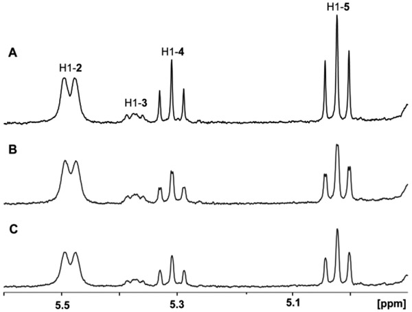
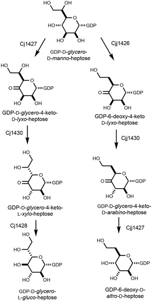
Campylobacter jejuni is a human pathogen and one of the leading causes of food poisoning in Europe and the United States. The outside of the bacterium is coated with a capsular polysaccharide that assists in the evasion of the host immune system. Many of the serotyped strains of C. jejuni contain a 6-deoxy-heptose moiety that is biosynthesized from GDP-d-glycero-d-manno-heptose by the successive actions of a 4,6-dehydratase, a C3/C5-epimerase, and a C4-reductase. We identified 18 different C3/C5-epimerases that could be clustered together into three groups at a sequence identity of >89%. Four of the enzymes from the largest cluster (from serotypes HS:3, HS:10, HS:23/36, and HS:41) were shown to only catalyze the epimerization at C3. Three enzymes from the second largest cluster (HS:2, HS:15, and HS:42) were shown to catalyze the epimerization at C3 and C5. Enzymes from the third cluster were not characterized. The three-dimensional structures of the epimerases from serotypes HS:3, HS:23/36, HS:15, and HS:41 were determined to resolutions of 1.5-1.9 Å. The overall subunit architecture places these enzymes into the diverse "cupin" superfamily. Within X-ray coordinate error, the immediate regions surrounding the active sites are identical, suggesting that factors extending farther out may influence product outcome. The X-ray crystal structures are consistent with His-67 and Tyr-134 acting as general acid/base catalysts for the epimerization of C3 and/or C5. Two amino acid changes (A76V/C136L) were enough to convert the C3-epimerase from serotype HS:3 to one that could now catalyze the epimerization at both C3 and C5.


Conflict of interest statement

The authors declare no competing conflicts of interests.

Figures

Figure 1.
Figure 1.
Sequence similarity networks (SSN) for the Cj1430-like epimerases from C. jejuni. (A) The closest 1000 sequences to Cj1430 from the HS:2 serotype at a sequence identity cutoff of 60%. The initial target protein from serotype HS:2 is shown in yellow and the related epimerases from the other serotyped strains of C. jejuni are shown in green. (B) SSN for the epimerases from 18 serotyped strains of C. jejuni at a sequence identity of 89%. The specific serotype is labeled in each circle. Yellow and green colors represent the C3/C5- and C3-epimerases that were tested for catalytic activity, whereas the gray color represent epimerases that have not been tested for catalytic activity.
Figure 2.
Figure 2.
1H NMR 400 MHz spectra of products formed from GDP-6-deoxy-4-keto-α-d-lyxo-heptose (2) after the addition of the C3/C5-epimerases from serotypes HS:2, HS:15, or HS:42. Reaction products include GDP-6-deoxy-4-keto-α-d-arabino-heptose (3), GDP-6-deoxy-4-keto-β-l-ribo-heptose (4) and GDP-6-deoxy-4-keto-β-l-xylo-heptose (5) formed by the catalytic activity of epimerases from serotype HS:2 (A), HS:15 (B) or HS:42 (C). In these experiments 4.0 μM of the epimerase was incubated with 4.0 mM of compound 2 for 30 min prior to the acquisition of the NMR spectrum of the products.
Figure 3.
Figure 3.
1H NMR 400 MHz spectra of product formed from GDP-6-deoxy-4-keto-α-d-lyxo-heptose (2) by the addition of C3-epimerases from serotype HS:3, HS:10, HS:23/36, or HS:41. The sole reaction product is GDP-6-deoxy-4-keto-α-d-arabino-heptose (3) formed by the catalytic activity of epimerases from HS:3 (A), HS:10 (B), HS:23/36 (C), or HS:41 (D). In these experiments 4.0 μM of the epimerase was incubated with 4.0 mM of compound 2 for 30 min prior to the acquisition of the NMR spectrum of the products.
Figure 4.
Figure 4.
1H NMR 400 MHz spectra of products formed from GDP-6-deoxy-4-keto-α-d-lyxo-heptose (2) after the addition of the epimerase from serotype HS:2 as a function of time. (A) Control reaction in absence of enzyme, (B) 15 min, (C) 45 min, (D) 75 min, (E) 105 min, and (F) 135 min. Relative integration values for shaded peaks are shown in parentheses.
Figure 5.
Figure 5.
Time course for the formation of reaction products from GDP-6-deoxy-4-keto-α-d-lyxo-heptose (2) after the addition of the Cj1430 epimerase from serotype HS:2. GDP-6-deoxy-4-keto-α-d-lyxo-heptose (2), black circle; GDP-6-deoxy-4-keto-α-d-arabino-heptose (3), red triangle; GDP-6-deoxy-4-keto-β-l-ribo-heptose (4), green square; and GDP-6-deoxy-4-keto-β-l-xylo-heptose (5), blue diamond.
Figure 6:
Figure 6:
Multiple sequence alignment for the epimerases from serotypes HS:2, HS:3, HS:10, HS:15, HS:23, HS:12, and HS:42. The residues highlighted in yellow illustrate those that are conserved in the C3-epimerases while another amino acid is conserved (shaded grey) is conserved in the C3/C5-epimerases. Two active site general acid/base groups (histidine and tyrosine) are highlighted in blue.
Figure 7.
Figure 7.
1H NMR 400 MHz spectra of products formed from GDP-6-deoxy-4-keto-α-D-lyxo-heptose (2) by the addition of wild-type and variant epimerases from serotype HS:3. Reaction products GDP-6-deoxy-4-keto-α-d-arabino-heptose (3), GDP-6-deoxy-4-keto-β-l-ribo-heptose (4) and GDP-6-deoxy-4-keto-β-l-xylo-heptose (5) formed by the catalytic activity of epimerases from HS:2 (A), HS:3 (B), A76V/C136L HS:3, (C) and A76V/A122S/C136L HS:3 (D). The epimerases (4 μM) were incubated with (2) for 15 h at 20 °C (pD 7.5).
Figure 8:
Figure 8:
Structure of the C-3 epimerase from C. jejuni serotype HS:3. Shown in (A) is a ribbon drawing of the dimer as observed in the asymmetric unit. Those secondary structural elements involved in domain swapping are highlighted in green. The bound ligands (GDP) are displayed in sphere representations. The arrow indicates the twofold rotational axis of the dimer. A closeup view of the active site, in stereo, is provided in (B). Those side chains colored in green belong to subunit A whereas those displayed in blue are contributed by subunit B. The dashed lines indicate possible interactions between the protein and the ligand within 3.2 Å. Water molecules are drawn as red spheres.
Scheme 1:
Scheme 1:
Structure of the repeating polysaccharide unit in the CPS from C. jejuni NCTC 11168.
Scheme 2:
Scheme 2:
Structures of heptoses previously identified in the capsular polysaccharides of C. jejuni (7).
Scheme 3.
Scheme 3.
Biosynthetic pathways for the formation of GDP-d-glycero-l-gluco-heptose from C. jejuni NCTC 11168 (13-15) and GDP-6-deoxy-d-altro-d-heptose from C. jejuni 81-176 (10-12).
Scheme 4:
Scheme 4:
Potential reaction products from the epimerization of GDP-6-deoxy-4-keto-α-d-lyxo-heptose (2) catalyzed by the various C3/C5-epimerases from C. jejuni.
Scheme 5:
Scheme 5:
Proposed mechanism of action.

References

    1. Heimesaat MM, Backert S, Alter T, and Bereswill S (2021) Human Campylobacteriosis-A serious infectious threat in a One Health Perspective. Curr. Top. Microbiol. Immunol 431, 1–23. - PubMed
    1. Burnham PM, and Hendrixson DR (2018) Campylobacter jejuni: collective components promoting a successful enteric lifestyle. Nat. Rev. Microbiol 16, 551–565. - PubMed
    1. Gaudet RG, and Gray-Owen SD (2016) Heptose sounds the alarm: Innate sensing of a bacterial sugar stimulates immunity. PLoS Pathog. 12, e1005807. - PMC - PubMed
    1. Kosma P (2008) Occurrence, synthesis and biosynthesis of bacterial heptoses. Curr. Org. Chem 12, 1021–1039.
    1. Karlyshev AV, Champion OL, Churcher C, Brisson JR, Jarrell HC, Gilbert M, Brochu D, St Michael F, Li JJ, Wakarchuk WW, Goodhead I, Sanders M, Stevens K, White B, Parkhill J, Wren BW, and Szymanski CM (2005) Analysis of Campylobacter jejuni capsular loci reveals multiple mechanisms for the generation of structural diversity and the ability to form complex heptoses. Mol. Microbiol 55, 90–103. - PubMed

Publication types

MeSH terms

LinkOut - more resources