. 2022 Dec;33(23-24):1293-1304.
doi: 10.1089/hum.2022.065. Epub 2022 Nov 1.

Preclinical Evaluation of Foamy Virus Vector-Mediated Gene Addition in Human Hematopoietic Stem/Progenitor Cells for Correction of Leukocyte Adhesion Deficiency Type 1

Affiliations

Preclinical Evaluation of Foamy Virus Vector-Mediated Gene Addition in Human Hematopoietic Stem/Progenitor Cells for Correction of Leukocyte Adhesion Deficiency Type 1

Richard H Smith et al. Hum Gene Ther. 2022 Dec.

Abstract

Ex vivo gene therapy procedures targeting hematopoietic stem and progenitor cells (HSPCs) predominantly utilize lentivirus-based vectors for gene transfer. We provide the first pre-clinical evidence of the therapeutic utility of a foamy virus vector (FVV) for the genetic correction of human leukocyte adhesion deficiency type 1 (LAD-1), an inherited primary immunodeficiency resulting from mutation of the β2 integrin common chain, CD18. CD34+ HSPCs isolated from a severely affected LAD-1 patient were transduced under a current good manufacturing practice-compatible protocol with FVV harboring a therapeutic CD18 transgene. LAD-1-associated cellular chemotactic defects were ameliorated in transgene-positive, myeloid-differentiated LAD-1 cells assayed in response to a strong neutrophil chemoattractant in vitro. Xenotransplantation of vector-transduced LAD-1 HSPCs in immunodeficient (NSG) mice resulted in long-term (∼5 months) human cell engraftment within murine bone marrow. Moreover, engrafted LAD-1 myeloid cells displayed in vivo levels of transgene marking previously reported to ameliorate the LAD-1 phenotype in a large animal model of the disease. Vector insertion site analysis revealed a favorable vector integration profile with no overt evidence of genotoxicity. These results coupled with the unique biological features of wild-type foamy virus support the development of FVVs for ex vivo gene therapy of LAD-1.

Keywords: foamy virus; hematopoietic stem and progenitor cells; leukocyte adhesion deficiency type 1; β2 integrin.


Conflict of interest statement

No competing financial interests exist.

Figures
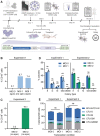
Figure 1.
Figure 1.
Experimental design and in vitro characterization of FVV transduction in CD34+ LAD-1 HSPCs. (A) A schematic representation of the experimental design is shown. The proviral structure of the FVV utilized in this study is depicted in the lower portion of the panel. The vector encodes a human CD18 transgene under the regulatory control of an MSCV promoter/enhancer and retains a truncated portion of the viral gag-pol-env coding sequences. The U3 region of the viral LTR bears a deletion conferring a SIN phenotype. Ψ, vector packaging signal. (B) Mobilized peripheral blood CD34+ HSPCs isolated from a severely affected LAD-1 patient were transduced overnight in FEP cell culture bags at an MOI of 1 TU per cell. Vector-encoded CD18 expression was measured 5 days post-transduction by flow cytometry. Cells were inoculated in either the presence (“spin”) or absence (“no spin”) of brief centrifugation (i.e., spinoculation). (C) CD34+ LAD-1 HSPCs were subjected to overnight transduction in FEP cell culture bags at an MOI of 2 FVV TU per cell. The percentage of CD18+ cells was determined 12 days post-transduction by flow cytometry. (D) The effect of FVV transduction on hematopoietic progenitor cell CFU activity is shown for each experiment. E, colony/burst-forming unit-erythroid; G, CFU-granulocyte; M, CFU-macrophage; GM, CFU-granulocyte macrophage; GEMM, CFU-granulocyte erythroid macrophage megakaryocyte. (E) Normalized CFU counts are graphed for each treatment group. The data in (D, E) are shown as mean ± SEM; ns, not significant by unpaired, two-tailed Student's t-test. CFU, colony-forming unit; FEP, fluoroethyl polymer; FVV, foamy virus vector; HSPCs, hematopoietic stem and progenitor cells; LAD-1, leukocyte adhesion deficiency type 1; LTR, long terminal repeat; MOI, multiplicity of infection; MSCV, murine stem cell virus; SEM, standard error of the mean; SIN, self-inactivating; TU, transducing unit.
Figure 2.
Figure 2.
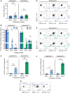
FVV-mediated CD18 expression partially rescues motility defects in myeloid-differentiated LAD-1 donor cells. (A) Migration of myeloid-differentiated LAD-1 cells in response to a chemotactic peptide gradient (gradient source: 10 nM fMLP) was captured using an EZ-TAXIScan device (depiction of the device chamber is adapted from Hattori et al). Two-dimensional migration coordinates of motile cells were plotted over time using ImageJ. Migration coordinates were used to calculate motility characteristics utilizing an application-specific software. (B) Still frame images of cellular migration patterns for each sample group at the 0-, 30-, and 60-minute time points in response to chemoattractant. The direction of migration is indicated by arrows. (C) Individual motility tracks of cells passing threshold are depicted from a common origin for visualization and comparison. The center of mass for each cell cluster is indicated by a red dot. (D–F) Quantification of motility parameters (velocity, accumulated migration distance, and Euclidean migration distance) in the presence of chemoattractant. Average cellular velocity was calculated using slices 2 through 8 of the final 25-slice image stack. The data in (D) through (F) are shown as mean ± SEM. Mock-transduced LAD-1, n = 16 tracks; CD18 LAD-1, n = 4 tracks; CD18+ LAD-1, n = 35 tracks; HD PMNs, n = 54 tracks. Statistical significance was evaluated using an unpaired, two-tailed Student's t-test. *p < 0.05; ***p < 0.001; ****p < 0.0001. fMLP, N-formylmethionine-leucyl-phenylalanine; HD, healthy donor; PMN, polymorphonuclear.
Figure 3.
Figure 3.
Characterization of LAD-1 HSPC engraftment, lineage reconstitution, and transgene marking in a mouse xenograft model. (A) LAD-1 HSPC engraftment within transplanted NSG mice was quantified by flow cytometry of murine bone marrow samples. The percentage of CD45+ human cells within the marrow is plotted for each mouse. (B) Percentages of human myeloid cells (CD13+), human T cells (CD3+), and human B cells (CD20+), observed within the bone marrow of transplanted NSG mice are indicated. (C) The percentage of human myeloid cells (CD45+CD13+) expressing the CD18 transgene is plotted for each transduction cohort. (D, E) Representative flow plots showing detection of CD13+ myeloid cells along with CD18+ or CD11b+ subpopulations. The complete gating strategies for flow cytometric analysis are shown in Supplementary Fig. S4. (F) The percentage of human myeloid cells expressing CD11b is shown. (G) CD18 and CD11b expression profile associated with the CD3+ T cell compartment of mouse #4013 (MOI = 1 cohort). The data in (A, B, C, F) are shown as mean ± SEM; *p < 0.05; **p < 0.01; ns, not significant, by unpaired, two-tailed Student's t-test.
Figure 4.
Figure 4.
Post-transplant histology and vector integration site analysis. (A) Representative images of H&E-stained tissue sections from mice that received either mock-transduced LAD-1 HSPCs or LAD-1 HSPCs transduced at an MOI of 1 or 2 FVV TU per cell. The image series labeled “MOI 0” is the nontransduced control arm derived from the MOI = 1 cohort (similar histological findings were observed in the MOI = 2 cohort). Magnification = 10 × . (B) Vector copy number per diploid human genome was determined by quantitative PCR analysis of genomic DNA extracted from murine bone marrow samples. (C) The average number of vector insertion sites per human chromosome is indicated. Insertions within genes are shown in dark blue, whereas insertions outside of genes are indicated in light blue. For integration events within genes, the percentage of insertion sites occurring within introns, exons, or promoter elements is depicted (inset). (D) Percentage of vector insertions observed as a function of distance from the nearest TSS. Proximity to oncogenes and nononcogenes is indicated. (E) Clonality profile of a subset of mice that received FVV-transduced LAD-1 HSPCs. The top 10 sites with the most abundant read counts (i.e., highly represented clones) are graphed as a percentage of total reads. The percentage of remaining reads is indicated in red. Total reads per mouse were as follows: mouse #4013, 513,913; mouse #4015, 457,976; mouse #4017, 516,268; mouse #4018, 1,501,949; mouse #4043, 172,634. The data in (B) are shown as mean ± SEM; *p < 0.05; ns, not significant, by unpaired, two-tailed Student's t-test. H&E, hematoxylin and eosin; PCR, polymerase chain reaction; TSS, transcription start site.

Similar articles

Cited by

References

    1. Vijver Evd, Maddalena A, Sanal O, et al. . Hematologically important mutations: Leukocyte adhesion deficiency (first update). Blood Cells Mol Dis 2012;48(1):53–61; doi:10.1016/j.bcmd.2011.10.004 - DOI - PMC - PubMed
    1. Takada Y, Ye X, Simon S. The integrins. Genome Biol 2007;8(5):215; doi:10.1186/gb-2007-8-5-215 - DOI - PMC - PubMed
    1. Fagerholm SC, Guenther C, Llort Asens M, et al. Beta2-integrins and interacting proteins in leukocyte trafficking, immune suppression, and immunodeficiency disease. Front Immunol 2019;10:254; doi:10.3389/fimmu.2019.00254 - DOI - PMC - PubMed
    1. Cavazzana M, Bushman FD, Miccio A, et al. . Gene therapy targeting haematopoietic stem cells for inherited diseases: progress and challenges. Nat Rev Drug Discov 2019;18(6):447–462; doi:10.1038/s41573-019-0020-9 - DOI - PubMed
    1. Ferrari G, Thrasher AJ, Aiuti A. Gene therapy using haematopoietic stem and progenitor cells. Nat Rev Genet 2021;22(4):216–234; doi:10.1038/s41576-020-00298-5 - DOI - PubMed

Publication types

Supplementary concepts

LinkOut - more resources