. 2022 Oct 11;22(20):3898-3909.
doi: 10.1039/d2lc00219a.

Bending stiffness of Candida albicans hyphae as a proxy of cell wall properties

Affiliations

Bending stiffness of Candida albicans hyphae as a proxy of cell wall properties

Elodie Couttenier et al. Lab Chip. .

Abstract

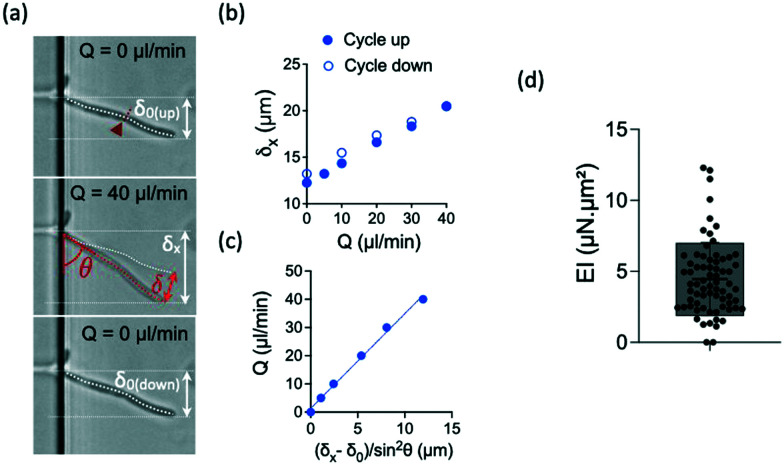
The cell wall is a key component of fungi. It constitutes a highly regulated viscoelastic shell which counteracts internal cell turgor pressure. Its mechanical properties thus contribute to define cell morphology. Measurements of the elastic moduli of the fungal cell wall have been carried out in many species including Candida albicans, a major human opportunistic pathogen. They mainly relied on atomic force microscopy, and mostly considered the yeast form. We developed a parallelized pressure-actuated microfluidic device to measure the bending stiffness of hyphae. We found that the cell wall stiffness lies in the MPa range. We then used three different ways to disrupt cell wall physiology: inhibition of beta-glucan synthesis, a key component of the inner cell wall; application of a hyperosmotic shock triggering a sudden decrease of the hyphal diameter; deletion of two genes encoding GPI-modified cell wall proteins resulting in reduced cell wall thickness. The bending stiffness values were affected to different extents by these environmental stresses or genetic modifications. Overall, our results support the elastic nature of the cell wall and its ability to remodel at the scale of the entire hypha over minutes.


Conflict of interest statement

There are no conflicts to declare.

Figures

Fig. 1
Fig. 1. Design of the chip and experimental procedure. (a) Scheme of the microfluidic device containing 2 main chambers connected by microchannels, a leftward seeding chamber and a rightward bending chamber. A long serpentine is used to increase the hydrodynamic resistance of the seeding compartment and consequently to reach an efficient positioning of the yeasts in front of the microchannels. (b) Fibronectin coating (in blue on the schemes) into the seeding chamber. The bending chamber remains uncoated, as assessed from the localization of the PLL-FITC coating replacing non fluorescent FN in mock experiments (image, obtained from fluorescence imaging). (c) Trapping of yeasts at the entrance of the microchannels, followed by (d) hyphal growth in the bending chamber where they are subjected to shear flow. Scale bars: 20 μm.
Fig. 2
Fig. 2. Forces exerted on a single hypha and deflection. (a) Scheme of hydrodynamic forces exerted on a hypha. Model considered in (top) versus model developed in this study, taking into account the variation along y of the hydrodynamic forces (bottom). The length l1 is deduced from Comsol simulations, see ESI Fig. S4. (b) Geometrical parameters characterizing a hypha of length l tilted by θ under the action of hydrodynamic forces.
Fig. 3
Fig. 3. Bending properties of hyphae from the reference strain SC5314. (a) Images of an hypha initially deflected by δ0 before (top), during (middle) and after (bottom) flow application. The initial tilted position of the hypha is indicated in the middle image. The amplitudes of the deflection δ0, δx and δ are indicated according to the nomenclature used in Fig. 2. The position of the septum is indicated by the red arrowhead and dashed line. (b) Example of curves obtained for this hypha. Top: deflection δx obtained during increasing (solid circles) and decreasing (open circles) flow rates Q. (c) Deflection data formatting to show the linear fit from which the bending stiffness EI will be deduced for each hypha (see eqn (4)). Only the data corresponding to increasing flow rate Q are considered. (d) Distribution (mean ± SD = 4.5 ± 2.5 μN μm2) of bending stiffness EI deduced from the slope of the linear variation shown in (c). n = 69 hyphae.
Fig. 4
Fig. 4. Consequences of an antifungal treatment using caspofungin at 1 μg mL−1 for 30 min on the reference strain SC5314. Graphs display mean (cross symbol), min, max, and median with interquartile range (25th percentile; 75th percentile) values. (a) Images of a hypha bending under flow before and after caspofungin treatment. The location of the septum is indicated by the red arrowheads and dashed lines. (b) Diameters. Left: distribution of diameters before (n = 18) and after (n = 21) antifungal treatment; p = 0.0081 (Mann–Whitney test); right: diameter ratio for paired data (n = 17) (c) bending stiffness EI. Left: distribution of the bending stiffness EI before (n = 18) and after (n = 21) antifungal treatment, p < 0.0001(Mann–Whitney test); right: distribution of EI ratio for paired data (n = 17).
Fig. 5
Fig. 5. Behavior of the reference strain SC5314 before and after an hyperosmotic shock provided by 2.5 M of sorbitol. Graphs display mean (cross symbol), min, max, and median with interquartile range (25th percentile; 75th percentile) values. (a) Left: distribution of diameters before (n = 10) and after (n = 13) sorbitol application, p < 0.0001 (Mann–Whitney test); right: diameter ratio for paired data (n = 10). (b) Left: distribution of the bending stiffness EI before (n = 8) and after (n = 12) sorbitol application, p < 0.0001 (Mann–Whitney test); right: distribution of EI ratio for paired data (n = 8).
Fig. 6
Fig. 6. Distribution of the bending stiffness EI of the reference strain SC5314 (WT, n = 69) and of the Δpga59Δpga62 mutant (ΔΔ, n = 26), p = 0.329 (Mann–Whitney test). Min, max, and median with interquartile range (25th percentile; 75th percentile) values are represented.

References

    1. Noble S. M. Gianetti B. A. Witchley J. N. Candida albicans cell-type switching and functional plasticity in the mammalian host. Nat. Rev. Microbiol. 2017;15(2):96–108. doi: 10.1038/nrmicro.2016.157. doi: 10.1038/nrmicro.2016.157. - DOI - DOI - PMC - PubMed
    1. Gupta R. Malik A. Rizvi M. Ahmed M. An Alarming Increase of Fungal Infections in Intensive Care Unit: Challenges in the Diagnosis and Treatment. J. Appl. Pharm. Sci. 2016;6(11):114–119. doi: 10.1016/j.semcdb.2011.08.010. doi: 10.7324/JAPS.2016.601118. - DOI - DOI
    1. Sudbery P. Gow N. Berman J. The distinct morphogenic states of Candida albicans. Trends Microbiol. 2004;12(7):317–324. doi: 10.1016/j.tim.2004.05.008. - DOI - PubMed
    1. Seman B. G. Moore J. L. Scherer A. K. Blair B. A. Manandhar S. Jones J. M. Wheeler R. T. Yeast and filaments have specialized, independent activities in a zebrafish model of Candida albicans infection. Infect. Immun. 2018;86(10):e00415-18. doi: 10.1128/IAI.00415-18. doi: 10.1128/IAI.00415-18. - DOI - DOI - PMC - PubMed
    1. Sudbery P. E. Growth of Candida albicans hyphae. Nat. Rev. Microbiol. 2011;9(10):737–748. doi: 10.1038/nrmicro2636. - DOI - PubMed

Publication types