. 2022 Oct;44(10):1329-1338.
doi: 10.1007/s00276-022-03016-4. Epub 2022 Sep 12.

The thoracoacromial trunk: a detailed analysis

Affiliations

The thoracoacromial trunk: a detailed analysis

Michał Bonczar et al. Surg Radiol Anat. 2022 Oct.

Abstract

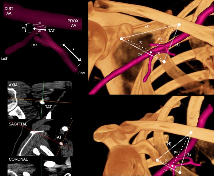
Purpose: The thoracoacromial trunk (TAT) originates from the second part of the axillary artery and curls around the superomedial border of the pectoralis minor, subsequently piercing the costocoracoid membrane. Knowledge about the location, morphology, and variations of the TAT and its branches is of great surgical importance due to its frequent use in various reconstructive flaps.

Methods: A retrospective study was conducted to establish anatomical variations, their prevalence, and morphometric data on TAT and its branches. The results of 55 consecutive patients who underwent neck and thoracic computed tomography angiography were analyzed. A qualitative evaluation of each TAT was performed.

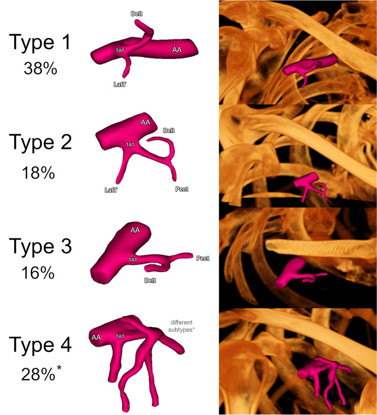
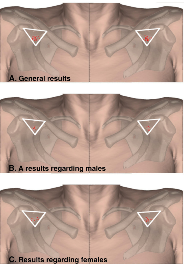
Results: A total of 15 morphologically different TAT variants were initially established. The median length of the TAT was set at 7.74 mm (LQ 3.50; HQ 13.65). The median maximum diameter of the TAT was established at 4.19 mm (LQ 3.86; HQ 4.90). The median TAT ostial area was set to 13.97 mm (LQ 11.70; HQ 18.86). To create a heat map of the most frequent location of the TAT, measurements of the relating structures were made.

Conclusion: In this study, the morphology and variations of the branching pattern of the TAT were presented, proposing a new classification system based on the four most commonly prevalent types. The prevalence of each branch arising directly from the TAT was also analyzed. It is hoped that the results of the present anatomical analysis can help to minimize potential complications when performing plastic or reconstructive procedures associated with TAT.

Keywords: Axillary artery; Perforator flaps; Subclavian artery; Thoracoacromial trunk.


Conflict of interest statement

The authors declare no competing interests.

The author(s) declares that they have no potential conflicts of interest with respect to the research, authorship, and/or publication of this article.

Figures

Fig. 1
Fig. 1
Measurement methodology presented in graphical form. To determine the TAT morphology, the multiplanar reconstruction of CT images was performed. PROX AA Proximal axillary artery; DIST AA distal axillary artery; TAT thoracoacromial trunk; Delt deltoid branch of TAT; LatT lateral thoracic artery; Pect pectoral branch of TAT; (1) Length of TAT; (2) maximal diameter of TAT; (3) TATs’ ostial area; (4) distance from the coracoid process to the clavicle, (5) distance from the coracoid process to the second rib, (6) distance from the clavicle process to the second rib, (7) distance from the coracoid process to the TAT, (8) distance from the clavicle to the TAT, and (9) distance from the second rib to the TAT. An asterisk (*) indicates the length of the corresponding consecutive Pect of TAT or its branches
Fig. 2
Fig. 2
Summary of the most common morphological types of TAT. Type 1: TAT arises from the second part of the axillary artery. First, the lateral thoracic artery branches out, and then the deltoid artery. Type 2: TAT arises from the second part of the axillary artery. The lateral thoracic, pectoral, and deltoid arteries branch out sequentially. Type 3: TAT arises from the second part of the axillary artery. First, the pectoral artery branches out, and then, the deltoid artery branches off. Type 4: Any other variation. An asterisk (*) indicate Type 4—Other, which is not an individual type but a separate set for the other TAT morphological types that do not fit Types 1–3
Fig. 3
Fig. 3
An anatomical map of the occurrence of TAT in the clavipectoral triangle. A general results. B results regarding males. C results regarding females

References

    1. Deng D, Xu F, Liu J, Li B, Li L, Liu J, Chen F. Clinical application of pedicled thoracoacromial artery perforator flaps for tracheal reconstruction. BMC Surg. 2020;20(1):299. doi: 10.1186/s12893-020-00972-9. - DOI - PMC - PubMed
    1. Geddes CR, Tang M, Yang D, Morris SF. An assessment of the anatomical basis of the thoracoacromial artery perforator flap. Can J Plast Surg. 2003;11(1):23–27. doi: 10.1177/229255030301100102. - DOI - PMC - PubMed
    1. Goishi K, Harada H, Keyama T, Tsuda T, Hashimoto I. Pectoralis major turnover flap based on thoracoacromial vessels for sternal dehiscence. Microsurgery. 2020;40(2):137–144. doi: 10.1002/micr.30509. - DOI - PubMed
    1. Hallock GG. The Island thoracoacromial artery muscle perforator flap. Ann Plast Surg. 2011;66(2):168–171. doi: 10.1097/SAP.0b013e3181dc83cd. - DOI - PubMed
    1. Iida T, Yoshimatsu H. Anatomical study and clinical application of free thoracoacromial artery true-perforator flap for reconstruction of the face. J Craniofac Surg. 2019;30(1):205–207. doi: 10.1097/SCS.0000000000004968. - DOI - PubMed

MeSH terms