. 2022 Sep 12;17(9):e0268664.
doi: 10.1371/journal.pone.0268664. eCollection 2022.

The PCI domains are "winged" HEAT domains

Affiliations

The PCI domains are "winged" HEAT domains

Eleanor Elise Paul et al. PLoS One. .

Abstract

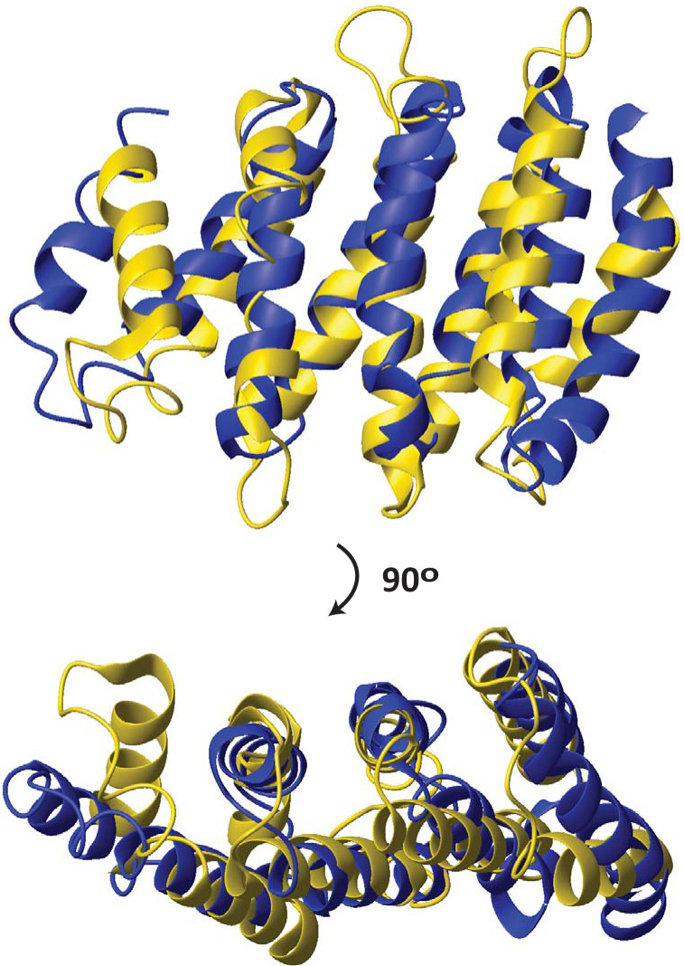
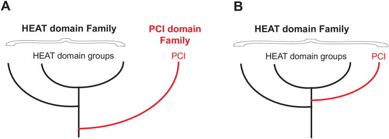
The HEAT domains are a family of helical hairpin repeat domains, composed of four or more hairpins. HEAT is derived from the names of four family members: huntingtin, eukaryotic translation elongation factor 3 (eEF3), protein phosphatase 2 regulatory A subunit (PP2A), and mechanistic target of rapamycin (mTOR). HEAT domain-containing proteins play roles in a wide range of cellular processes, such as protein synthesis, nuclear transport and metabolism, and cell signaling. The PCI domains are a related group of helical hairpin domains, with a "winged-helix" (WH) subdomain at their C-terminus, which is responsible for multi-subunit complex formation with other PCI domains. The name is derived from the complexes, where these domains are found: the 26S Proteasome "lid" regulatory subcomplex, the COP9 signalosome (CSN), and eukaryotic translation initiation factor 3 (eIF3). We noted that in structure similarity searches using HEAT domains, sometimes PCI domains appeared in the search results ahead of other HEAT domains, which indicated that the PCI domains could be members of the HEAT domain family, and not a related but separate group, as currently thought. Here, we report extensive structure similarity analysis of HEAT and PCI domains, both within and between the two groups of proteins. We present evidence that the PCI domains as a group have greater structural similarity with individual groups of HEAT domains than some of the HEAT domain groups have among each other. Therefore, our results indicate that the PCI domains have evolved from a HEAT domain that acquired a WH subdomain. The WH subdomain in turn mediated self-association into a multi-subunit complex, which eventually evolved into the common ancestor of the Proteasome lid/CSN/eIF3.


Conflict of interest statement

The authors have declared that no competing interests exist.

Figures

Fig 1
Fig 1. Structure alignment between the HEAT domain of CTF3 and the PCI domain of RPN12.
DALI [27]-based structure alignment between the HEAT domain of S. cerevisiae CTF3 (6wuc.pdb, chain B) and the PCI domain of human RPN12 (5l4k.pdb, chain P). The structures are shown in ribbon. CTF3 is colored gold; RPN12 is colored blue. Only the aligned portions of the two structures are shown.
Fig 2
Fig 2. Possible alternatives for the evolution of PCI domains.
A. PCI domains (red) diverged from HEAT domains before the last common ancestor of all HEAT domains and constitute a separate family of domains, as currently thought. B. The PCI domains are members of the HEAT domain family that acquired a WH subdomain after the last common HEAT domain ancestor.
Fig 3
Fig 3. Structure similarity scores among groups of HEAT and PCI domains.
Average pairwise DALI [27] Z-scores +/- standard deviation (SD) between members of individual HEAT and PCI domain groups. Groups are labeled and color-coded. The Z-scores of each group with the other five groups are shown on the Y-axis, with slight offset along the X-axis, and color-coded. Classic HEAT domains (Group 1) are navy. MA3 domains (Group 2) are red. MIF4G domains (Group 3) are grey. W2 domains (Group 4) are orange. Outlier HEAT domains (Group 5) are light blue. PCI domains are green. Intragroup Z-scores are not shown.
Fig 4
Fig 4. Structure-based sequence alignment of hairpins from HEAT and PCI domains.
Eight hairpins per group were used for the alignment. Alignment viewer (https://alignmentviewer.org/) with Clustal residue coloring scheme was used, instead of conservation, because there were no detectable sequence conservation patterns, besides hydrophobic side chains at buried positions. Secondary structure is shown above the alignment: H, helix; L, loop. Positions of intra-hairpin contacts are marked with a “*” above the alignment. Note that side chains at the same position in the alignment may contact both the other helix in the hairpin and an adjacent hairpin, or contact one or the other in different hairpins, due to variations in inter-helix angles and orientations. Positions with hydrophobic side chains in >50% of sequences are labeled with “Φ”; positions with hydrophobic side chains in >75% of the sequences are labeled with “Φ” in bold. Gaps are marked with “-“. Positions of deleted amino-acid sequences in the interhelix loop are marked with “‥”.
Fig 5
Fig 5. Possible evolutionary tree of HEAT and PCI domains.
The dendrogram is based on average structure similarity Z scores from the DALI server [27] between individual groups of domains and branches, using intact HEAT domains and WH-less PCI domains. The outliers, UTP10 and the FAT domain of mTOR, which had been operationally grouped together as Group 5 in Fig 3, Table 1, and Table in S1 Table, had slightly lower structure similarity between each other (Z score 5.7) than their highest average Z scores with other groups (UTP10, Z score 6.0 with Group 1; and the mTOR FAT domain, Z score 5.9 with Group 6). Therefore, these two structures were not considered as a group in building the dendrogram and were added separately, instead, and are shown in smaller font.

Similar articles

Cited by

References

    1. Andrade MA, Perez-Iratxeta C, Ponting CP. Protein repeats: structures, functions, and evolution. J Struct Biol. 2001;134(2–3):117–31. Epub 2001/09/12. doi: 10.1006/jsbi.2001.4392 . - DOI - PubMed
    1. Andrade MA, Petosa C, O’Donoghue SI, Muller CW, Bork P. Comparison of ARM and HEAT protein repeats. J Mol Biol. 2001;309(1):1–18. Epub 2001/08/09. doi: 10.1006/jmbi.2001.4624 . - DOI - PubMed
    1. Groves MR, Barford D. Topological characteristics of helical repeat proteins. Curr Opin Struct Biol. 1999;9(3):383–9. Epub 1999/06/11. doi: 10.1016/s0959-440x(99)80052-9 . - DOI - PubMed
    1. Yoshimura SH, Hirano T. HEAT repeats—versatile arrays of amphiphilic helices working in crowded environments? J Cell Sci. 2016;129(21):3963–70. Epub 2016/11/02. doi: 10.1242/jcs.185710 . - DOI - PubMed
    1. Andrade MA, Bork P. HEAT repeats in the Huntington’s disease protein. Nat Genet. 1995;11(2):115–6. Epub 1995/10/01. doi: 10.1038/ng1095-115 . - DOI - PubMed

Publication types

MeSH terms