. 2022 Sep 12;18(9):e1010811.
doi: 10.1371/journal.ppat.1010811. eCollection 2022 Sep.

SIRT5 is a proviral factor that interacts with SARS-CoV-2 Nsp14 protein

Affiliations

SIRT5 is a proviral factor that interacts with SARS-CoV-2 Nsp14 protein

Marius Walter et al. PLoS Pathog. .

Abstract

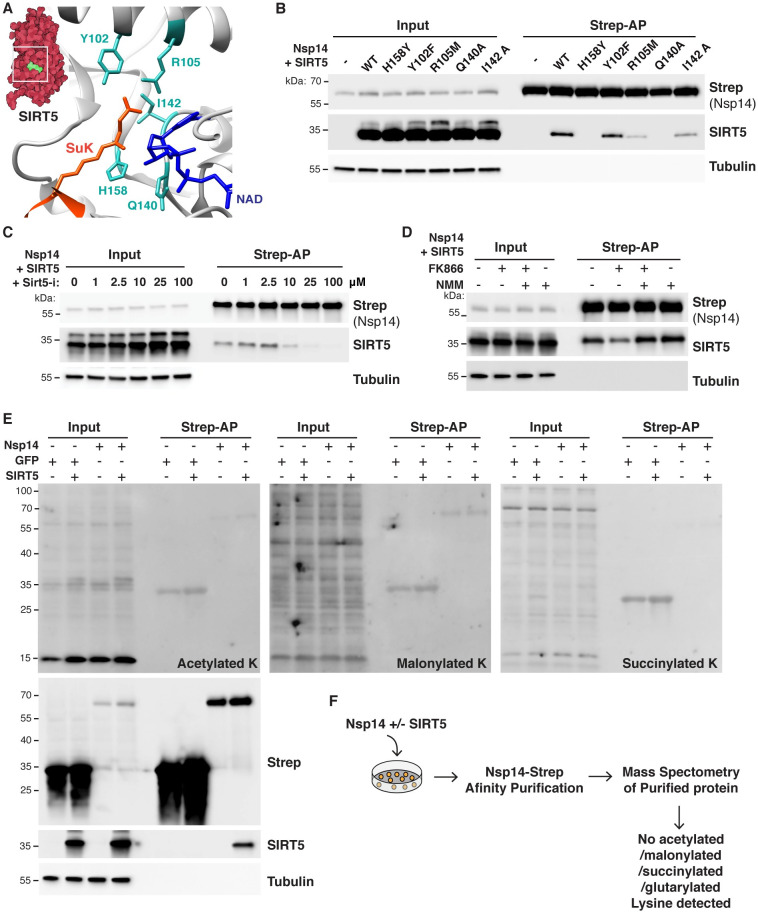
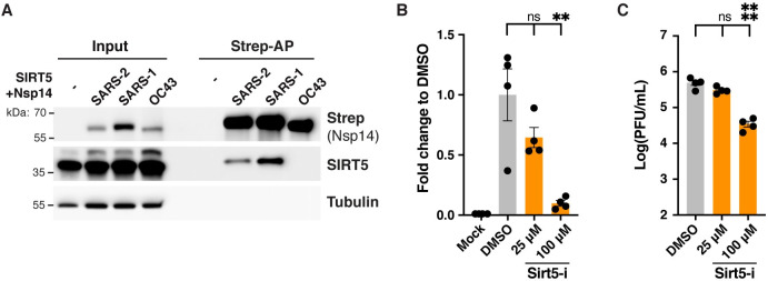
SARS-CoV-2 non-structural protein Nsp14 is a highly conserved enzyme necessary for viral replication. Nsp14 forms a stable complex with non-structural protein Nsp10 and exhibits exoribonuclease and N7-methyltransferase activities. Protein-interactome studies identified human sirtuin 5 (SIRT5) as a putative binding partner of Nsp14. SIRT5 is an NAD-dependent protein deacylase critical for cellular metabolism that removes succinyl and malonyl groups from lysine residues. Here we investigated the nature of this interaction and the role of SIRT5 during SARS-CoV-2 infection. We showed that SIRT5 interacts with Nsp14, but not with Nsp10, suggesting that SIRT5 and Nsp10 are parts of separate complexes. We found that SIRT5 catalytic domain is necessary for the interaction with Nsp14, but that Nsp14 does not appear to be directly deacylated by SIRT5. Furthermore, knock-out of SIRT5 or treatment with specific SIRT5 inhibitors reduced SARS-CoV-2 viral levels in cell-culture experiments. SIRT5 knock-out cells expressed higher basal levels of innate immunity markers and mounted a stronger antiviral response, independently of the Mitochondrial Antiviral Signaling Protein MAVS. Our results indicate that SIRT5 is a proviral factor necessary for efficient viral replication, which opens novel avenues for therapeutic interventions.


Conflict of interest statement

The Krogan Laboratory has received research support from Vir Biotechnology, F. Hoffmann-La Roche, and Rezo Therapeutics. Nevan J. Krogan has financially compensated consulting agreements with the Icahn School of Medicine at Mount Sinai, New York, Maze Therapeutics, Interline Therapeutics, Rezo Therapeutics, GEn1E Lifesciences, Inc. and Twist Bioscience Corp. He is on the Board of Directors of Rezo Therapeutics and is a shareholder in Tenaya Therapeutics, Maze Therapeutics, Rezo Therapeutics, and Interline Therapeutics.

Figures

Fig 1
Fig 1. SARS-CoV-2 Nsp14 interacts with human SIRT5.
A. Cartoon representation of the protein structure of Nsp14/Nsp10 (PDB 7N0B) and SIRT5 (PDB 3YIR) shows the Nsp14 N-terminal ExoN domain and C-terminal MTase domain. B. Affinity-purification of Nsp14-strep and co-purification of endogenous SIRT5 after transfection in HEK293T cells, as shown by western blot. C. Immunofluorescence of transfected Nsp14-Strep and endogenous SIRT5 in A549 cells. D. CETSA in HEK293T cells transfected with Nsp14-step and/or SIRT5, showing an increase in the stability of SIRT5 and Nsp14 by western blot. E. Western blot showing the absence of SIRT5 in SIRT5-KD HEK293T cells. F. Strep-tag affinity-purification or Flag-tag immunoprecipitation, followed by western blot, after transfection with Nsp14-strep, Nsp10-flag and SIRT5 expression constructs. SIRT5 does not interact with Nsp10. 0.5 μg of each construct or of empty control plasmids were transfected in SIRT5-KD HEK293T cells in a six-well plate. G. Strep-tag affinity-purification and western blot after transfection of Nsp14-strep, SIRT5 and increasing concentrations of Nsp10-tag indicate competitive binding of SIRT5 and Nsp10. 0.5 μg of Nsp14-strep and SIRT5 plasmid were used in a 6-well plate, with 0, 0.5, 1 or 2 μg of Nsp10-Flag.
Fig 2
Fig 2. SIRT5 catalytic activity is necessary to interact with Nsp14.
A. Cartoon representation of the protein structure of SIRT5 catalytic site in complex with cofactor NAD and succinylated lysine substrate (SuK), showing conserved residues mutated in panel B. B. Strep-tag affinity-purification and western blot after transfection of Sirt5-KD HEK293T cells with Nsp14-strep and SIRT5 catalytic mutants, showing that the interaction with Nsp14 is lost in several mutants. C. Strep-tag affinity-purification and western blot after transfection with Nsp14-strep and SIRT5, in SIRT5-KD HEK293T cells incubated with increasing concentrations of SIRT5 inhibitor Sirt5-i. High concentrations of Sirt5-i prevent the interaction. D. Strep-tag affinity-purification and western blot after transfection with Nsp14-strep and SIRT5, in SIRT5-KD HEK293T cells incubated with NAMPT FK866 inhibitor (low cellular NAD), FK866 and NMN, or NMN alone (high cellular NAD). SIRT5 binding strength correlated with NAD levels. E. Pan-acetylation, malonylation and succinylation in SIRT5-KD HEK293T total or Strep-purified proteins, after transfection with Nsp14-Strep, GFP-strep control and/or SIRT5. No specific lysine modifications could be detected. F. Summary of mass spectrometry experiments. Nsp14-strep proteins purified from SIRT5-KD HEK293T, with or without co-transfection with SIRT5, were analyzed by mass spectrometry. No acetylation, malonylation, succinylation or glutarylation modifications could be detected.
Fig 3
Fig 3. SARS-CoV-2 Nsp14 interacts with human SIRT1.
A. Co-purification of endogenous sirtuins SIRT1, 2, 3, 6 and 7 after transfection of Nsp14-strep in HEK293T cells, as shown by western blot. Loading and purification controls are the same as in Fig 1B. B. Strep-tag affinity-purification and western blot after transfection of HEK293T cells with Nsp14-strep and SIRT1 WT and H355Y catalytic mutant, showing that the interaction with Nsp14 is lost in H355Y mutant. C. Strep-tag affinity-purification and western blot after transfection with Nsp14-strep and SIRT1, in HEK293T cells incubated with increasing concentrations of SIRT1 inhibitor Ex-527. High concentrations of Ex-527 prevent the interaction. D-E. Strep-tag affinity-purification and western blot after transfection with Nsp14-strep and SIRT1, in HEK293T cells incubated with increasing concentrations of SIRT1 specific activator SRT1720 (D) or non-specific activator resveratrol (E). Both drugs were cytotoxic at high concentrations and the apparent decrease in SIRT1 binding correlated with a similar decrease in the input lanes. F. In vitro desuccinylation activity of purified SIRT5 incubated with increasing concentrations of Nsp14, showing no effect. G. In vitro methyltransferase activity of purified Nsp14 incubated with increasing concentrations of SIRT5, showing no specific effect. Unmethylated GpppG cap-analog was used as a substrate.
Fig 4
Fig 4. SIRT5 is a proviral factor.
A. Western blot showing the absence of SIRT5 and SIRT1 in SIRT5- and SIRT1-KO A549-ACE2 cells, after CRISPR knockout. B. Decrease in cell-associated viral mRNA levels in SIRT5- and SIRT1-KO cells infected with SARS-Cov-2 for 3 days at MOI = 0.1 or MOI = 1, as shown by RT-qPCR. Data show fold-changes compared to WT levels at MOI = 0.1. n = 4. C. Decrease in viral titers in SIRT5- and SIRT1-KO cells infected with SARS-Cov-2 for 3 days at MOI = 1, as shown by plaque assay. n = 4. D. Absence of cytotoxicity in A549-ACE2 cells treated with Sirt5-i and Ex-527 inhibitor, as measured by flow cytometry. n = 4. E. Decrease in cell-associated viral mRNA levels in A549-ACE2 cells infected with SARS-Cov-2 for 3 days at MOI = 0.1, and treated with SIRT5 and SIRT1 inhibitors Sirt5-i and Ex-527, as shown by RT-qPCR. Data show fold-change compared to DMSO-treated levels. n = 6. F. Decrease in viral titers in A549-ACE2 cells infected with SARS-Cov-2 for 3 days at MOI = 0.1, and treated with SIRT5 and SIRT1 inhibitors Sirt5-i and Ex-527, as shown by plaque assay. n = 9. G/H. Same as E. (with n = 4), and F. (n = 6), using Calu3 cells. B-H. Data show mean and standard error of the mean (SEM) between biological replicates. RT-qPCR results were internally normalized with GAPDH and ACTIN reference genes. Viral titers after plaque assay are expressed in log-transformed PFU (plaque-forming unit) per mL of supernatant. Asterisks summarize the results of one-way ANOVAs followed by Holm–Šidák multiple comparisons test (on log-transformed data for plaque assays) *: p < 0.05, **: p < 0.01, ***: p < 0.001, ****: p < 0.0001.
Fig 5
Fig 5. SIRT5 proviral activity is partially independent from the interaction with Nsp14.
A. Strep-tag affinity-purification and western blot after transfection of SIRT5-KD HEK293T cells with SIRT5 and Nsp14-strep from different coronaviruses, showing that the interaction with SIRT5 is specific to SARS-like coronaviruses. B. Decrease in supernatant-associated viral mRNA levels in HCT-8 cells infected with HCoV-OC43 for 5 days at MOI = 0.1, and treated with SIRT5 inhibitor Sirt5-i, as shown by RT-qPCR. Data show fold-change compared to DMSO-treated levels. n = 4. C. Decrease in viral titers in HCT-8 cells infected with HCoV-OC43 for 5 days at MOI = 0.1, and treated with SIRT5 inhibitors Sirt5-i, as shown by plaque assay. n = 4. B-C. Data show mean and SEM between biological replicates. Asterisks summarize the results of one-way ANOVAs followed by Holm–Šidák multiple comparisons test (on log-transformed data for plaque assays) *: p < 0.05, **: p < 0.01, ***: p < 0.001, ****: p < 0.0001.
Fig 6
Fig 6. SIRT5-KO cells mount a stronger innate immune response.
RNA-seq analysis of WT and SIRT5-KO A549-ACE2 cells infected or mock-infected for 3 days with SARS-CoV-2 at MOI = 1. n = 4. A. Volcano plots showing differentially expressed genes between the different conditions. Highlighted genes display a q-value q<0.05 (green), log2 fold-change >1 (orange), or both (purple). Left panel: SIRT5-KO vs WT in mock-infected cells. Middle: Infected vs mock-infected WT cells. Right: Infected vs mock-infected SIRT5-KO cells. B. Normalized gene count of SARS-CoV-2. C-D. Unsupervised clustering of the 3221 genes differentially expressed between at least two of the four conditions (q<0.01). C: heatmap of normalized expression. D. Z-scores of differentially expressed genes as grouped by clustering. Colored lines represent the quantification of an individual gene whereas solid black lines show the cluster Tukey boxplot. E. Enrichment analysis of biological gene sets in the identified gene clusters (C and D).
Fig 7
Fig 7. SIRT5-KO cells express a higher basal level of viral restriction factors.
A. Expression heatmap of interferon-stimulated genes and other restriction factors, showing that mock-infected SIRT5-KO cells express higher basal levels of restriction factors, and that antiviral responses are stronger in SIRT5-KO cells. Data show mean log2 fold-change, compared to mock-infected WT, and the q-value between mock-infected WT and SIRT5-KO cells. Only genes differentially expressed between at least two conditions were included in the analysis (q<0.01). B. RT-qPCR confirmation of restriction factors upregulated in non-infected SIRT5-KO cells (n = 8). Data show fold-changes compared to WT levels after normalization with ACTIN. Data show mean and SEM between replicates. p-values after unpaired two-tailed t-test are shown and asterisks summarize the results. *: p < 0.05, **: p < 0.01, ***: p < 0.001, ****: p < 0.0001.
Fig 8
Fig 8. SIRT5 proviral activity is independent of the MAVS signaling pathway.
A. Western blot showing 90% reduction of MAVS in A549-A/T cells transduced with a CRISPR lentivirus against MAVS. A549-A/T cells stably co-express ACE2 and TMPRSS2. B. Viral titers in MAVS-KD cells treated with DMSO or SIRT5 inhibitor Sirt5-i, after infection with SARS-Cov-2 for 3 days at MOI = 0.1, as shown by plaque assay. Sirt5-i had a similar effect in WT and MAVS-KO, suggesting that SIRT5 function is independent of MAVS. n = 9. C. RT-qPCR of GFP or Nsp14 after doxycycline induction. WT and SIRT5-KO A549-ACE2 cells were stably transduced with doxycycline-inducible constructs for GFP and Nsp14. After doxycycline treatment for 48 hours at 100ng/mL, GFP was strongly overexpressed, but Nsp14 failed to be expressed. Data show fold-changes compared to the first column (WT cells transduced with GFP without doxycycline), after normalization with ACTIN. n = 4. D. Hypotheses for the role of the SIRT5/Nsp14 interaction during SARS-CoV-2 infection. In model 1, Nsp14 could enhance SIRT5 activity, which would decrease innate immune responses and favor viral replication. In model 2, Nsp14 could redirect SIRT5 to novel targets, potentially in the replication-transcription complex, where SIRT5 could deacylate other viral proteins. In model 3, the Nsp14/SIRT5 complex could be primarily involved in mRNA cap methylation. Absence or inhibition of SIRT5 would lead to incomplete cap methylation and stronger immune recognition of viral mRNA. B-C. Data show mean and SEM between biological replicates. Asterisks summarize the results of one-way ANOVAs followed by Holm–Šidák multiple comparisons test (on log-transformed data for plaque assays). *: p < 0.05, **: p < 0.01, ***: p < 0.001, ****: p < 0.0001.

Update of

References

    1. Gordon DE, Jang GM, Bouhaddou M, Xu J, Obernier K, White KM, et al. A SARS-CoV-2 protein interaction map reveals targets for drug repurposing. Nature. 2020;583: 459–468. doi: 10.1038/s41586-020-2286-9 - DOI - PMC - PubMed
    1. Gordon DE, Hiatt J, Bouhaddou M, Rezelj VV, Ulferts S, Braberg H, et al. Comparative host-coronavirus protein interaction networks reveal pan-viral disease mechanisms. Science. 2020;370. doi: 10.1126/science.abe9403 - DOI - PMC - PubMed
    1. Li J, Guo M, Tian X, Wang X, Yang X, Wu P, et al. Virus-Host Interactome and Proteomic Survey Reveal Potential Virulence Factors Influencing SARS-CoV-2 Pathogenesis. Med (N Y). 2021;2: 99–112.e7. doi: 10.1016/j.medj.2020.07.002 - DOI - PMC - PubMed
    1. Laurent EMN, Sofianatos Y, Komarova A, Gimeno JP. Global BioID-based SARS-CoV-2 proteins proximal interactome unveils novel ties between viral polypeptides and host factors involved in multiple COVID19 …. BioRxiv. 2020. Available: https://www.biorxiv.org/content/10.1101/2020.08.28.272955v1.abstract - DOI
    1. Minskaia E, Hertzig T, Gorbalenya AE, Campanacci V, Cambillau C, Canard B, et al. Discovery of an RNA virus 3’->5’ exoribonuclease that is critically involved in coronavirus RNA synthesis. Proc Natl Acad Sci U S A. 2006;103: 5108–5113. - PMC - PubMed