. 2022 Sep 12;17(9):e0273076.
doi: 10.1371/journal.pone.0273076. eCollection 2022.

Critical evaluation of an autologous peripheral blood mononuclear cell-based humanized cancer model

Affiliations

Critical evaluation of an autologous peripheral blood mononuclear cell-based humanized cancer model

Harinarayanan Janakiraman et al. PLoS One. .

Abstract

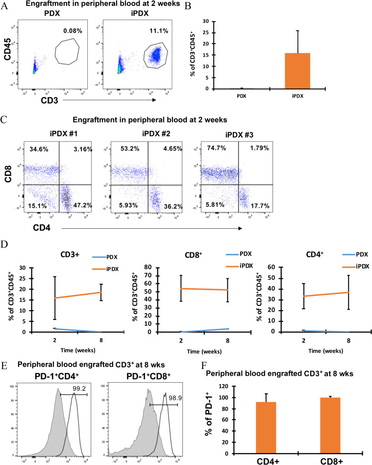
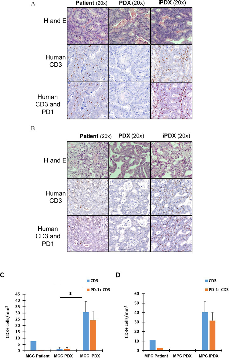
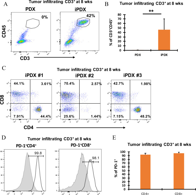
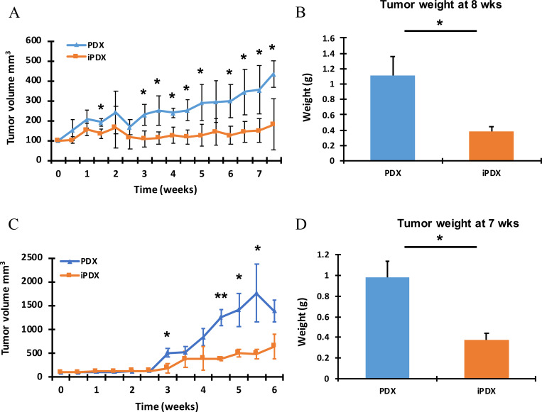
The use of humanized mouse models for oncology is rapidly expanding. Autologous patient-derived systems are particularly attractive as they can model the human cancer's heterogeneity and immune microenvironment. In this study, we developed an autologous humanized mouse cancer model by engrafting NSG mice with patient-derived xenografts and infused matched peripheral blood mononuclear cells (PBMCs). We first defined the time course of xenogeneic graft-versus-host-disease (xGVHD) and determined that only minimal xGVHD was observed for up to 8 weeks. Next, colorectal and pancreatic cancer patient-derived xenograft bearing NSG mice were infused with 5x10⁶ human PBMCS for development of the humanized cancer models (iPDX). Early after infusion of human PBMCs, iPDX mice demonstrated engraftment of human CD4+ and CD8+ T cells in the blood of both colorectal and pancreatic cancer patient-derived models that persisted for up to 8 weeks. At the end of the experiment, iPDX xenografts maintained the features of the primary human tumor including tumor grade and cell type. The iPDX tumors demonstrated infiltration of human CD3+ cells with high PD-1 expression although we observed significant intra and inter- model variability. In summary, the iPDX models reproduced key features of the corresponding human tumor. The observed variability and high PD-1 expression are important considerations that need to be addressed in order to develop a reproducible model system.


Conflict of interest statement

The authors have declared that no competing interests exist.

Figures

Fig 1
Fig 1. Effective engraftment of human PBMC derived T cells in MCC iPDX mouse peripheral blood.
(A) Representative flow cytometry plots are shown for MCC model CD3+CD45+ T lymphocytes from PDX mice (left), iPDX mice (middle). (B) Bar graph of the average % of CD3+CD45+ cells (right). Data from peripheral blood samples collected at 2 weeks. (C) Flow cytometry plots for CD4+ and CD8+ T lymphocytes from all iPDX mice (n = 3). Data from peripheral blood samples collected at 2 weeks. (D) The change in the average % of human T lymphocytes from early (2 week) to late (7 weeks) engraftment are shown for MCC CD3+CD45+ T lymphocytes (left), CD8+ T lymphocytes (middle) and CD4+ T lymphocytes (right). (E) PD-1+CD4+ T lymphocytes (left) PD-1+CD8+ T lymphocytes (right). T lymphocytes from healthy human donor PB (grey histogram, left) and iPDX PB (empty histogram, right). (F) Bar graph showing average percentage of PD-1+CD4+ and CD8+ T lymphocytes. Data from peripheral blood samples collected at 8 weeks. The values represent the percentage of human CD3, CD4 and CD8 population in iPDX mice peripheral blood. Average of per group (n = 3) is shown.
Fig 2
Fig 2. Comparison of histopathologic features of the corresponding human tumor to the xenograft models.
Histopathological evaluation of (A) MCC and (B) MPC models by Hematoxylin and Eosin staining (top row), human CD3 (middle row), and PD-1 and CD3 dual staining (bottom row) comparing the corresponding human tumor, PDX and iPDX tumors at 20X magnification. (C) and (D) Quantification of CD3 and PD-1 expression in the human cancer, MCC and MPC models. Data from samples collected at the end of the experiment at 7–8 weeks. Values represent mean of three animals per group ± SD. *p < 0.05.
Fig 3
Fig 3. Identification and phenotypic characterization of MCC iPDX tumor derived human CD3+ T cells.
(A) Representative flow cytometry dot plots showing CD3+CD45+ tumor infiltrating lymphocytes (TILs) (top left); (B) Bar graph of average values (top right). Data from samples collected at 8 weeks. (C) CD4+ and CD8+ TILs from 3 individual MCC iPDX mouse tumors (bottom panel). Data from samples collected at 8 weeks. (D) PD-1+CD4+ TILs (left) and PD-1+CD8+ TILs (middle) from a representative MCC iPDX mouse tumor. T lymphocytes from non-cancer human donor PB obtained from phlebotomy lab (grey histogram, left) and TILs from iPDX tumor (empty histogram, right). (E) Bar graph showing average percentage of PD-1+CD4+ and CD8+ T lymphocytes from 3 individual MCC iPDX mouse tumors. Data from samples collected at 8 weeks. Values represent mean of three animals per group ± SD. **p < 0.005.
Fig 4
Fig 4. iPDX demonstrate reduced tumor growth compared with the corresponding PDX.
Tumor (A) Average tumor volume (mm3) measured over time in the MCC model. (B) Final tumor weight (g) average in the MCC model. Data from samples collected at 8 weeks. (C) Average tumor volume (mm3) measured over time in the MPC model. (D) Final tumor weight (g) average in the MPC model. Data from samples collected at 7 weeks. Values represent mean of three animals per group ± SD. *p < 0.05. **p < 0.005.

References

    1. Topalian SL, Hodi FS, Brahmer JR, Gettinger SN, Smith DC, McDermott DF, et al. Safety, activity, and immune correlates of anti-PD-1 antibody in cancer. N Engl J Med. 2012;366(26):2443–54. doi: 10.1056/NEJMoa1200690 - DOI - PMC - PubMed
    1. Kieler M, Unseld M, Bianconi D, Prager G. Challenges and Perspectives for Immunotherapy in Adenocarcinoma of the Pancreas: The Cancer Immunity Cycle. Pancreas. 2018;47(2):142–57. doi: 10.1097/MPA.0000000000000970 - DOI - PubMed
    1. Gadgeel S, Rodríguez-Abreu D, Speranza G, Esteban E, Felip E, Dómine M, et al. Updated Analysis From KEYNOTE-189: Pembrolizumab or Placebo Plus Pemetrexed and Platinum for Previously Untreated Metastatic Nonsquamous Non-Small-Cell Lung Cancer. J Clin Oncol. 2020;38(14):1505–17. doi: 10.1200/JCO.19.03136 - DOI - PubMed
    1. Schmid P, Cortes J, Pusztai L, McArthur H, Kümmel S, Bergh J, et al. Pembrolizumab for Early Triple-Negative Breast Cancer. N Engl J Med. 2020;382(9):810–21. doi: 10.1056/NEJMoa1910549 - DOI - PubMed
    1. Mosely SI, Prime JE, Sainson RC, Koopmann JO, Wang DY, Greenawalt DM, et al. Rational Selection of Syngeneic Preclinical Tumor Models for Immunotherapeutic Drug Discovery. Cancer Immunol Res. 2017;5(1):29–41. doi: 10.1158/2326-6066.CIR-16-0114 - DOI - PubMed

Publication types

Substances