. 2023 Apr;19(4):1258-1276.
doi: 10.1080/15548627.2022.2123639. Epub 2022 Sep 22.

Apicoplast biogenesis mediated by ATG8 requires the ATG12-ATG5-ATG16L and SNAP29 complexes in Toxoplasma gondii

Affiliations

Apicoplast biogenesis mediated by ATG8 requires the ATG12-ATG5-ATG16L and SNAP29 complexes in Toxoplasma gondii

Jiawen Fu et al. Autophagy. 2023 Apr.

Abstract

In apicomplexan parasites, the macroautophagy/autophagy machinery is repurposed to maintain the plastid-like organelle apicoplast. Previously, we showed that in Toxoplasma and Plasmodium, ATG12 interacts with ATG5 in a non-covalent manner, in contrast to the covalent interaction in most organisms. However, it remained unknown whether apicomplexan parasites have functional orthologs of ATG16L1, a protein that is essential for the function of the covalent ATG12-ATG5 complex in vivo in other organisms. Furthermore, the mechanism used by the autophagy machinery to maintain the apicoplast is unclear. We report that the ATG12-ATG5-ATG16L complex exists in Toxoplasma gondii (Tg). This complex is localized on isolated structures at the periphery of the apicoplast dependent on TgATG16L. Inducible depletion of TgATG12, TgATG5, or TgATG16L caused loss of the apicoplast and affected parasite growth. We found that a putative soluble N-ethylmaleimide sensitive factor attachment protein receptor (SNARE) protein, synaptosomal-associated protein 29 (TgSNAP29, Qbc SNARE), is required to maintain the apicoplast in T. gondii. TgSNAP29 depletion disrupted TgATG8 localization at the apicoplast. Additionally, we identified a putative ubiquitin-interacting motif-docking site (UDS) of TgATG8. Mutation of the UDS site abolished TgATG8 localization on the apicoplast but not lipidation. These findings suggest that the TgATG12-TgATG5-TgATG16L complex is required for biogenesis of the apicoplast, in which TgATG8 is translocated to the apicoplast via vesicles in a SNARE -dependent manner in T. gondii.Abbreviations: AID: auxin-inducible degron; CCD: coiled-coil domain; HFF: human foreskin fibroblast; IAA: indole-3-acetic acid; LAP: LC3-associated phagocytosis; NAA: 1-naphthaleneacetic acid; PtdIns3P: phosphatidylinositol-3-phosphate; SNARE: soluble N-ethylmaleimide sensitive factor attachment protein receptor; UDS: ubiquitin-interacting motif-docking site; UIM: ubiquitin-interacting motif.

Keywords: Apicoplast; T. gondii; TgATG12–TgATG5-TgATG16L; TgATG8; TgSNAP29; ubiquitin-interacting motif.


Conflict of interest statement

No potential conflict of interest was reported by the author(s).

Figures
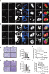
Figure 1.
Figure 1.
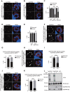
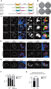
TgATG12 and TgATG5 are recruited to the periphery of the apicoplast and required for the survival of the tachyzoites. (A) The subcellular localization of TgATG12. (B) The subcellular localization of TgATG5. SmFP-HA-3AID-TgATG12 and SmFP-HA-3AID-TgATG5 were stained with anti-HA monoclonal antibodies (green). TgSAG2 (parasite surface protein) and TgCPN60 (apicoplast-resident protein) were stained with polyclonal antibodies (red). TgATRX1 and TgAPT1 were used as additional apicoplast markers. SmFP-HA-3AID-TgATG12 and SmFP-HA-3AID-TgATG5 parasites were transfected with plasmids expressing TgATRX1 and TgAPT1. TgATRX1 and TgAPT1 were stained with anti-MYC and anti-V5 monoclonal antibodies (red). Scale bars: 2 μm (left panels); 0.2 μm (zoomed panels). (C and E) Plaque assays were performed by infecting human foreskin fibroblast cells with SmFP-HA-3AID-TgATG12 (C) or SmFP-HA-3AID-TgATG5 (E) parasites for 8 d in the presence or absence of NAA. The total sizes of the ten biggest plaques were measured from three independent wells, and statistical differences were analyzed using unpaired t-tests and are shown in the graphs;***P=0.0002 (TgATG12) and ***P=0.0001 (TgATG5). (D and F) The growth of SmFP-HA-3AID-TgATG12 and SmFP-HA-3AID-TgATG5 parasites was monitored in co-culture with GFP-expressing wild-type parasites in the presence or absence of 1-naphthaleneacetic acid (NAA). The percentage of GFP-expressing parasites was determined by flow cytometry every generation. WT, wild type.
Figure 2.
Figure 2.
TgATG16L forms a complex with TgATG12–TgATG5 and is required for the survival of tachyzoites. (A) Co-immunoprecipitation of 4FLAG-TgATG16L and 4 HA-TgATG16L. HEK293T cells were transiently transfected with plasmids expressing 4FLAG-TgATG16L and 4 HA-TgATG16. The empty vector of pFlag-CMV-4 was used as a negative control. The cell lysates were then subjected to immunoprecipitation and blotted with anti-Flag and anti-HA antibodies. (B) Co-immunoprecipitation of 4FLAG-tagged TgATG16L and HA-tagged TgATG12 and TgATG5. The cells were transfected with plasmids expressing 4FLAG-TgATG16L and HA-TgATG12 and TgATG5. A plasmid expressing HA-eGFP was used as the negative control. The cell lysates were analyzed as indicated in A. (C) Co-immunoprecipitation of the N terminally truncated version of TgATG16L and HA-tagged TgATG5. The cells were transfected and the lysates were analyzed as indicated in A. (D) Immunofluorescence analysis of the colocalization of SmFP-HA-3AID-TgATG12 or -TgATG5 (red) with SmFP-MYC-tagged TgATG16 (green). Scale bars: 2 μm (left panels); 0.2 μm (zoomed panels). (E) Subcellular localization of TgATG16L. The sample was transfected with the identical plasmids and stained in the same way as in Figure 1 Scale bars: 2 μm (left panels); 0.2 μm (zoomed panels). (F) Plaque assays were performed by infecting human foreskin fibroblast cells with SmFP-HA-3AID-TgATG16L parasites for 8 d in the presence or absence of 1-naphthaleneacetic acid (NAA). The total sizes of the ten biggest plaques were quantified from three independent replicates. Statistical significance was determined using unpaired t-test; ***P = 0.0002. (G) the growth of SmFP-HA-3AID-TgATG16L parasites was monitored in co-culture with GFP-expressing wild-type parasites in the presence or absence of NAA. The percentage of GFP-expressing parasites was determined by flow cytometry every generation. IB, immunoblot; IP, immunoprecipitation; WT, wild type.
Figure 3.
Figure 3.
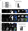
The TgATG2–TgATG5-TgATG16L complex is required for apicoplast inheritance. (A and B) SmFP-HA-3AID-TgATG12 and SmFP-HA-3AID-TgATG5 cell lines were cultured with or without indole-3-acetic acid (IAA) for 24 h, and stained with antibodies against TgSAG2 (red) and TgCPN60 (green). (C) The abnormal parasitophorous vacuoles (PVs) in which at least one parasite had lost its apicoplast (abnormal) or all parasites contained the apicoplast (normal) were counted. (D) SmFP-HA-3AID-TgATG16L cells were treated under the same conditions as in A and stained with antibodies against TgSAG2 (red) and TgCPN60 (green). (E) The abnormal and normal PVs were counted from D. (F), H and J) SmFP-HA-3AID-TgATG12 (F), SmFP-HA-3AID-TgATG5 (H), and SmFP-HA-3AID-TgATG16L (J) cell lines were transfected with a plasmid expressing MYC-tagged TgATRX1 and selected with pyrimethamine. The parasites were then treated under the same conditions as in A, and TgSAG2 (red) and TgATRX1 (green) were stained to monitor apicoplast inheritance. Scale bars: 2 μm. (G, I and K) Abnormal and normal PVs were counted from F, H, and J. (L) SmFP-HA-3AID-TgATG16L and SmFP-HA-3AID-TgATG12 parasites were cultured for 3 d with 1-naphthaleneacetic acid (NAA), and maturation of TgCPN60 was examined by immunoblotting. M, the proteolytic form of TgCPN60; Pre, the precursor of TgCPN60; WT, wild type. Data are the mean ± SEM from three independent slides (at least 100 PVs were counted for each slide).The statistical difference in the number of abnormal PVs was analyzed by two-way ANOVA (****P<0.0001).
Figure 4.
Figure 4.
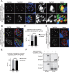
TgATG16L is required for apicoplast targeting of TgATG8. (A) The formation of the lipidated form of TgATG8 (TgATG8-II) in the SmFP-HA-3AID-TgATG16L parasites was detected using anti-V5 antibodies. SmFP-HA-3AID-TgATG16 parasites were transfected with a plasmid expressing 3×V5-TgATG8 under the control of its endogenous promoter and selected with pyrimethamine. The parasites were then treated with indole-3-acetic acid (IAA) for 72 h and subjected to western blot analysis. (B) The lipidation of 3×V5-TgATG8 was quantified from three biological repeats; statistical differences were analyzed using unpaired t-tests.***P=0.0004. (C) The SmFP-HA-3AID-TgATG16L parasites were transfected with a plasmid expressing TgATG8 derived from the endogenous promoter. SmFP-HA-3AID-TgATG16L was stained with an anti-HA antibody (green). TgATG8 fused with SmFP-MYC tag was stained with anti-MYC monoclonal antibody (red). (D) The apicoplast-associated localization of TgATG8 in TgATG16L-deficient parasites. SmFP-HA-3AID-TgATG16 parasites were transfected with a plasmid expressing SmFP-MYC-TgATG8 under the control of its endogenous promoter and selected with pyrimethamine. The parasites were then treated with IAA for 24 h and stained with anti-TgCPN60 and anti-MYC antibodies. Scale bars: 2 μm (C,D); 0.2 μm (C, zoomed image). (E and F) The parasite surface (red) and TgATG8 (green) were stained to calculate the percentage of abnormal parasitophorous vacuoles (PVs), in which TgATG8 did not accumulate as a normal apicoplast-like shape in the SmFP-HA-3AID-TgATG16L parasites treated with IAA for 24 h.
Figure 5.
Figure 5.
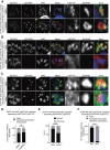
Accumulation of TgATG12–TgATG5-TgATG16L at the apicoplast depends on TgATG16L. (A) SmFP-MYC-TgATG12 parasites were treated with a PI3K inhibitor (LY294002), and then the localization of SmFP-MYC-TgATG12 (green) was observed by staining with anti-MYC antibodies. The apicoplast was labeled with anti-TgCPN60 (red). (B) Examination of TgATG12 localization in SmFP-HA-3AID-TgATG16L-depleted parasites. SmFP-MYC-TgATG12 was inserted at the N terminus of the TgATG12 locus using the CRISPR/Cas9 method in the SmFP-HA-3AID-TgATG16L parasites. SmFP-MYC-TgATG12 was stained in red and SmFP-HA-3AID-TgATG16L was labeled in green. (C) Examination of the association of SmFP-MYC-TgATG12 (green) with the apicoplast in SmFP-HA-3AID-TgATG16L-depleted parasites. TgCPN60 was stained as an apicoplast marker (red). (D) Quantification of parasites that contained apicoplast-associated SmFP-MYC-TgATG12 puncta at the apicoplast from A. Data were analyzed by two-way ANOVA; ns, not significant (P>0.05). (E) Quantification of the apicoplast inheritance in the SmFP-HA-3AID-TgATG16L parasites expressing SmFP-MYC-TgATG12 after IAA treatment. Data were analyzed by two-way ANOVA; ****P<0.0001. PVs, parasitophorous vacuoles. (F) Quantification of the apicoplast association with SmFP-MYC-TgATG12 from C.
Figure 6.
Figure 6.
Accumulation of TgATG12–TgATG5-TgATG16L at the apicoplast requires the coiled-coil domain. (A) Schematic diagram of the SmFP-MYC-tagged TgATG16L mutants used to transfect SmFP-HA-3AID-TgATG16L parasites. (1) Wild-type TgATG16L, (2) TgATG16LΔCCD, (3) TgATG16LΔWD, (4) TgATG16L with mutated potential lipid-binding sites at the N terminus, or (5) TgATG16L with mutated potential lipid-binding sites and ΔIS. (B) Plaque assays were performed with the TgATG16L mutants. The total sizes of the ten biggest plaques were quantified from three independent replicates. Plaque assays were performed with the prepared parasite strains expressing different versions of TgATG16L mutants in the presence or absence of indole-3-acetic acid (IAA) for 8 d. Data were analyzed for statistical significance by unpaired t-test. **P =0.0025 (SmFP-HA-3AID-TgATG16L parasites); **P=0.0016 (TgATG16LΔCCD mutant); ns, not significant (P>0.05). (C) The localization of the TgATG16L mutants was observed by immunofluorescence staining. TgATG16L mutants were stained with anti-MYC antibody (green), and the apicoplast was labeled with anti-TgCPN60 antibody (red). (D) TgATG16L association with the apicoplast was counted from G. Data were analyzed by two-way ANOVA. *P=0.0295; ns, not significant (P>0.05). (E) Rescue of apicoplast loss by expression of TgATG16L mutants in the SmFP-HA-3AID-TgATG16L-depleted parasites. The data were analyzed for statistical significance by an unpaired t-test. ****P<0.0001 (SmFP-HA-3AID-TgATG16L parasites); **P=0.0041 (TgATG16LΔCCD mutant); ns, not significant (P>0.05). Scale bars: 5 μm (A); 2 μm (C, D and G).
Figure 7.
Figure 7.
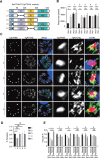
A Qbc SNARE, TgSNAP29, is required for apicoplast inheritance. (A) The molecular architecture of TgSNAP29. SNAP-25 has two splice variants, SNAP-25a and SNAP-25b. SNAP25a is shown in the figure. (B) Plaque assays were performed by infecting human foreskin fibroblast cells with TgSNAP29-mAID-SmFP-HA parasites for 8 d in the presence or absence of indole-3-acetic acid (IAA). (C) The subcellular localization of TgSNAP29. The sample was transfected with the same plasmids and stained in the same way as in Figure 1A. (D) TgSNAP29-mAID-SmFP-HA cells were cultured with or without IAA for 24 h, and stained with antibodies against TgSAG2 (red) and TgCPN60 (green) to monitor apicoplast inheritance. (E) Apicoplast inheritance in TgSNAP29-depleted parasites stably expressing TgATRX1. The parasites were cultured with the presence of IAA for 24 h, and stained with antibodies against TgSAG2 (red) and TgATRX1 (green). (F and G) The normal and abnormal parasitophorous vacuoles (PVs) were counted from E and F, respectively. Data are mean ± SEM from three independent slides, and at least 100 PVs were counted for each slide. The statistical difference in the number of abnormal PVs was analyzed by two-way ANOVA. ****P<0.0001 (F); ****P<0.0001 (G). ns, not significant; WT, wild type.
Figure 8.
Figure 8.
TgSNAP29 is required for apicoplast targeting of TgATG8. (A) Immunofluorescence detection of SmFP-MYC-TgATG8 localization in TgSNAP29-mAID-SmFP-HA cells. TgATRX1 was used as an apicoplast marker. TgSNAP29-mAID-SmFP-HA cells were transfected with a plasmid expressing 3×V5-tagged TgATG8 and selected with pyrimethamine. The parasites were then transiently transfected with a plasmid expressing TgATRX1 and cultured with or without indole-3-acetic acid (IAA) for 24 h, and stained with antibodies against V5 (green) and MYC (red). (B and C) The parasite surface (red) and TgATG8 (green) were stained to calculate the percentage of abnormal parasitophorous vacuoles (PVs), in which TgATG8 did not accumulate as a normal apicoplast-like shape in the TgSNAP29-mAID-SmFP-HA parasites treated with IAA for 24 h. (D and E) Immunofluorescence detection of SmFP-MYC-TgATG8 localization (green) and TgSAG2 (red) in the 12HA-AID*-TgVAMP4-2 parasites treated with IAA for 24 h. The percentage of abnormal PVs in which TgATG8 did not accumulate as the normal apicoplast-like shape in D was calculated. ****P<0.0001 (two-way ANOVA). (F) Co-immunoprecipitation of 3Flag-tagged TgAVAMP4-2 and 3HA-tagged TgSNAP29. HEK293T cells were transiently transfected with the plasmids expressing 3Flag-tagged TgAVAMP4-2 and 3HA-tagged TgSNAP29. The cell lysates were then subjected to immunoprecipitation and blotted with anti-Flag and anti-HA antibodies. IB, immunoblot. Scale bars: 2 μm (left panels); 0.2 μm (zoomed panels).
Figure 9.
Figure 9.
A UDS-like site of TgATG8 is critical for the proper localization at the apicoplast. (A) Alignment of the ubiquitin-interacting motif (UIM)-docking site (UDS) sequence of TgATG8 and its orthologs. (B) the three-dimensional structure of TgATG8 (PDB: 3VXW), highlighting the opposed positions of the LIR/AIM docking site (LDS, orange) and UDS (blue). (C) Localization of TgATG8 mutants at the apicoplast. The 9 HA-AID*-TgATG8 parasites were transfected with plasmids expressing the mutated versions of TgATG8 under the control of the native promoter, and parasites stably expressing these mutants were selected with pyrimethamine. The 9 HA-AID* tag was stained with HA antibody (red), and the TgATG8 mutants fused with the SmFP-MYC tag were stained with MYC antibody (green). Scale bars: 2 μm. (D) the parasites as labeled in the figure were cultured with or without IAA for 24 h, and stained with antibodies against TgSAG2 (green) and TgCPN60 (red). Scale bars: 2 μm. (E) the abnormal parasitophorous vacuoles (PVs) in the parasites expressing different TgATG8 mutants were counted from D. Data are mean ± SEM from three independent slides (at least 100 PVs were counted for each slide). The statistical difference in the number of abnormal PVs was analyzed by two-way ANOVA. ****p < 0.0001 (-); ****p < 0.0001 (Y85A); ns, not significant. (F) Plaque assays were performed by infecting human foreskin fibroblast cells with related parasites for 8 d in the presence or absence of indole-3-acetic acid (IAA). (G) the lipidation of TgATG8 mutants was examined in a reconstituted system established previously. The HEK 293T cells were transfected with plasmids expressing the codon-optimized versions of the related proteins. The lipidation of the mutants was detected by western blotting. (H) the lipidation of TgATG8 mutants was detected in the parasites. Wild-type RH strain parasites were stably transfected with plasmids containing the mutants and selected with pyrimethamine. Then, the lipidation of the mutants in T. gondii was examined by western blotting. (I) a proposed model of TgATG8 association with the apicoplast. The TgAtg12–tgatg5-TgATG16 L complex localizes close to the apicoplast and probably binds with lipids on the membrane of the vesicular structure through its coiled-coil domain. TgATG8 could be lipidated on an unknown vesicular structure with the assistance of the TgAtg12–tgatg5-TgATG16 L complex. Then, a vesicular fusion process mediates the translocation of TgATG8 to the apicoplast.

Similar articles

Cited by

References

    1. Ohsumi Y. Historical landmarks of autophagy research. Cell Res. 2014. Jan;24(1):9–23. - PMC - PubMed
    1. Lynch-Day MA, Klionsky DJ. The Cvt pathway as a model for selective autophagy. FEBS Lett. 2010 Apr 2;584(7):1359–1366. DOI:10.1016/j.febslet.2010.02.013. - DOI - PMC - PubMed
    1. Mizushima N, Yoshimori T, Ohsumi, Y. The role of Atg proteins in autophagosome formation. Annu Rev Cell Dev Biol. 2011;27(1):107–132. - PubMed
    1. Nakatogawa H. Mechanisms governing autophagosome biogenesis. Nat Rev Mol Cell Biol. 2020 May 5;21(8):439–458. DOI:10.1038/s41580-020-0241-0. - DOI - PubMed
    1. Kuma A, Mizushima N, Ishihara N, et al. Formation of the approximately 350-kDa Apg12-Apg5.Apg16 multimeric complex, mediated by Apg16 oligomerization, is essential for autophagy in yeast. J Biol Chem. 2002 May 24 277;(21)18619–18625. DOI:10.1074/jbc.M111889200. - DOI - PubMed

Publication types