

Evaluating differences in Young's Modulus of regenerated and uninjured mouse digit bone through microCT density-based calculation and nanoindentation testing
- PMID: 36095912
- PMCID: PMC9947921
- DOI: 10.1016/j.jbiomech.2022.111271
Evaluating differences in Young's Modulus of regenerated and uninjured mouse digit bone through microCT density-based calculation and nanoindentation testing
Abstract
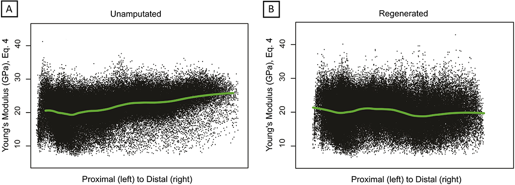
The mouse digit tip amputation model is an excellent model of bone regeneration, but its size and shape present an obstacle for biomechanical testing. As a result, assessing the structural quality of the regenerated bone in this model has focused on mineral density and bone architecture analysis. Here we describe an image-processing based method for assessment of mechanical properties in the regenerated digit by using micro-computed tomography mineral density data to calculate spatially discrete Young's modulus values throughout the entire distal third phalange. Further, we validate this method through comparison to nanoindentation-measured values for Young's modulus. Application to a set of regenerated and unamputated digits shows that regenerated bone has a lower Young's modulus compared to the uninjured digit, with a similar trend for experimental hardness values. Importantly, this method heightens the utility of the digit regeneration model, allows for more impactful treatment evaluation using the model, and introduces an analysis platform that can be used for other bones that do not conform to a standard long-bone model.
Keywords: Bone; Digit regeneration; Micro-computed tomography; Nanoindentation; Young’s modulus.
Copyright © 2022 Elsevier Ltd. All rights reserved.
Conflict of interest statement
Declaration of Competing Interest The authors declare that they have no known competing financial interests or personal relationships that could have appeared to influence the work reported in this paper.
Figures


Similar articles
-
Age-Dependent Changes in Bone Architecture, Patterning, and Biomechanics During Skeletal Regeneration.Front Cell Dev Biol. 2021 Oct 13;9:749055. doi: 10.3389/fcell.2021.749055. eCollection 2021. Front Cell Dev Biol. 2021. PMID: 34722531 Free PMC article.
-
A new approach to analyzing regenerated bone quality in the mouse digit amputation model using semi-automatic processing of microCT data.Bone. 2021 Mar;144:115776. doi: 10.1016/j.bone.2020.115776. Epub 2020 Dec 2. Bone. 2021. PMID: 33276153 Free PMC article.
-
The elastic properties of trabecular and cortical bone tissues are similar: results from two microscopic measurement techniques.J Biomech. 1999 Apr;32(4):437-41. doi: 10.1016/s0021-9290(98)00177-8. J Biomech. 1999. PMID: 10213035
-
Young's modulus of trabecular bone at the tissue level: A review.Acta Biomater. 2018 Sep 15;78:1-12. doi: 10.1016/j.actbio.2018.08.001. Epub 2018 Aug 4. Acta Biomater. 2018. PMID: 30081232 Review.
-
Anisotropy, Anatomical Region, and Additional Variables Influence Young's Modulus of Bone: A Systematic Review and Meta-Analysis.JBMR Plus. 2023 Oct 31;7(12):e10835. doi: 10.1002/jbm4.10835. eCollection 2023 Dec. JBMR Plus. 2023. PMID: 38130752 Free PMC article. Review.
Cited by
-
Age-Dependent Changes in Bone Architecture, Patterning, and Biomechanics During Skeletal Regeneration.Front Cell Dev Biol. 2021 Oct 13;9:749055. doi: 10.3389/fcell.2021.749055. eCollection 2021. Front Cell Dev Biol. 2021. PMID: 34722531 Free PMC article.
References
-
- Borgens RB, 1982. Mice regrow the tips of their foretoes. Science 217, 747–750. - PubMed
-
- Brockes JP, Kumar A, 2005. Appendage regeneration in adult vertebrates and implications for regenerative medicine. Science 310, 1919–1923. - PubMed
-
- Bryant SV, Endo T, Gardiner DM, 2002. Vertebrate limb regeneration and the origin of limb stem cells. Int. J. Dev. Biol 46, 887–896. - PubMed
-
- Currey J, 2002. Bones: Structure and Mechanics. Princeton University Press.
Publication types
MeSH terms
Grants and funding
LinkOut - more resources
Full Text Sources
Medical
Research Materials
