. 2022 Sep:479:153318.
doi: 10.1016/j.tox.2022.153318. Epub 2022 Sep 9.

Effect of heated tobacco products and traditional cigarettes on pulmonary toxicity and SARS-CoV-2-induced lung injury

Affiliations

Effect of heated tobacco products and traditional cigarettes on pulmonary toxicity and SARS-CoV-2-induced lung injury

Han-Hsing Tsou et al. Toxicology. 2022 Sep.

Abstract

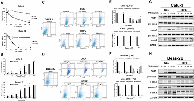
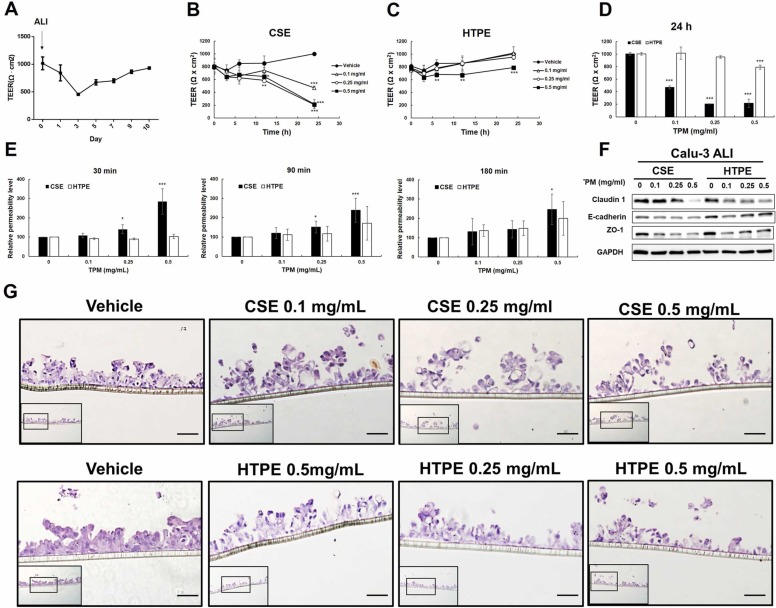
Cigarette smoke (CS) significantly contributes to the development of chronic obstructive pulmonary disease (COPD). Heated tobacco products (HTPs), newly developed cigarette products, have been proposed as an alternative for safe cigarette smoking. Although it is plausible to think that replacing traditional cigarettes with HTPs would lower the risks of COPD, this notion requires confirmation by further investigations from sources independent of the tobacco industry. COPD is characterized by an ongoing inflammatory process in the lungs, and the renin-angiotensin system (RAS) has been implicated in the pathogenesis of COPD. Angiotensin-converting enzyme-2 (ACE2) functions as a negative regulator of RAS and has been suggested as a cellular receptor for the causative agent of SARS-CoV-2. It has been shown that smoking is most likely associated with the negative progression and adverse outcomes of SARS-CoV-2. In this study, we found that cigarette smoke extracts from traditional cigarettes (CSE) caused higher cytotoxicity and higher oxidative stress levels than extracts from HTPs (HTPE) in two lung cell lines (Calu-3 and Beas-2B). CSE and HTPE induced RAS activation, MAPK activation, and NF-kB inflammatory pathway activation, resulting in the production of inflammatory cytokines. Furthermore, CSE and a high dose of HTPE reduced tight junction proteins, including claudin 1, E-cadherin, and ZO-1, and disrupted lung epidermal tight junctions at the air-liquid interface (ALI). Finally, CSE and HTPE enhanced the spike protein S1-induced lung injury response. Together, these results suggest that HTPE induced similar lung pathogenesis relevant to COPD and SARS-CoV-2-induced lung injury caused by CSE.

Keywords: Air-liquid-interface (ALI); Angiotensin-converting enzyme-2 (ACE2); Heated tobacco products; Lung injury; Pulmonary toxicity; SARS-CoV-2; Spike protein.


Conflict of interest statement

Declaration of Competing Interest The authors declare that they have no known competing financial interests or personal relationships that could have appeared to influence the work reported in this paper.

Figures

ga1
Graphical abstract
Fig. 1
Fig. 1
Cytotoxicity and apoptosis pathway in Calu-3 cells or Beas-2B cells treated with CSE or HTPE. Cytotoxicity in Calu-3 cells Beas-2B cells was analyzed using (A) MTT or (B) LDH assays after 24 h treatment with CSE (0–1 mg/mL, upper panel) or HTPE (0–1 mg/mL, lower panel). (C-F) Flow cytometric analyses with annexin-V-FITC and PI staining in Calu-3 or Beas-2B treated with CSE (0–1 mg/mL) or HTPE (0–1 mg/mL) for 24 h. (C-D) Apoptosis is determined by staining with annexin-V+PI. Q1: necrotic, AV − /PI + ; Q2: late apoptotic, AV + /PI + ; Q3: live, AV − /PI − ; Q4: early apoptotic, AV + /PI − . (E-F) The percentage of cells in each group in Calu-3 (E) or Beas-2B (F). Values are presented as mean ± SD. Student’s t-tests were used to determine statistical significance, and two-tailed p-values were shown. *p < 0.05, ***p < 0.005 compared with control group. (G-H) Western blot analysis of apoptotic pathway in (G) Calu-3 or (H) Beas-2B treated with CSE (0–0.5 mg/mL) or HTPE (0–0.5 mg/mL) for 24 h. pro-cas. 3/9: proform of caspase 3/9; c-cas. 3/9: cleavage form of caspase 3/9. A minimum of three independent experiments were performed.
Fig. 2
Fig. 2
The effect of CSE and HTPE on RAS, the production of ROS, and NF-kB inflammatory pathways in Calu-3 and Beas-2B cells. Calu-3 or Beas-2B were treated with CSE (0–0.5 mg/mL) or HTPE (0–0.5 mg/mL) for 24 h. (A) The secretion of angiotensin II (Ang. II) in Calu-3 (upper panel) or Beas-2B (lower panel) was analyzed using the Angiotensin II ELISA kit (Sigma). (B-C) Western blot analysis of ACE2 and downstream MAPK pathway including p-JNK, JNK, p-Erk, and Erk, and NF-kB inflammatory pathways including p-NF-kB, NF-kB, and IkB in (B) Calu-3 or (C) Beas-2B. (D) the production of intracellular ROS in Calu-3 (upper panel) or Beas-2B (lower panel) using DCF assays. (E-J) The secretion of IL-6, IL-1β, and TNFα in (E-G) Calu-3 or (H-J) Beas-2B cells was analyzed using IL-6, IL-1β, and TNFα ELISA kit (Invitrogen). Values are presented as mean ± SD. Student’s t-tests were used to determine statistical significance, and two-tailed p-values were shown. *p < 0.05, **p < 0.01, ***p < 0.005 compared with control group. A minimum of three independent experiments were performed.
Fig. 3
Fig. 3
The effect of CSE and HTPE on the expression of tight junction proteins and epithelial barrier function. Calu-3 or Beas-2B were treated with CSE (0–0.5 mg/mL) or HTPE (0–0.5 mg/mL) for 24 h. (A-B) Western blot analysis of tight junction proteins including claudin-1, E-cadherin, and ZO-1 in (A) Calu-3 or (B) Beas-2B (C&D) Immunofluorescent staining assay for tight junction proteins including claudin-1, E-cadherin, ZO-1 in (C) Calu-3 or (D) Beas-2B treated with CSE (0.25 mg/mL) or HTPE (0.25 mg/mL) for 24 h. Scale bar = 50 µm. A minimum of three independent experiments were performed.
Fig. 4
Fig. 4
The effect of CSE and HTPE on the epithelial barrier function of Calu-3 cultured in air-liquid interface (ALI). (A) The establishment of the ALI lung epithelial cell model consists of two stages: submerged culture (2–3 days) and ALI culture (10 days). Measurement of transepithelial electrical resistance (TEER) on day 1 to day 10 of ALI culture. (B-C) On day 10 of ALI culture, Calu-3 was treated with CSE (0–0.5 mg/mL, B) or HTPE (0–0.5 mg/mL, C) for 3–24 h, followed by measurement of TEER. (D-E) After 24 h-treatment of CSE or HTPE for 24 h, Calu-3 was analyzed by measurement of (D) TEER and (E) permeability assay using fluorescein isothiocyanate (FITC)-conjugated dextran (4 kDa, Sigma) at 30, 90 or 120 min. Values are presented as mean ± SD. Student’s t-tests were used to determine statistical significance, and two-tailed p-values were shown. *p < 0.05, **p < 0.01, ***p < 0.005 compared with control group. (F) Western blot analysis of tight junction proteins including claudin-1, E-cadherin, and ZO-1 in Calu-3 cells cultured in ALI and treated with CSE (0–0.5 mg/mL) or HTPE (0–0.5 mg/mL) for 24 h. (G) Tissue morphology following exposure to CSE or HTPE in Calu-3 cultured in ALI. After 24 h treatment of CSE or HTPE in Calu-3 in ALI, tissues were fixed, paraffin-embedded, sectioned and stained by H&E to visualize tissue morphology. Representative images of control treatments, CSE-exposed cells, and HTPE-exposed cells were shown. Scale bar, 50 µm. A minimum of three independent experiments were performed.
Fig. 5
Fig. 5
The effect of CSE and HTPE on SARS-CoV-2 spike protein S1-induced lung injury responses. To mimic SARS-CoV-2 infection and study SARS-CoV-2 induced cellular injury, we stimulated Calu-3 cells with SARS-CoV2 spike protein S1 (aa 14–683) (Invitrogen # RP-87681, 10 ng/mL) for 0, 4, and 24 h after treatment of CSE or HTPE for 24 h. Since spike protein S1 was fused to His-Avi Tag at C-terminus, cells were stimulated with the peptide encoding the His-Avi Tag (His-Avi as a negative control). (A) The secretion of angiotensin II (Ang. II) was analyzed using the Angiotensin II ELISA kit (Sigma). (B) Western blot analysis of ACE2 and downstream MAPK pathway including p-JNK, p-ERK, and NF-kB inflammatory pathways including p-NF-kB, NF-kB, and Ik-B in Calu-3. (C-E) The secretion of IL-6, IL-1β, and TNFα in Calu-3 was analyzed using the IL-6, IL-1β, and TNFα ELISA kit (Invitrogen). Values are presented as mean ± SD. Student’s t-tests were used to determine statistical significance, and two-tailed p-values were shown. *p < 0.05, **p < 0.01, ***p < 0.005 compared with cells without spike protein. #p < 0.05, ##p < 0.01, ###p < 0.005 compared with cells treated with CSE or HTPE in the absence of spike protein. A minimum of three independent experiments were performed.

Similar articles

Cited by

References

    1. Aghapour M., Raee P., Moghaddam S.J., Hiemstra P.S., Heijink I.H. Airway epithelial barrier dysfunction in chronic obstructive pulmonary disease: role of cigarette smoke exposure. Am. J. Respir. Cell Mol. Biol. 2018;58:157–169. - PubMed
    1. Aokage T., Tsukahara K., Fukuda Y., Tokioka F., Taniguchi A., Naito H., Nakao A. Heat-not-burn cigarettes induce fulminant acute eosinophilic pneumonia requiring extracorporeal membrane oxygenation. Respir. Med. Case Rep. 2019;26:87–90. - PMC - PubMed
    1. Barnes P.J., Burney P.G., Silverman E.K., Celli B.R., Vestbo J., Wedzicha J.A., Wouters E.F. Chronic obstructive pulmonary disease. Nat. Rev. Dis. Prim. 2015;1:15076. - PubMed
    1. Benigni A., Cassis P., Remuzzi G. Angiotensin II revisited: new roles in inflammation, immunology and aging. EMBO Mol. Med. 2010;2:247–257. - PMC - PubMed
    1. Biondi-Zoccai G., Sciarretta S., Bullen C., Nocella C., Violi F., Loffredo L., Pignatelli P., Perri L., Peruzzi M., Marullo A.G.M., De Falco E., Chimenti I., Cammisotto V., Valenti V., Coluzzi F., Cavarretta E., Carrizzo A., Prati F., Carnevale R., Frati G. Acute effects of heat-not-burn, electronic vaping, and traditional tobacco combustion cigarettes: the Sapienza University of Rome-Vascular Assessment of Proatherosclerotic Effects of Smoking ( SUR - VAPES) 2 Randomized Trial. J. Am. Heart Assoc. 2019;8 - PMC - PubMed

Publication types