Review
. 2022 Dec 1;14(12):a041264.
doi: 10.1101/cshperspect.a041264.

Structure and Function of the Nuclear Pore Complex

Affiliations
Review

Structure and Function of the Nuclear Pore Complex

Stefan Petrovic et al. Cold Spring Harb Perspect Biol. .

Abstract

The nucleus, a genome-containing organelle eponymous of eukaryotes, is enclosed by a double membrane continuous with the endoplasmic reticulum. The nuclear pore complex (NPC) is an ∼110-MDa, ∼1000-protein channel that selectively transports macromolecules across the nuclear envelope and thus plays a central role in the regulated flow of genetic information from transcription to translation. Its size, complexity, and flexibility have hindered determination of atomistic structures of intact NPCs. Recent studies have overcome these hurdles by combining biochemical reconstitution and docking of high-resolution structures of NPC subcomplexes into cryo-electron tomographic reconstructions with biochemical and physiological validation. Here, we provide an overview of the near-atomic composite structure of the human NPC, a milestone toward unlocking a molecular understanding of mRNA export, NPC-associated diseases, and viral host-pathogen interactions, serving as a paradigm for studying similarly large complexes.


Figures

Figure 1.
Figure 1.
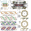
The nuclear pore complex (NPC) linker scaffold. (A) Schematic cutaway representation of the NPC architecture. (B) Near-atomic composite structure of the human NPC symmetric core generated by quantitatively docking nucleoporin and nucleoporin complex crystal and single particle cryo–electron microscopy (cryo-EM) structures into an ∼12-Å cryo–electron tomographic (cryo-ET) map of the intact human NPC (Mosalaganti et al. 2022; Petrovic et al. 2022). The nuclear envelope is shown as a gray isosurface and the nucleoporin structures are displayed in cartoon representation colored according to the legend. (C) Schematic layer-by-layer representation of a cross-section view of three spokes of the NPC inner ring showing the network of linker–scaffold interactions. (D) Schematic representation of the top views of human NPC cytoplasmic and nuclear outer rings and corresponding side views of two spokes, illustrating the reticulated head-to-tail arrangement of the coat nucleoporin complexes (CNCs) cross-linked by trans-spoke NUP93–NUP205 interactions. (Figure adapted from Petrovic et al. 2022, with permission from the authors.)
Figure 2.
Figure 2.
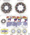
Dilation of the nuclear pore complex (NPC) and lateral channel formation. (A) Cytoplasmic views of the near-atomic composite structure of constricted and dilated human NPCs (Bley et al. 2022; Mosalaganti et al. 2022). The nuclear envelope is shown as a gray isosurface and nucleoporins are shown in cartoon representation. (B) Close-up views of the interface between two spokes, separated by a dashed line, in the constricted and dilated human NPCs. Nucleoporins are shown in surface representation, with the inner ring spokes uniformly colored in pale blue. (C) Schematic representation of the NPC symmetric core cross section, illustrating inner nuclear membrane integral membrane protein (INM-IMP) passage through the lateral channels. Top views of the NPC symmetric core illustrate that lateral channels can accommodate freely diffusing small, folded pore-facing domains or unstructured linkers tethering larger folded domains or karyopherin-binding nuclear localization sequences (NLSs; classical Kapα/Kapβ-mediated import shown). (Figure adapted from Petrovic et al. 2022, with permission from the authors.)
Figure 3.
Figure 3.
Architecture of the cytoplasmic face of the human nuclear pore complex (NPC). (A) Cross-sectional view of the near-atomic composite structure of the human NPC generated by docking nucleoporin and nucleoporin complex crystal and single particle cryo-EM structures into an ∼12-Å cryo-ET map of the intact human NPC (Bley et al. 2022; Mosalaganti et al. 2022). The nuclear envelope is shown as a gray isosurface, the inner ring is displayed in cartoon representation uniformly colored pale blue, and the nuclear and cytoplasmic face structures are displayed in cartoon representation colored according to the legend. (B) Architecture of the cytoplasmic face of the human NPC (Bley et al. 2022). Flexibly attached NUP358 domains protrude into the cytoplasm. (C) Schematic representation of a single cytoplasmic face protomer, illustrating the attachment of a NUP358 pentameric bundle held together by interactions between oligomerization elements (OEs). Four NUP358NTD copies wrap around the tandem arranged Y-shaped CNC stalks, generating a binding site for a fifth copy. (D) Cartoon representations of crystal structures of NUP358 complexes: NUP358NTD (PDB ID 7MNL), NUP358ZnF7•Ran(GDP) (PDB ID 7MNU), NUP358RanBD-IV•Ran(GTP) (PDB ID 7MNZ) (Bley et al. 2022), NUP358E3•SUMO-RanGAP•UBC9 (PDB ID 1Z5S) (Reverter and Lima 2005), NUP358CTD (PDB ID 4I9Y) (Lin et al. 2013). (Figure adapted from Bley et al. 2022, with permission from the authors.)
Figure 4.
Figure 4.
mRNP export and remodeling at the cytoplasmic face of the nuclear pore complex (NPC). (A) Cartoon representation and corresponding schematic conceptualization of a spoke of the NPC cytoplasmic face illustrating the relative positions of the cytoplasmic filament nucleoporins, with the cytoplasmic filament nucleoporin complex (CFNC) anchored by NUP93R1 and a pentameric bundle of NUP358 bound to the stalk of tandem-arranged Y-shaped coat nucleoporin complexes (CNCs). (B) Cartoon representations of crystal structures of human cytoplasmic filament complexes: DDX19(ADP)•GLE1CTD•NUP42GBM (PDB ID 6B4I) (Lin et al. 2018), DDX19(ATP)• GLE1CTD•NUP42GBM (PDB ID 6B4J) (Lin et al. 2018), DDX19(ATP)•U10RNA (PDB ID 3FHT) (von Moeller et al. 2009), DDX19(ATP) (PDB ID 6B4K) (Lin et al. 2018), DDX19(ADP)•NUP214NTD (PDB ID 3FMO) (Napetschnig et al. 2009), NUP214NTD (PDB ID 2OIT) (Napetschnig et al. 2007), NUP88NTD•NUP98APD (PDB ID 7MNI) (Bley et al. 2022), RAE1•NUP98GLEBS (PDB ID 3MMY) (Ren et al. 2010). (C) Schematic representation of the maturation, export, and remodeling of mRNPs by the DDX19 helicase cycle. In the nucleus, mRNA is transcribed by RNA polymerase II, followed by 5′ capping, recruitment of the cap-binding complex (CBC), deposition of exon junction complexes (EJCs) at splice sites, loading of the transcription-export (TREX) complex in a splicing-dependent manner, 3′ polyadenylation, and deposition of polyadenylate-binding nuclear protein 1 (PABPN1). ATP-hydrolyzing DEAD-box helicase UAP56 may facilitate loading of export factors P15•TAP to produce an export-competent mRNP. The exported mRNP is remodeled at the cytoplasmic face of the NPC, where P15•TAP is removed in a DDX19-dependent manner: (1) ATP-bound DDX19 cycles between autoinhibited and closed conformations. (2) Upon binding RNA, DDX19 adopts a closed, catalytically active conformation that is incompatible with GLE1 binding but could strip P15•TAP from mRNA. (3) ATP hydrolysis by DDX19 triggers RNA release, converting DDX19 to an autoinhibited ADP-bound conformation in which the autoinhibitory α-helix binds between amino- and carboxy-terminal RecA domains. (4) GLE1 binding destabilizes the autoinhibited conformation. (5) NUP214 binding converts DDX19 to an open conformation, promoting nucleotide exchange. (6) Nucleotide exchange displaces NUP214, priming the DDX19(ATP)•GLE1•NUP42 complex to restart the cycle (Lin et al. 2018). (Figure adapted from Bley et al. 2022, with permission from the authors.)
Figure 5.
Figure 5.
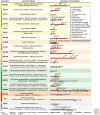
Nucleoporin-associated diseases. An overview of human nucleoporin diseases and viral virulence factors targeting the nuclear pore complex (NPC). Domain structures of human nucleoporins drawn as horizontal boxes with residue numbers indicated and their observed or predicted folds colored according to the legend. NUP160: dilated cardiomyopathy (Tarazón et al. 2012), steroid-resistant nephrotic syndrome (SRNS) (Braun et al. 2018; Zhao et al. 2019), B-cell acute lymphoblastic leukemia (B-ALL) (Harvey et al. 2010); NUP75: congestive heart failure (Satoh et al. 2007), SRNS (Braun et al. 2018); SEC13: breast cancer (Li and Liu 2021), gastric cancer (Mottaghi-Dastjerdi et al. 2015); NUP107: cardiac arrhythmia (Guan et al. 2019), SRNS (Braun et al. 2018; Guan et al. 2019), microcephaly (Rosti et al. 2017), ovarian dysgenesis (Weinberg-Shukron et al. 2015), breast cancer (Li and Liu 2021), brain cancer, glioblastoma multiforme (Hodgson et al. 2009), dedifferentiated liposarcoma (Wang et al. 2012); NUP133: SRNS (Braun et al. 2018), breast cancer (Hernández et al. 2007); NUP43: aortic dilatation (Haskell et al. 2017); NUP37: breast cancer (Li and Liu 2021), atrial fibrillation (Haskell et al. 2017), SRNS (Braun et al. 2018); NUP205: congenital heart disease associated with situs inversus and heterotaxy (Chen et al. 2019), SRNS (Braun et al. 2016); NUP188: congenital heart disease associated with heterotaxy (Fakhro et al. 2011), mitral valve prolapse (Haskell et al. 2017), Sandestig–Stefanova syndrome (Muir et al. 2020; Sandestig et al. 2020), B-ALL (Nowak et al. 2010); NUP93: dilated cardiomyopathy (Tarazón et al. 2012), SRNS (Braun et al. 2016; Sandokji et al. 2019), Down syndrome (Lima et al. 2011); NUP155: atrial fibrillation (Oberti et al. 2004; Zhang et al. 2008). (Continued) ALADIN: Triple A syndrome (Huebner et al. 2004); NUP62: ischemic cardiomyopathy (Chahine et al. 2015), primary biliary cirrhosis (Wesierska-Gadek et al. 1996), amyotrophic lateral sclerosis (ALS) (Aizawa et al. 2019), infantile striatonigral degeneration (Basel-Vanagaite et al. 2006), protein degradation induced by poliovirus (Gustin and Sarnow 2001) and rhinovirus (Gustin and Sarnow 2002) infections, hyperphosphorylation induced by cardiovirus infection (Porter and Palmenbert 2009), protein mislocalization induced by HIV-1 infection (Monette et al. 2011); NUP98: acute myeloid leukemia (Borrow et al. 1996; Nakamura et al. 1996; Xu and Powers 2009; Köhler and Hurt 2010; Gough et al. 2011), acute megakaryoblastic leukemia without Down syndrome (Roussy et al. 2018; Lalonde et al. 2021), protein degradation induced by poliovirus infection (Park et al. 2008), liver cancer (Singer et al. 2012); RAE1: acute myeloid leukemia (Roussy et al. 2018; Lalonde et al. 2021), breast cancer (Funasaka et al. 2011), herpesviruses (Gong et al. 2016), influenza A (Satterly et al. 2007), vesicular stomatitis virus (VSV) (Faria et al. 2005; Ren et al. 2010), and SARS-CoV2 (Oh et al. 2019) virulence factor binding; NUP88: fetal akinesia deformation sequence (Miorin et al. 2020), prostate, ovarian, breast, hepatocellular, colon, and lung cancers, and mesotheliomas and lymphomas (Bonnin et al. 2018); NUP214: acute febrile encephalopathy (Martinez et al. 1999; Gould et al. 2000), acute myeloid leukemia (von Lindern et al. 1992; Kraemer et al. 1994), breast cancer (Hernández et al. 2007), hyperphosphorylation induced by cardiovirus infection (Porter and Palmenberg 2009), protein degradation induced by rhinovirus infection (Ghildyal et al. 2009); GLE1: ALS (Kaneb et al. 2015), lethal congenital contracture syndrome (Nousiainen et al. 2008), lethal arthrogryposis with anterior horn cell disease (Paakkola et al. 2018); NUP358: acute myelomonocytic leukemia (Paakkola et al. 2018), dilated cardiomyopathy (Tarazón et al. 2012), ischemic cardiomyopathy (Lim et al. 2014), acute necrotizing encephalopathy (Neilson et al. 2009; Sell et al. 2016), colorectal cancer (Gylfe et al. 2013), protein degradation induced by rhinovirus infection (Ghildyal et al. 2009), HIV-1 virulence factor binding (Di Nunzio et al. 2012); NUP153: dilated cardiomyopathy and ischemic cardiomyopathy (Tarazón et al. 2012), cardiac arrhythmia (Nanni et al. 2016), protein degradation induced by poliovirus and rhinovirus (Gustin and Sarnow 2002) infections, hyperphosphorylation induced by cardiovirus infection (Porter and Palmenberg 2009), HIV-1 virulence factor binding (Matreyek et al. 2013), autoimmune liver disease/rheumatic disease (Enarson et al. 2004); TPR: gastric, lung, bone, thyroid, and colorectal cancers (Soman et al. 1991; Greco et al. 1997; Yu et al. 2000), autoimmune liver disease/rheumatic disease (Enarson et al. 2004); NDC1: dilated cardiomyopathy and ischemic cardiomyopathy (Tarazón et al. 2012); POM210: congenital heart disease (Chen et al. 2019), breast cancer (Curtis et al. 2012; Amin et al. 2021), primary biliary cirrhosis (Courvalin et al. 1990), colorectal cancer (Landi et al. 2012), and cervical cancer (Rajkumar et al. 2011). (fs) Frameshift, (*) nonsense mutation, (Δ) deletion.

Similar articles

Cited by

References

    1. Ader C, Frey S, Maas W, Schmidt HB, Görlich D, Baldus M. 2010. Amyloid-like interactions within nucleoporin FG hydrogels. Proc Natl Acad Sci 107: 6281–6285. 10.1073/pnas.0910163107 - DOI - PMC - PubMed
    1. Ahn JH, Davis ES, Daugird TA, Zhao S, Quiroga IY, Uryu H, Li J, Storey AJ, Tsai YH, Keeley DP, et al. 2021. Phase separation drives aberrant chromatin looping and cancer development. Nature 595: 591–595. 10.1038/s41586-021-03662-5 - DOI - PMC - PubMed
    1. Aizawa H, Yamashita T, Kato H, Kimura T, Kwak S. 2019. Impaired nucleoporins are present in sporadic amyotrophic lateral sclerosis motor neurons that exhibit mislocalization of the 43-kDa TAR DNA-binding protein. J Clin Neurol 15: 62–67. 10.3988/jcn.2019.15.1.62 - DOI - PMC - PubMed
    1. Akey CW. 1995. Structural plasticity of the nuclear pore complex. J Mol Biol 248: 273–293. - PubMed
    1. Akey CW, Radermacher M. 1993. Architecture of the Xenopus nuclear pore complex revealed by three-dimensional cryo-electron microscopy. J Cell Biol 122: 1–19. 10.1083/jcb.122.1.1 - DOI - PMC - PubMed

Publication types

LinkOut - more resources