. 2022 Sep 12;5(11):e202201557.
doi: 10.26508/lsa.202201557. Print 2022 Nov.

α-Actinin-4 recruits Shp2 into focal adhesions to potentiate ROCK2 activation in podocytes

Affiliations

α-Actinin-4 recruits Shp2 into focal adhesions to potentiate ROCK2 activation in podocytes

Chien-Chun Tseng et al. Life Sci Alliance. .

Abstract

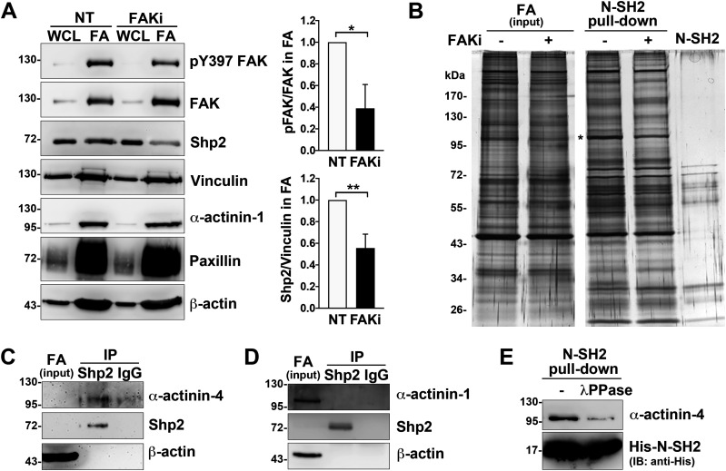
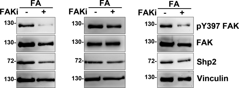
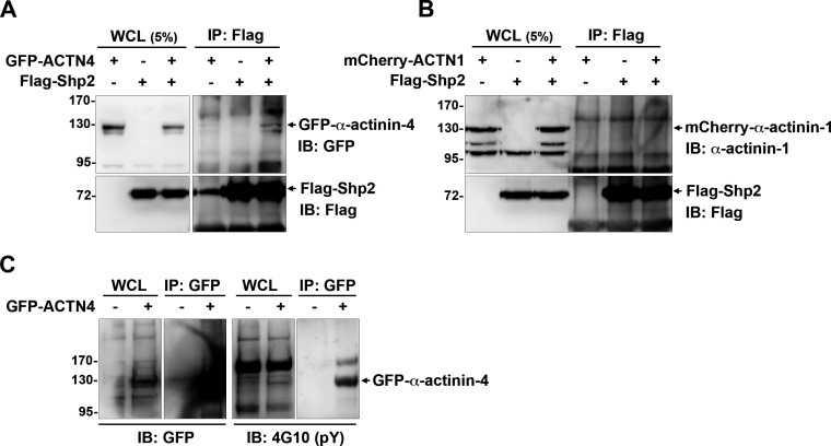
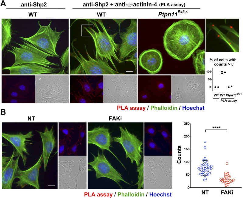
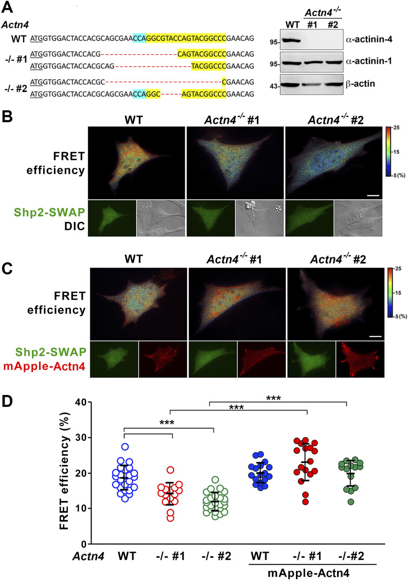
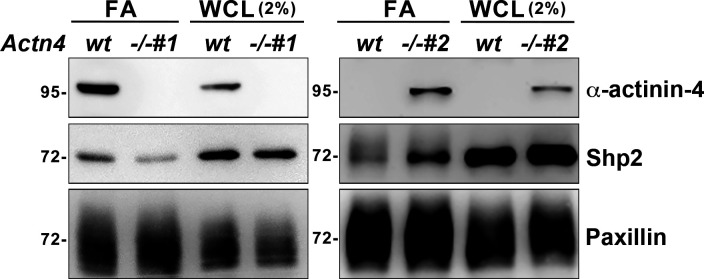
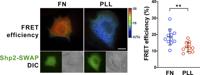
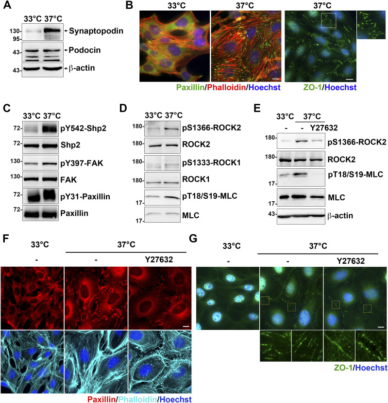
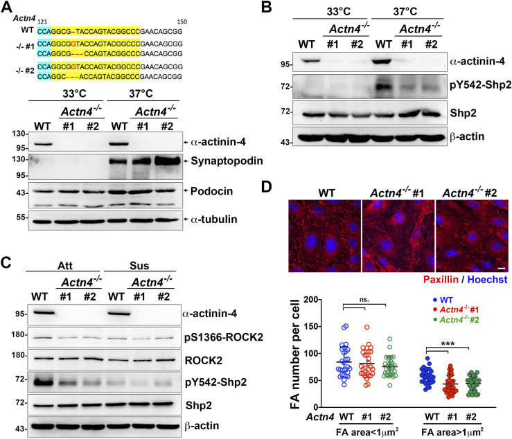
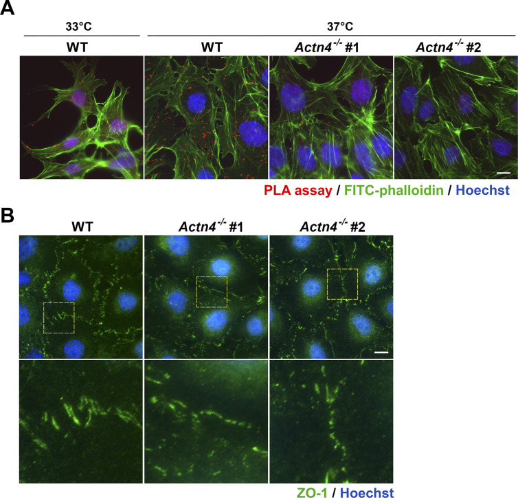
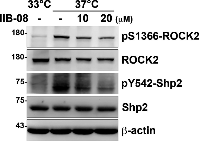
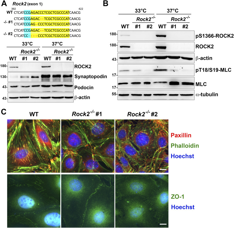
Cell-matrix adhesions are mainly provided by integrin-mediated focal adhesions (FAs). We previously found that Shp2 is essential for FA maturation by promoting ROCK2 activation at FAs. In this study, we further delineated the role of α-actinin-4 in the FA recruitment and activation of Shp2. We used the conditional immortalized mouse podocytes to examine the role of α-actinin-4 in the regulation of Shp2 and ROCK2 signaling. After the induction of podocyte differentiation, Shp2 and ROCK2 were strongly activated, concomitant with the formation of matured FAs, stress fibers, and interdigitating intracellular junctions in a ROCK-dependent manner. Gene knockout of α-actinin-4 abolished the Shp2 activation and subsequently reduced matured FAs in podocytes. We also demonstrated that gene knockout of ROCK2 impaired the generation of contractility and interdigitating intercellular junctions. Our results reveal the role of α-actinin-4 in the recruitment of Shp2 at FAs to potentiate ROCK2 activation for the maintenance of cellular contractility and cytoskeletal architecture in the cultured podocytes.


Conflict of interest statement

The authors declare that they have no conflict of interest.

Figures

Figure 1.
Figure 1.. Identification of Shp2-interacting proteins in FAs.
MEFs seeded on FN-coated dishes were treated with or without FAK inhibitor 14 (15 μM) for 90 min for isolation of FAs as described in the Materials and Methods section. (A) Whole-cell lysate (WCL; 1%) and isolated FA fractions (FA) were subjected to Western analysis as indicated. Relative FAK Y397 phosphorylation and levels of Shp2 versus vinculin in FA fractions from four independent experiments were measured. (B) FA fractions were incubated with 50 μg of His-tagged Shp2 N-SH2 recombinant protein followed by pulled-down with Ni-beads, SDS–PAGE, and sliver staining. By mass spectrometry analysis, the band indicated by * was identified as α-actinin-4 and α-actinin-1. (C, D) FA fractions isolated from non-treated MEFs were immunoprecipitated with anti-Shp2 antibody followed by Western blotting analysis as indicated. The concentration of Shp2 and α-actinin-4 in the IP input sample might be too low to be detected here. (E) FA fractions were incubated with or without 400 U of λPPase for 30 min before pull-down with His-N-SH2 proteins and Ni-beads. The levels of pulled-down α-actinin-4 was detected by Western blotting as indicated. Data are mean ± SD. *P < 0.05, **P < 0.01 (two-tailed, paired t test). Source data are available for this figure.
Figure S1.
Figure S1.. Inhibition of FAK reduces Shp2 recruitment in FAs.
MEFs seeded on FN-coated dishes were treated with or without FAK inhibitor 14 (15 μM) for 90 min for isolation of FAs as Fig 1A. The isolated FA fractions (FA) were subjected to Western analysis as indicated. Relative FAK Y397 phosphorylation and levels of Shp2 versus vinculin in FA fractions were measured and showed in Fig 1A.
Figure S2.
Figure S2.. Interaction of exogenous Shp2 and α-actinin-4 expressed in HEK293T cells.
(A, B) HEK293T cells seeded were co-transfected with the expression constructs of flag-Shp2 and GFP-α-actinin-4 (A) or mCherry-α-actinin-1 (B). Cells were harvested for immunoprecipitation with anti-flag, and the immunoprecipitated proteins were analyzed by Western blot as indicated. (C) HEK293T cells were transiently transfected with the expression construct of GFP-α-actinin-4. Cells were harvested for immunoprecipitation with the anti-GFP antibody (GFP-trap) and then subjected to Western blot analysis with anti-pY (4G10) and anti-GFP antibodies.
Figure 2.
Figure 2.. Shp2 interacts with α-actinin-4 at FAs.
(A) Wild-type (WT) and Ptpn11Ex3−/− MEFs were seeded on FN-coated coverslips for 2 h and fixed for in situ proximity ligation assay (PLA) with anti-Shp2 plus anti-α-actinin-4 antibodies (red). Reaction with anti-Shp2 antibody serves as a negative control. After reaction, cells were stained with FITC-phalloidin (green) and Hoechst (blue) for F-actin and nucleus, respectively. Scatter dot plots show the percentage of cells with PLA count >5 in each independent experiments. (B) MEFs seeded FN-coated coverslips were treated with or without FAK inhibitor 14 (20 μM) for PLA assay. Scatter dot plots (mean ± SD) show PLA counts from more than 36 cells in three independent experiments; each dot represents one single cell. ****P < 0.0001 (Mann–Whitney U test). Scale bars, 10 μm.
Figure 3.
Figure 3.. Loss of α-actinin-4 reduces Shp2 activation.
(A) The modified DNA sequence of Actn4 in two selected Actn4−/− MEF clones. The gRNA targeted region is highlighted in yellow; PAN site is shown in light blue, and modified DNA sequence is shown in red. The expression of α-actinin-4 in these clones was checked by Western blotting. (B) The FRET efficiency images of a wild-type or Actn4−/− MEF transfected with Shp2 FRET reporter (Shp2-SWAP). (C) The expression construct of mApple-Actn4 (red) was co-transfected with Shp2-SWAP into MEF cells for FRET imaging analysis. (D) Scatter dot plots (mean ± SD) of the Shp2-SWAP FRET efficiency. The sample numbers are 24 and 17 for WT 16 and 18 for Actn4−/− #1 and 25 and 15 and 24 for Actn4−/− #2 cells in the absent or present of mApple-Actn4 co-transfection, respectively. Differences between continuous variables were compared using the Mann–Whitney U test. ***P < 0.0005. Scale bars, 10 μm. Source data are available for this figure.
Figure S3.
Figure S3.. Loss of α-actinin-4 reduced Shp2 FA targeting in MEFs.
The FA fractions were isolated from wild-type and Actn4−/− MEFs seeded on FN-coated dishes. Whole-cell lysate (WCL; 2%) and enriched FA fractions were subjected to Western blot analysis as indicated.
Figure S4.
Figure S4.. Integrin-mediated Shp2 activation in MEFs.
MEFs were transfected with Shp2 FRET reporter (Shp2-SWAP) and then replated on fibronectin (FN) or poly–L-lysine (PLL)–coated glass bottom dishes for FRET imaging. Scatter dot plots show the Shp2-SWAP FRET efficiency of each individual cells. Data are expressed as mean ± SD from more than 10 cells in two independent experiments. Differences between continuous variables were compared using the Mann–Whitney U test. **P < 0.005. Scale bar, 10 μm.
Figure 4.
Figure 4.. The contractile signals in cultured podocytes.
The conditional immortalized mouse podocytes cultured at 33°C in the present of INFγ (proliferative) or at 37°C in the absent of INFγ (differentiated) for 10–14 d on the type-IV collagen–coated substrates. (A) Podocytes were harvested for Western blot analysis with antibodies specific for podocyte marker proteins. (B) Podocytes seeded on type-IV collagen–coated glass coverslips were fixed for immunofluorescence staining with anti-paxillin and anti-ZO-1 antibodies for detecting FA and intercellular junctions and phalloidin and Hoechst for F-actin and DNA, respectively. (C, D) Protein extracts form podocytes were subjected to Western blot analysis with antibodies as indicated. (E, F, G) Podocytes were serum-starved for 24 h and then treated with or without 20 μM of Y27632 for 1 h. Cells were harvested for Western blotting analysis or fixed for immunofluorescence staining as indicated. Scale bars, 10 μm. Source data are available for this figure.
Figure 5.
Figure 5.. α-Actinin-4 is required for Shp2 activation underlying adhesion signaling in podocytes.
(A) The modified DNA sequence of two selected Actn4−/− podocyte clones are showed. The gRNA targeted region is highlighted in yellow; the PAN site is shown in light blue, and modified DNA sequence is shown in red. Podocytes maintained at permission (33°C) or differentiation (37°C) conditions for 14 d were harvested for Western blotting analysis as indicated. (B) The phosphorylation status of Shp2 at Y542 in these clones were also detected. (C) Podocytes were induced for differentiation and then maintained in attached (Att) or suspension for 30 min (Sus) before harvested for Western blotting analysis as indicated. (D) Podocytes were induced for differentiation and seeded on collagen type-IV–coated glass coverslips for immunofluorescence staining with anti-paxillin antibody and Hoechst for detecting FA and DNA, respectively. Scatter dot plots of the numbers of small FA (area < 1 μm2) and matured FA (area > 1 μm2) were shown. Data are expressed as mean ± SD from 30 representative cells in three independent experiments. Differences between continuous variables were compared using two-tail unpaired student t test. ***P < 0.001. ns, not statistically significant. Bars, 10 μm. Source data are available for this figure.
Figure S5.
Figure S5.. The Actn4−/− podocytes.
(A) Wild-type and Actn4−/− podocytes maintained at permission (33°C) or differentiation (37°C) conditions on type-IV collagen–coated glass coverslips were fixed for in situ proximity ligation assay with anti-Shp2 plus anti-α-actinin-4 antibodies (red). After reaction, cells were stained with FITC-phalloidin (green) and Hoechst (blue) for F-actin and nucleus, respectively. (B) Differentiated podocytes were fixed for immunofluorescence staining with anti-ZO-1 antibody and Hoechst for detecting intercellular junctions and DNA, respectively. Scale bar, 10 μm.
Figure S6.
Figure S6.. Inhibition of Shp2 reduced ROCK2 activation in podocytes.
Undifferentiated cells (33°C) and differentiated (37°C) podocytes were serum-starved for 24 h and then treated with or without IIB-08 (10 or 20 μM) for 24 h. Cells were harvested for Western blot analysis as indicated.
Figure 6.
Figure 6.. The crucial role of ROCK2 in the maintenance of cellular contractility and cytoskeletal architecture in podocytes.
(A) The modified DNA sequence of Rock2 in two Rock2−/− podocyte clones are showed. The gRNA targeted region is highlighted in yellow; PAN site is shown in light blue, and modified DNA sequence is shown in red. Podocytes maintained at permission (33°C) or differentiation (37°C) conditions for 14 d were harvested for Western blotting analysis as indicated. (B) The phosphorylation status of myosin light chain in these clones were also detected. (C) Podocytes were induced for differentiation and seeded on type-IV collagen–coated glass coverslips for immunofluorescence staining with anti-paxillin and anti-ZO-1 antibodies for detecting FA and intercellular junctions and phalloidin and Hoechst for F-actin and DNA, respectively. Scale bars, 10 μm. Source data are available for this figure.

Similar articles

Cited by

References

    1. Asanuma K, Kim K, Oh J, Giardino L, Chabanis S, Faul C, Reiser J, Mundel P (2005) Synaptopodin regulates the actin-bundling activity of alpha-actinin in an isoform-specific manner. J Clin Invest 115: 1188–1198. 10.1172/jci200523371 - DOI - PMC - PubMed
    1. Chuang HH, Liang SW, Chang ZF, Lee HH (2013) Ser1333 phosphorylation indicates ROCKI activation. J Biomed Sci 20: 83. 10.1186/1423-0127-20-83 - DOI - PMC - PubMed
    1. Chuang HH, Yang CH, Tsay YG, Hsu CY, Tseng LM, Chang ZF, Lee HH (2012) ROCKII Ser1366 phosphorylation reflects the activation status. Biochem J 443: 145–151. 10.1042/bj20111839 - DOI - PubMed
    1. Connelly JT, Gautrot JE, Trappmann B, Tan DWM, Donati G, Huck WT, Watt FM (2010) Actin and serum response factor transduce physical cues from the microenvironment to regulate epidermal stem cell fate decisions. Nat Cell Biol 12: 711–718. 10.1038/ncb2074 - DOI - PubMed
    1. Dandapani SV, Sugimoto H, Matthews BD, Kolb RJ, Sinha S, Gerszten RE, Zhou J, Ingber DE, Kalluri R, Pollak MR (2007) α-Actinin-4 is required for normal podocyte adhesion. J Biol Chem 282: 467–477. 10.1074/jbc.m605024200 - DOI - PubMed

Publication types