. 2022 Sep 12;19(1):224.
doi: 10.1186/s12974-022-02585-w.

Chronic suppurative otitis media causes macrophage-associated sensorineural hearing loss

Affiliations

Chronic suppurative otitis media causes macrophage-associated sensorineural hearing loss

Anping Xia et al. J Neuroinflammation. .

Abstract

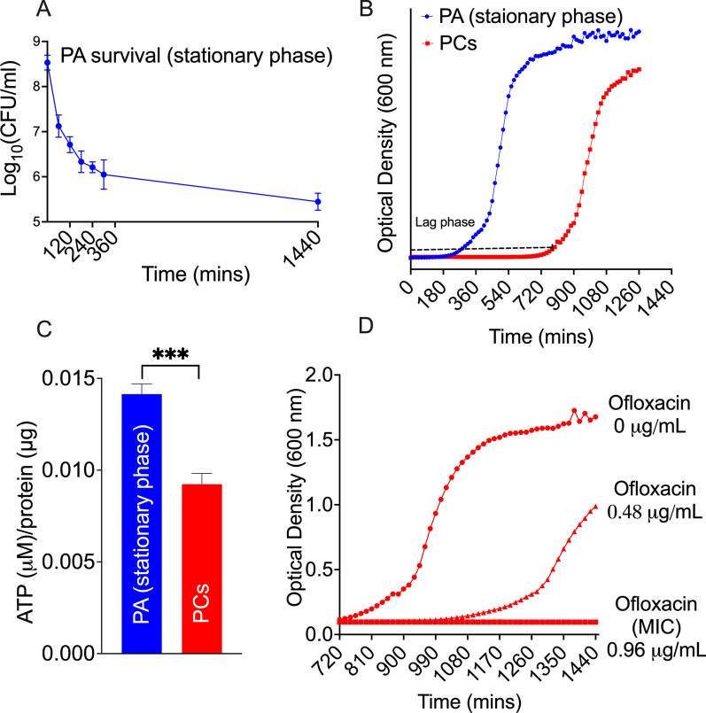
Background: Chronic suppurative otitis media (CSOM) is the most common cause of permanent hearing loss in children in the developing world. A large component of the permanent hearing loss is sensory in nature and our understanding of the mechanism of this has so far been limited to post-mortem human specimens or acute infection models that are not representative of human CSOM. In this report, we assess cochlear injury in a validated Pseudomonas aeruginosa (PA) CSOM mouse model.

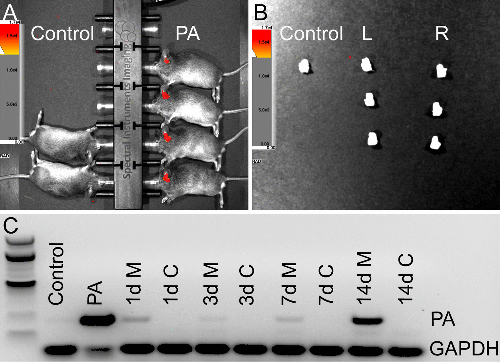
Methods: We generated persisters (PCs) and inoculated them into the mouse middle ear cavity. We tracked infection with IVIS and detected PA using RT-PCR. We assessed cochlear damage and innate immunity by Immunohistochemistry. Finally, we evaluated cytokines with multiplex assay and quantitative real-time PCR.

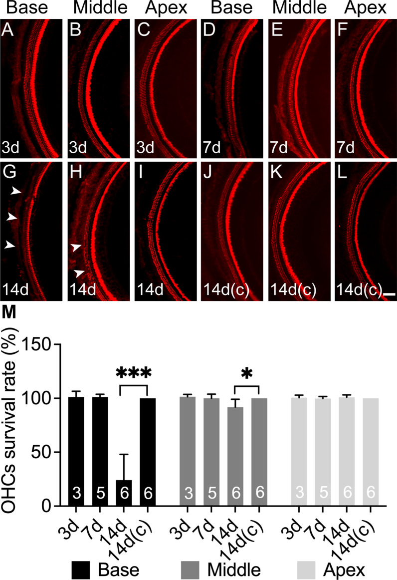
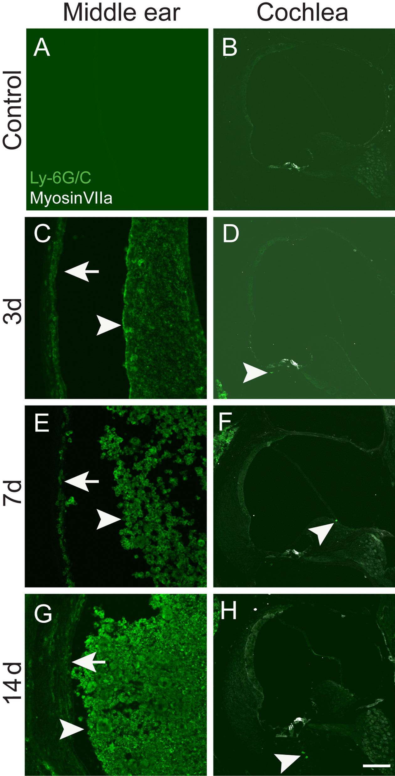
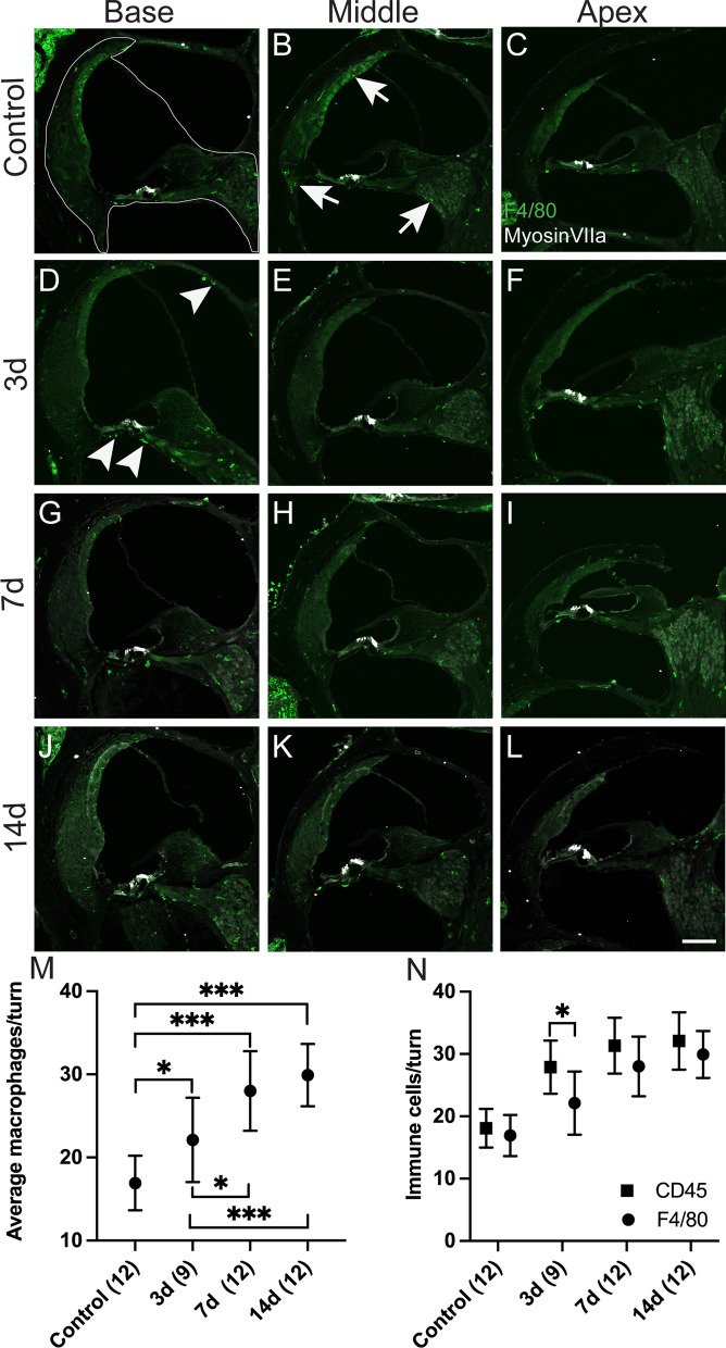
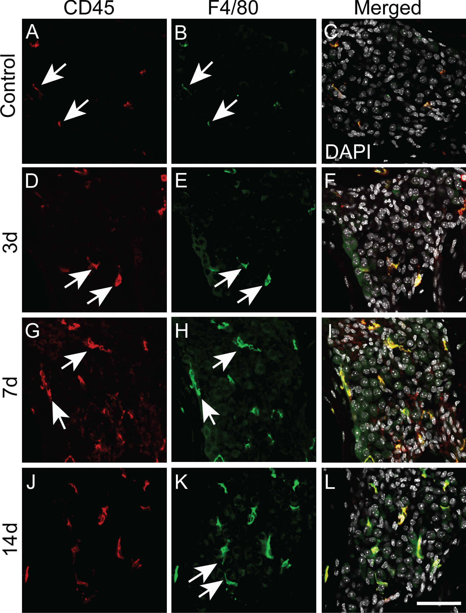
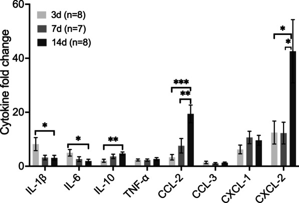
Results: We observed outer hair cell (OHC) loss predominantly in the basal turn of the cochlear at 14 days after bacterial inoculation. Macrophages, not neutrophils are the major immune cells in the cochlea in CSOM displaying increased numbers and a distribution correlated with the observed cochlear injury. The progression of the morphological changes suggests a transition from monocytes into tissue macrophages following infection. We also show that PA do not enter the cochlea and live bacteria are required for cochlear injury. We characterized cytokine activity in the CSOM cochlea.

Conclusions: Taken together, this data shows a critical role for macrophages in CSOM-mediated sensorineural hearing loss (SNHL).

Keywords: CSOM; Cytokines; HC loss; Macrophages; PA; SNHL.


Conflict of interest statement

The authors declare no relevant conflicts of interest.

Figures

Fig. 1
Fig. 1
Characterization of persister cells (PCs). A Time-dependant survival curve for PA stationary phase after treatment with ofloxacin (5 μg/mL). The survival curve demonstrates rapid killing from 0 to 5 h (predominantly metabolically active cells) and plateauing of survival (PCs) from 5 to 24 h. B Growth curve of ofloxacin-generated PCs compared to stationary phase PA. PCs displayed delayed awakening on growth curve compared to stationary phase PA, consistent with the PC phenotype. Upon awakening, the growth rate of the PC phenotype matches that of the stationary phase PA. C Intracellular ATP level comparison between PCs compared to stationary phase PA. PCs contained significantly less intracellular ATP following normalization to protein levels compared to stationary phase PA. Due to lower metabolic activity, PC exhibit lower ATP level. D The MIC of the surviving PCs was determined by performing growth curves for serial dilutions of ofloxacin (0, 0.48 and 0.96 µg/mL) showing that the PCs are not resistant (MIC < 2 µg/mL)
Fig. 2
Fig. 2
OHC loss occurs in CSOM. AC Whole mount immunostaining showed no HC loss.in the base, middle and apex regions of the cochlea at 3 days (AC), 7 days (DF) and 14 days (GI) compared to 14 days control (JL). The normal condition displays 3 rows of OHCs on the left and 1 row of IHC on the right of the pictures. CSOM cochlea displayed OHC loss at 14 days (GI) in base (G, arrowheads) and small areas of absence in middle (H, arrowheads), and no OHC in the apex (I). M OHC survival rate was significantly lower in the base (P < 0.001) and middle (P < 0.05) at 14 days compared to control (M). The mouse number for each group is presented in the column (M). Red: myosin VIIa. The data represent mean ± SD. Scale bar = 100 µm
Fig. 3
Fig. 3
PA is not detected in CSOM cochlea. A IVIS revealed the presence of PA (red) in CSOM ears but not in the ears of control mice at 7 days. IVIS was performed with 15 mice for each group. B After the cochleae were dissected from the same mice in A, PA was not detected in the inner ears (absence of red) of control or either the infected ear (L) or non-infected ear (R). C RT-PCR for the PAO1 O-acetylase gene showed detection of PA in the CSOM middle ears at 1 days (1d M), 3 days (3d M), 7 days (7d M) and 14 days (14d M) but not in the CSOM cochleae at 1 days (1d C), 3 days (3d C), 7 days (7d C) 14 days (14d C) or control cochlea (control). There were 3 mice at each time point. GAPDH was used as housekeeping gene
Fig. 4
Fig. 4
Neutrophils are present in the CSOM middle ear. Staining with the neutrophil-specific marker Ly-6G/C was performed on cryosections of middle ears and cochleae from the same mouse. The CSOM middle ear displayed abundant neutrophils at 3 days, 7 days and 14 days in the effusion (arrowheads to right in C, E and G) and middle ear mucosa (arrows to left in C, E and G), whereas a few limited neutrophils were present in the CSOM cochleae (arrowheads in D, F and H). Top row (A, B) is control. There were 3 mice at each time point. Scale bar = 100 µm
Fig. 5
Fig. 5
Macrophage numbers are significantly increases in the CSOM cochlea. Cochlear cryosections were stained with the macrophage-specific marker F4/80 (green) and were counted in the region of the spiral ligament, the organ of Corti, spiral ganglion neurons, and the scala media as outlined (A-dotted). Myosin VIIa staining (white) labels HCs in the cochlea. Counts were performed in all cochlear turns (base, middle, apex) in control mice (AC) and CSOM cochleae at 3d (DF), 7d (GI), and 14 DAYS (JL). Arrows in the control group (B) show macrophages in the stria vascularis, spiral ligament and spiral ganglion neuron area. Arrowheads in CSOM at 3d (D) show macrophages also present in the basilar membrane, scala vestibuli. M Statistical analysis of the macrophages per turn, revealed significant macrophage elevation in the CSOM cochlea at all time points compared to control mice. N F4/80-labeled macrophages compared with CD45 positive immune cells showing all similar numbers in all time points except at 3 days. Number of mice per group is in parentheses alongside the timepoint. The data represent mean ± SD. Scale bar = 100 µm
Fig. 6
Fig. 6
Macrophage morphological changes in the region of spiral ganglion neuron (SGN) in CSOM. Macrophages in cochlear cryosections were labeled with the pan-leukocyte marker CD45 and F4/80. Representative images are shown. Macrophages (arrows) were small with few projections in the control group (AC, arrows), and they were bigger and round at 3 days (D-F, arrows). Macrophages were larger and irregularly shaped at 7 days (GI, arrows), and had fine, dendritic projections at 14 days (JL, arrows). Scale bar = 50 µm. n = 3 per group
Fig. 7
Fig. 7
Selected cytokines are upregulated in CSOM. Cochlear selected cytokine expression was assessed via quantitative PCR with normalization to GAPDH and β-actin at 3 days, 7 days and 14 days, and fold-change was quantified relative to control cochlea. The pro-inflammatory cytokines interleukin-1β (IL-1β) and IL-6 displayed increased expression at 3 days compared to 14 days. The anti-inflammatory cytokine IL-10 displayed increased expression at 14 days compared to 3 days. The chemokines CCL-2 and CXCL-2 displayed increased expression at 14 days compared to 7 days and 3 days, respectively. TNF-α, CCL-3 and CXCL-1 remained unchanged in these time points. The mouse number is shown in brackets. The data represent mean ± SEM

Similar articles

Cited by

References

    1. Monasta L, Ronfani L, Marchetti F, Montico M, Vecchi Brumatti L, Bavcar A, et al. Burden of disease caused by otitis media: systematic review and global estimates. PLoS ONE. 2012;7(4):e36226. doi: 10.1371/journal.pone.0036226. - DOI - PMC - PubMed
    1. Mittal R, Lisi CV, Gerring R, Mittal J, Mathee K, Narasimhan G, et al. Current concepts in the pathogenesis and treatment of chronic suppurative otitis media. J Med Microbiol. 2015;64(10):1103–1116. doi: 10.1099/jmm.0.000155. - DOI - PMC - PubMed
    1. Mittal R, Kodiyan J, Gerring R, Mathee K, Li JD, Grati M, et al. Role of innate immunity in the pathogenesis of otitis media. Int J Infect Dis. 2014;29:259–267. doi: 10.1016/j.ijid.2014.10.015. - DOI - PMC - PubMed
    1. Santa Maria PL, Kaufman AC, Bacacao B, Thai A, Chen X, Xia A, et al. Topical Therapy Failure in Chronic Suppurative Otitis Media is Due to Persister Cells in Biofilms. Otol Neurotol. 2021. - PubMed
    1. Conlon BP, Rowe SE, Gandt AB, Nuxoll AS, Donegan NP, Zalis EA, et al. Persister formation in Staphylococcus aureus is associated with ATP depletion. Nat Microbiol. 2016;1:16051. doi: 10.1038/nmicrobiol.2016.51. - DOI - PubMed

LinkOut - more resources