. 2022 Sep 12;13(1):4733.
doi: 10.1038/s41467-022-32246-8.

Reactivating hippocampal-mediated memories during reconsolidation to disrupt fear

Affiliations

Reactivating hippocampal-mediated memories during reconsolidation to disrupt fear

Stephanie L Grella et al. Nat Commun. .

Abstract

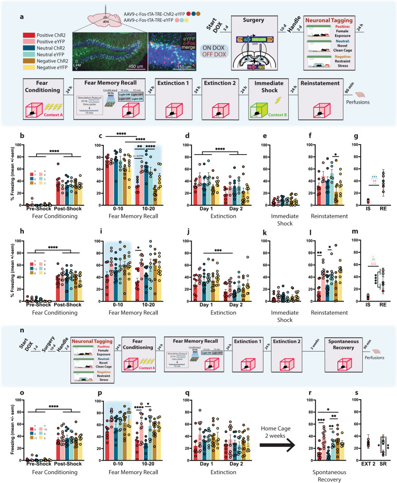
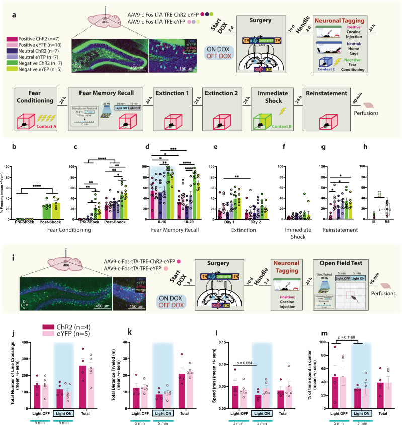
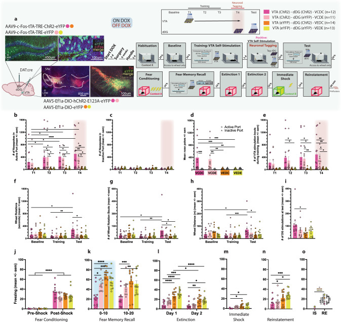
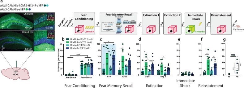
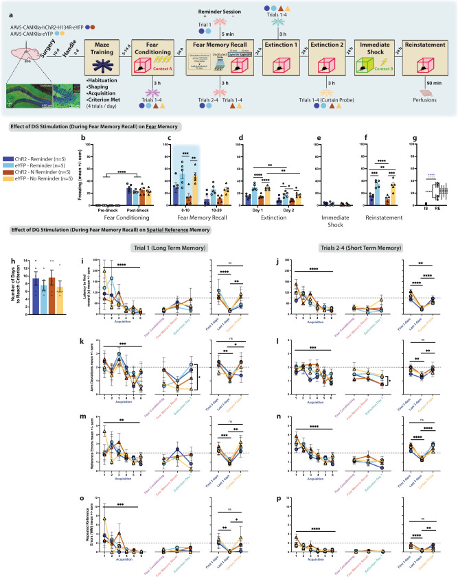
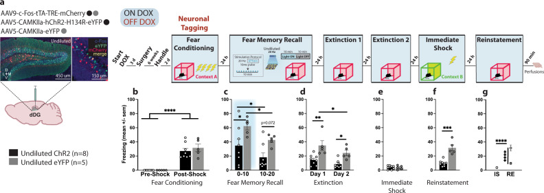
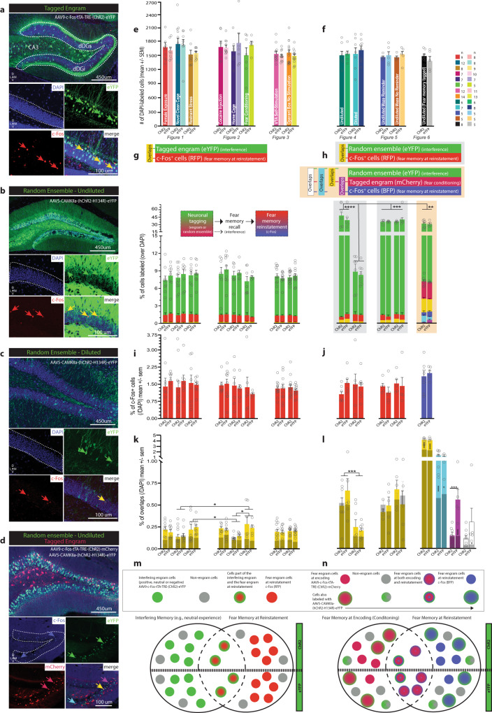
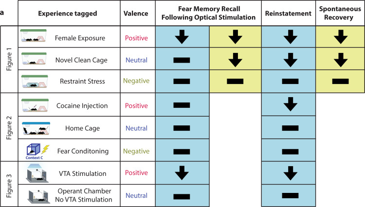
Memories are stored in the brain as cellular ensembles activated during learning and reactivated during retrieval. Using the Tet-tag system in mice, we label dorsal dentate gyrus neurons activated by positive, neutral or negative experiences with channelrhodopsin-2. Following fear-conditioning, these cells are artificially reactivated during fear memory recall. Optical stimulation of a competing positive memory is sufficient to update the memory during reconsolidation, thereby reducing conditioned fear acutely and enduringly. Moreover, mice demonstrate operant responding for reactivation of a positive memory, confirming its rewarding properties. These results show that interference from a rewarding experience can counteract negative affective states. While memory-updating, induced by memory reactivation, involves a relatively small set of neurons, we also find that activating a large population of randomly labeled dorsal dentate gyrus neurons is effective in promoting reconsolidation. Importantly, memory-updating is specific to the fear memory. These findings implicate the dorsal dentate gyrus as a potential therapeutic node for modulating memories to suppress fear.


Conflict of interest statement

The authors declare no competing interests.

Figures

Fig. 1
Fig. 1. Artificial reactivation of hippocampal-mediated memories during fear memory reconsolidation reduces fear acutely and enduringly.
a Viral strategy and experimental design. dDG cells encoding positive, neutral, and negatively-valenced behavioral epochs were tagged off-DOX (orange). Mice were FC (context A) and 24 h later given a recall session during which cells previously tagged were artificially reactivated in either the first (F10), or last half (L10) of the session. Across next 2 days, mice were given 2 EXT sessions. The following day, to reinstate fear responding, mice received an IS (context B) and the next day were tested for RE. b Mice showed greater freezing post-shock (three-way RM ANOVA: F(1,33) = 448.3, P < 0.0001). c During recall, L10 mice in positive (P = 0.022) and negative (P < 0.0001) ChR2-groups showed less freezing compared to neutral-ChR2 mice but not eYFP-controls (three-way RM ANOVA: F(2,34) = 4.665, P = 0.0162, Time × Valence × Virus). Freezing declined faster for both groups (P < 0.0001). df No group differences during EXT, or IS. At RE, negative-ChR2 mice showed less freezing than neutral-eYFP mice (p = 0.0449). g eYFP controls froze more than experimental mice at RE (three-way RM ANOVA: F(1,34) = 5.704, P = 0.0226, Virus × Day); F(1,34) = 3.969, P = 0.0282, Virus × Valence). h A separate cohort was FC demonstrating greater freezing post-shock (three-way RM ANOVA: F(1,48) = 761.4, P < 0.0001). i With F10 stimulation, only positive-ChR2 mice showed reduced fear in the last half of the session compared to eYFP controls (three-way RM ANOVA: F(1,48) = 7.737; P = 0.0077). j Neutral-ChR2 mice extinguished most rapidly (three-way RM ANOVA: F(1,48) = 57.75, P < 0.0001, Time; F(1,48) = 10.99; P = 0.0017). k No group differences seen during IS. l During RE, both positive (P = 0.0016) and neutral (P = 0.0031) ChR2-groups showed less fear compared to eYFP controls, while negative-ChR2 mice did not (two-way ANOVA: F(1,48) = 26.97, P < 0.0001). m Control mice froze more than experimental mice at RE (three-way RM ANOVA: F(1,48) = 24.66, P < 0.0001, Day × Virus). n A separate cohort was tested on SR. o Mice demonstrated greater freezing post-shock (three-way RM ANOVA: F(1,46) = 685.2, P < 0.0001). p Positive (P = 0.0251) and neutral (P = 0.0266) ChR2 mice showed reduced fear in the last half of recall compared to eYFP controls (three-way RM ANOVA: F(1,46) = 16.75, P = 0.0002, Time; F(1,46) = 28.15, P < 0.0001). q No group differences observed during EXT. r In a test for SR, both positive (P = 0.0004, P = 0.0122) and neutral (P = 0.0070, P = 0.0011) mice showed less freezing compared to eYFP controls and compared to negative-ChR2 mice (two-way ANOVA: F(2,46) = 6.894, P = 0.0024, Valence × Virus). s Positive and neutral ChR2 mice continued to exhibit less fear 2 weeks after EXT compared to both positive and neutral-eYFP mice, and both negative groups (three-way RM ANOVA: F(2,46) = 3.784, P = 0.0301, Valence × Virus; F(1,46) = 7.859, P = 0.0074, Day). Data represented as means ± s.e.m. *P < 0.05, **P < 0.01, ***P < 0.005, ****P < 0.00. dDG dorsal dentate gyrus, DOX doxycycline, EXT extinction, IS immediate shock, RE reinstatement, SR spontaneous recovery. Source data provided as a Source Data file.
Fig. 2
Fig. 2. Valence matters: Artificial reactivation of a neutral home cage experience during reconsolidation is not sufficient to disrupt a  fear memory.
a Viral strategy and experimental design. dDG cells encoding positive, neutral, and negatively-valenced behavioral epochs were tagged off-DOX (orange). b Negative groups were initially FC in an alternate context (Context C). These mice showed greater freezing post-shock (two-way RM ANOVA: F(1,10) = 168.4, P < 0.0001). c The next day all mice were FC. While all mice showed increased freezing post-shock (three-way RM ANOVA: F(1,37) = 295, P < 0.0001), mice in the negative groups demonstrated higher freezing pre-shock (Negative: vs. Positive P < 0.0001, vs. Neutral P < 0.0001) (two way RM ANOVA: F(2,40) = 27.55, P < 0.0001), and post-shock (three-way RM ANOVA: F(2,37) = 25.25, P < 0.0001). d During recall, negative groups continued to exhibit increased freezing (three-way RM ANOVA: F(2,37) = 3.286, P < 0.0486, Valence × Virus × Time). Negative-ChR2 mice froze more than neutral-ChR2 mice in the first (P = 0.0086) and last 10 min (P < 0.0001) and more than positive-ChR2 mice at both time points (P = 0.255, P < 0.0001). Positive (P = 0.0072) and neutral (P = 0.0002) ChR2 mice showed faster decline in freezing compared to eYFP controls. e During EXT1, positive and neutral-ChR2 mice froze less than other groups (three-way ANOVA: F(2,37) = 4.107, P = 0.0245, Valence × Day; F(2,37) = 6.841, P = 0.0128, Virus × Day). f No group differences observed during IS. g During RE, positive ChR2-mice showed reduced fear compared to eYFP controls (P = 0.0249) and both negative-ChR2 (P = 0.0147) and eYFP (P = 0.0498) groups (two-way ANOVA: F(1,37) = 5.923, P = 0.0199, Virus; F(2,37) = 3.440, P = 0.0426). h Positive-ChR2 mice froze less than other groups except neutral-ChR2 mice at RE compared to IS (three-way RM ANOVA: F(1,37) = 5.912, P = 0.0200, Day × Virus). i Viral strategy and experimental design. dDG cells encoding acute cocaine-exposure (positive) were tagged off-DOX (orange) and 24 h later mice were placed in the OF (10 min) where tagged dDG cells were reactivated in the last 5 min. No group differences in locomotor j line crossings k distance traveled and l speed m or anxiety measures (percentage time spent in center). Data represented as means ± s.e.m. *P < 0.05, **P < 0.01, ***P < 0.005, ****P < 0.00. dDG dorsal dentate gyrus, DOX doxycycline, EXT extinction, OF open field, IS immediate shock, RE reinstatement. Source data provided as a Source Data file.
Fig. 3
Fig. 3. Mice will perform an operant response for artificial reactivation of a positive memory.
a Viral strategy and experimental design. DAT-Cre mice were given access to a wheel that produced no consequences (baseline and training). They were then trained to nose poke for 500 ms bursts of optogenetic VTA stimulation (T1–T3). dDG cells encoding this positive experience were tagged off-DOX (orange, T4). The next day, mice were again given access to the wheel, where spinning it now produced artificial reactivation of the tagged dDG cells responsive to VTA self-stimulation. 24 h later, mice underwent the same experimental procedure as previous experiments. Mice were injected with either ChR2 (VC) or eYFP (VE) in the VTA, and also injected with either ChR2 (DC) or eYFP (DE) in the dDG. b In the active port, VCDC & VCDE mice nose-poked more than VEDC & VEDE mice (two-way RM ANOVA: F(9,138) = 2.179, P = 0.0270, Time × Group) and increased responding across days whereas c, no effects were seen for the inactive port. d Summary of nose poke behavior across days (two-way RM ANOVA: F(3,46) = 6.102, P = 0.0014, Noseport × Group). e Active port nose pokes resulted in VTA stimulation bouts, which were observed significantly more in VCDC and VCDE groups (two-way RM ANOVA: F(9,138) = 2.472, P = 0.0120, Time × Group). f We found VCDC mice spun the wheel longer during the test compared to baseline (P = 0.0230) and training (P = 0.0029) and also longer than VEDC mice during the test (P = 0.0264), demonstrating they will perform an operant response for access to a positive memory (two-way RM ANOVA: F(6,92) = 2.233, P = 0.0467, Time × Group). g We found a similar result for number of wheel spins (At test - VCDC vs. VEDC: P = 0.0108, VCDC vs. VEDE: P = 0.0133; VCDC - from baseline to test: P = 0.0148, from training to test: P = 0.0032) (two-way RM ANOVA: F(6,92) = 2.513, P = 0.0269, Time × Group). h and wheel distance (At test - VCDC vs. VEDC: P = 0.0246; VCDC - from baseline to test: P = 0.0177, from training to test: P = 0.0009) (two-way RM ANOVA: F(6,92) = 2.575, P = 0.0237, Time × Group). i VCDC mice produced more dDG stimulations at test than VEDC mice (P = 0.0318) (one-way ANOVA: F(3,46) = 2.836, P = 0.0484). j Mice showed post-shock freezing following FC (three way RM ANOVA: F(1,92) = 386.4, P < 0.0001, Time). k During recall, stimulation produced decreases in freezing in the VCDC group compared to other groups (vs. VCDE: P = 0.0112, VEDC: P < 0.0001, VEDE: P = 0.0004) persisting into the last half of the session (vs. VEDC: P = 0.0001, VEDE: P = 0.0027) (three-way RM ANOVA: F(1,46) = 11.65, P = 0.0013, dDG Virus × VTA Virus; F(1,46) = 9.841, P = 0.003, Time). l During EXT, VCDC mice showed less freezing compared to VEDC (Day 1: P < 0.0001, Day 2: P = 0.0090) and VEDE (Day 1: P < 0.0001, Day 2: P = 0.0144) controls (three way RM ANOVA: F(1,46) = 8.526, P = 0.0054, VTA Virus × Day; F(1,46) = 5.896, P = 0.0191, dDG Virus × Day). m These same differences were seen during IS (VCDC vs. VEDC: P = 0.0107; VCDC vs. VEDE: P = 0.0201) (two-way ANOVA: F(1,46) = 10.70, P = 0.002, VTA Virus). n During RE, VCDC mice showed reduced fear compared to VEDC (P = 0.0405) and VEDE controls (P = 0.0007). VCDE mice, which differed from VEDE mice by their VTA self-stimulation experience alone, also showed less freezing (P = 0.0199) (two-way ANOVA: F(1,46) = 16.78, P = 0.0002, VTA Virus). o VCDC and VCDE mice froze less than VTA-eYFP controls (VEDC & VEDE mice) at RE (three-way RM ANOVA: F(1,46) = 7.879, P = 0.0073, VTA Virus × Day). Data represented as means ± s.e.m. *P < 0.05, **P < 0.01, ***P < 0.005, ****P < 0.00. dDG dorsal dentate gyrus, DOX doxycycline, EXT extinction, IS immediate shock, RE reinstatement, VTA ventral tegmental area. Source data provided as a Source Data file.
Fig. 4
Fig. 4. Activation of randomly labeled dDG is also sufficient to disrupt fear reconsolidation.
a Viral strategy and experimental design. We used a virus with a constitutive promoter to randomly label dDG neurons not tied to the encoding of a behavioral epoch. Mice were injected with either undiluted or diluted virus. The experimental procedure was similar to the previous experiments with the exception of the neuronal tagging component. b During FC, all mice froze post-shock (three-way RM ANOVA: F(1,23) = 447.9, P < 0.0001). c During recall, stimulation produced decreases in freezing in both the undiluted (P = 0.0106) and diluted (P = 0.0369) ChR2-groups compared to eYFP-controls (three-way RM ANOVA: F(1,23) = 25.67, P < 0.0001, Virus). In the latter half of the session, mice in the undiluted ChR2 continued to show decreased freezing compared to eYFP-controls (P = 0.0300). d During EXT, there was an overall decrease in freezing across days (three-way RM ANOVA: F(1,23) = 4.312, P = 0.0492) and a reduction in freezing in ChR2-groups compared to eYFP-controls (three-way ANOVA: F(1,23) = 20.88, P = 0.0001). More specifically, in the undiluted groups on day 1 (P = 0.0394). e No group differences were seen during IS. f During RE, mice in both the undiluted (P = 0.0004) and diluted (P = 0.0304) ChR2-groups froze less compared to eYFP-controls (two-way ANOVA: F(1,23) = 25.15, P < 0.0001). g They also froze less at RE compared to IS (three-way RM ANOVA: F(1,23) = 27.08, P < 0.0001, Virus × Day). Data represented as means ± s.e.m. *P < 0.05, **P < 0.01, ***P < 0.005, ****P < 0.00. dDG dorsal dentate gyrus, EXT extinction, IS immediate shock, RE reinstatement. Source data are provided as a Source Data file.
Fig. 5
Fig. 5. dDG interference is specific to the fear memory and does not affect other hippocampal-mediated memories.
a Viral strategy and experimental design. We randomly labeled a dDG ensemble and then trained water-restricted mice to obtain water from one of the arms in an 8-arm radial maze (4 trials/day). Following criterion, they were FC and 3 h later tested on spatial memory. The next day they received a recall test during which labeled cells were stimulated. Half the mice received one reminder session (trial 1, T1) 5 min prior to recall, and trials 2–4 (T2–4) 3 h after recall. The other half did not receive a reminder session, and instead all 4 regular trials 3 h after recall. Spatial performance was tested the next day, 3 h before the first EXT session. The next day mice got a second EXT session and 3 h later a curtain probe. The next day, mice underwent IS and 24 h later a RE test. Effect of dDG Stimulation on Fear Memory: b During FC, all mice froze post-shock (three-way RM ANOVA: F(1,16) = 377.1, P < 0.0001). c During recall, stimulation decreased freezing in the No Reminder-ChR2 group (P = 0.0027) compared to eYFP controls (three-way RM ANOVA: F(1,16) = 26.11, P = 0.0001, Time; F(1,16) = 24.75, P = 0.0001, Virus). d Freezing decreased across EXT days and in ChR2 groups compared to eYFP controls (three-way RM ANOVA: F(1,16) = 6.008, P = 0.0261, Time × Virus). More specifically, in No Reminder groups on day 1 (P < 0.0001) and in both Reminder (P = 0.0279) and No Reminder (P = 0.0395) groups on day 2. e No group differences seen during IS. f During RE, mice in both Reminder (P = 0.0008) and No Reminder (P = 0.0003) ChR2 groups froze less compared to eYFP controls (two-way ANOVA: F(1,16) = 53.27, P < 0.0001). g and at RE compared to IS (three-way RM ANOVA: F(1,16) = 66.81, P < 0.0001, Virus × Day). Effect of dDG Stimulation on the Spatial Memory: h Mice reached criterion in 5–14 days. Dependent measures: i, j latency to find reward, k, l arm-deviations, m, n reference errors, and o, p repeated reference errors. During acquisition (left panels, first 3 and last 3 days of training), for T1 (long-term memory) and T2–4 (short-term memory) all mice improved across training (three-way RM ANOVAs. Latency - T1: F(5,48) = 8.053, P < 0.0001, T2–4: F(5,48) = 19.66, P < 0.0001; Arm-Deviations - T1: F(5,48) = 6.817, P < 0.0001, T2–4; F(5,48) = 5.186, P = 0.0007; Reference Errors - T1: F(5,48) = 3.921, P = 0.0046, T2–4: F(5,48) = 14.94, P < 0.0001; Repeated Reference Errors - T1: F(5,48) = 6.392, P = 0.0001, T2–4: F(5,48) = 9.848, P < 0.0001). We compared spatial performance after FC (purple), before (Reminder groups only) and after recall (pink), and before EXT1 (mint) (middle panels). We only saw differences for arm-deviations. For T1, both ChR2 and eYFP-mice in the Reminder groups had higher arm-deviations during the first trial prior to EXT1 (three-way RM ANOVA: F(1,24) = 4.741, P = 0.0395). As differences were not between ChR2 and eYFP mice, this suggests the Reminder session, and not the reconsolidation-based manipulation, briefly affected this measure. Briefly since, on T2–4: mice in the ChR2 groups (Reminder and No Reminder) demonstrated better performance (fewer arm-deviations) compared to eYFP groups on EXT 1, suggesting the manipulation improved performance on the maze despite whether a reminder session was given (three-way RM ANOVA: F(1,24) = 6.819, P = 0.0153). For the curtain probe, extra-maze cues were not visible. We compared performance on this test to the first and last 3 days of acquisition (peach) (right panels). All mice performed as poorly as the start of training (three-way RM ANOVAs. Latency - T1: F(2,24) = 15.16, P < 0.0001, T2–4: F(2,24) = 43.93, P < 0.0001; Arm-Deviations - T1: F(2,24) = 5.671, P = 0.0096, T2–4; F(2,24) = 8.610, P = 0.0015; Reference Errors - T1: F(2,24) = 13.80, P = 0.0001, T2–4: F(2,24) = 6.009, P < 0.0077, Time × Reminder; Repeated Reference Errors - T1: F(2,24) = 7.927, P = 0.0023, T2–4: F(2,24) = 5.862, P = 0.0085, Time × Virus). Dotted lines: criterion required for 2 consecutive days. Data represented as means ± s.e.m. *P < 0.05, **P < 0.01, ***P < 0.005, ****P < 0.00. dDG dorsal dentate gyrus, CP curtain probe, EXT extinction, F3 first 3 days, IS immediate shock, L3 last 3 days, RE reinstatement. Source data provided as a Source Data file.
Fig. 6
Fig. 6. A tale of two memories.
a Viral strategy and experimental design. To assess whether our reconsolidation-based manipulation was able to alter the original fear engram, we combined two viral strategies. All mice were injected with c-Fos-tTA-TRE-mCherry to tag dDG cells encoding the FC epoch. Mice were also injected with either undiluted AAV5-CaMKIIa-hChR2-H134R-eYFP or undiluted AAV5-CaMKIIa-eYFP to randomly label dDG neurons. Mice then underwent the same experimental protocol as before. b All mice froze post-shock (two-way RM ANOVA: F(1,11) = 85.26, P < 0.0001). c During recall, stimulation produced decreases in freezing (0–10) (P = 0.0392) in ChR2-mice compared to eYFP controls (two-way RM ANOVA: F(1,11) = 20.32, P = 0.0009, Time; F(1,11) = 6.468, P = 0.0273, Virus). d These differences persisted in EXT (Day 1: P = 0.0016; Day 2: P = 0.0127) (two-way RM ANOVA: F(1,11) = 15.05, P = 0.0026, Time; F(1,11) = 14.26, P = 0.0031, Virus). e No group differences were observed during IS. f During reinstatement, mice in the ChR2 group continued to show decreased of freezing (Unpaired t-test: t(11) = 5.768, P = 0.0001, two-tailed). g They also froze less at RE compared to IS (two-way RM ANOVA: F(1,11) = 22.40, P = 0.0006, Virus × Day). Data represented as means ± s.e.m. *P < 0.05, **P < 0.01, ***P < 0.005, ****P < 0.00. dDG: dorsal dentate gyrus, DOX doxycycline, EXT extinction, IS immediate shock, RE reinstatement. Source data are provided as a Source Data file.
Fig. 7
Fig. 7. Perturbing dDG neurons during fear memory reconsolidation rewrites original fear memory but does not bias it in the direction of the interfering cellular ensemble.
Representative dDG images (20×) of a a tagged engram (dDGs: suprapyramidal layer; dDGi: infrapyramidal layer). b Randomly-labeled ensemble- undiluted. c -diluted. DAPI (blue, enclosed within white dotted lines), eYFP (green), c-Fos (red), overlaps (yellow). d Both, a tagged engram (mCherry, red) and randomly-labeled ensemble (undiluted) (eYFP, green). c-Fos (blue), mCherry-eYFP overlaps (yellow), mCherry-c-Fos overlaps (magenta), eYFP-c-Fos overlaps (cyan). e DAPI-labeled cells across experiments—where engrams were tagged. f —where ensembles were labeled randomly. g Percentage of cells (/DAPI) labeled with eYFP (green, tagged engram), RFP (c-Fos+, red, fear memory at RE), or both (overlaps, yellow) corresponding to graph above. h Left & Middle: Percentage of cells (/DAPI) labeled with eYFP (green, random ensemble), RFP (c-Fos+, red, fear memory at RE), or both (overlaps, yellow) corresponding to graph above. Percentage of dDG cells labeled with diluted virus similar to size of tagged engram and significantly lower than dDG cells labeled with undiluted virus (two-way ANOVA: F(4,54) = 27.55, P < 0.0001). Right: Percentage of cells (/DAPI) labeled with eYFP (green, random ensemble), mCherry (tagged FC engram), or both (overlaps, yellow), BFP (c-Fos+, blue, fear memory at RE), eYFP and BFP (overlaps, cyan), and mCherry and BFP (overlaps, magenta) corresponding to graphs above. i, j Percentage of c-Fos+ cells (/DAPI) labeled with RFP or BFP enlarged. k Percentage of overlaps (/DAPI) corresponding to graphs above, enlarged and set against chance (grey). Higher overlap between fear memory at RE and engrams associated with negative experiences compared to engrams associated with neutral experiences despite viral condition (restraint stress vs. home cage: P = 0.0461, FC vs. home cage: P = 0.0121, FC vs. novel clean cage: P = 0.0018) (two-way ANOVA: F(7,131) = 3.427, P = 0.0021). Overlaps not significantly higher than chance suggesting manipulation did not bias fear engram toward interfering engram even when fear was significantly reduced. l Percentage of overlaps (/DAPI) corresponding to graphs above, enlarged and set against chance (grey). Left & Middle: Overlaps higher when mice injected with undiluted compared to diluted virus (three-way RM ANOVA: F(1,26) = 7.356, P = 0.0117, Dilution × Chance), a direct result of more cells labeled. Not significantly greater than chance suggesting randomly labeled cells did not disproportionately include fear engram RE cells involved (those cells did not become ensuing fear engram). Right: Percentage of cells part of original fear memory and part of fear memory at RE significantly lower in ChR2-mice (magenta). Reconsolidation-based manipulation caused orthogonal disengagement of these ensembles, separating memories to degree of overlap expected in differentially-valenced memories (P = 0.0005) or simply chance levels (P < 0.0001) (two-way RM ANOVA: F(1,11) = 15.70, P = 0.0022, Virus × Chance). Proportion of randomly labeled cells/activated (green) also part of original fear engram (cherry) was above chance (yellow, ChR2: P < 0.0001, eYFP: P < 0.0001) (two-way RM ANOVA: F(1,11) = 155.3, P < 0.0001). However, proportion of cells randomly labeled/activated (green) also part of the fear engram at RE (blue) was also above chance (cyan, ChR2: P < 0.0001, eYFP: P = 0.0103) (two-way RM ANOVA: F(1,11) = 48.46, P < 0.0001. m, n Schematic depicting ensemble dynamics described in k, l. N values listed in panel (f). Data represented as means ± s.e.m. *P < 0.05, **P < 0.01, ***P < 0.005, ****P < 0.00. dDG: dorsal dentate gyrus, FC fear conditioning, RE reinstatement. Source data provided as a Source Data file.
Fig. 8
Fig. 8. The significance of valence.
a Reactivation of a competing positive memory via optical stimulation is sufficient to update a fear memory during reconsolidation. Reactivation of an engram associated with a negative experience, or a neutral experience with the exception of exposure to a novel clean cage, was not able to diminish freezing when assessed immediately after stimulation, upon stress-induced reinstatement, or spontaneous recovery. Source data are provided as a Source Data file.

Similar articles

Cited by

References

    1. Maren S, Phan KL, Liberzon I. The contextual brain: Implications for fear conditioning, extinction, and psychopathology. Nat. Rev. Neurosci. 2013;14:417–428. doi: 10.1038/nrn3492. - DOI - PMC - PubMed
    1. Marek R, Strobel C, Bredy TW, Sah P. The amygdala and medial prefrontal cortex: Partners in the fear circuit: Amygdala and mPFC. J. Physiol. 2013;591:2381–2391. doi: 10.1113/jphysiol.2012.248575. - DOI - PMC - PubMed
    1. Milad MR, et al. Presence and acquired origin of reduced recall for fear extinction in PTSD: Results of a twin study. J. Psychiatr. Res. 2008;42:515–520. doi: 10.1016/j.jpsychires.2008.01.017. - DOI - PMC - PubMed
    1. Sripada, R. K., Garfinkel, S. N. & Liberzon, I. Avoidant symptoms in PTSD predict fear circuit activation during multimodal fear extinction. Front. Hum. Neurosci. 10.3389/fnhum.2013.00672 (2013). - PMC - PubMed
    1. Jovanovic T, et al. Posttraumatic stress disorder may be associated with impaired fear inhibition: Relation to symptom severity. Psychiatry Res. 2009;167:151–160. doi: 10.1016/j.psychres.2007.12.014. - DOI - PMC - PubMed

Publication types

LinkOut - more resources