Review
. 2023 Feb;21(2):87-96.
doi: 10.1038/s41579-022-00789-8. Epub 2022 Sep 12.

Lassa fever - the road ahead

Affiliations
Review

Lassa fever - the road ahead

Robert F Garry. Nat Rev Microbiol. 2023 Feb.

Abstract

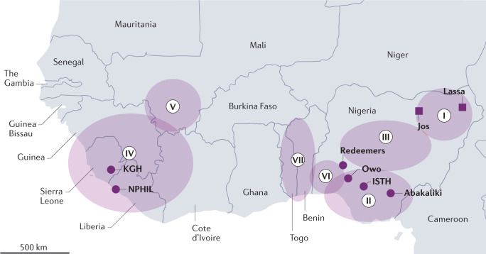
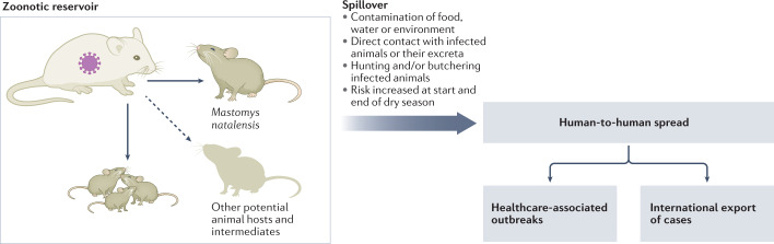
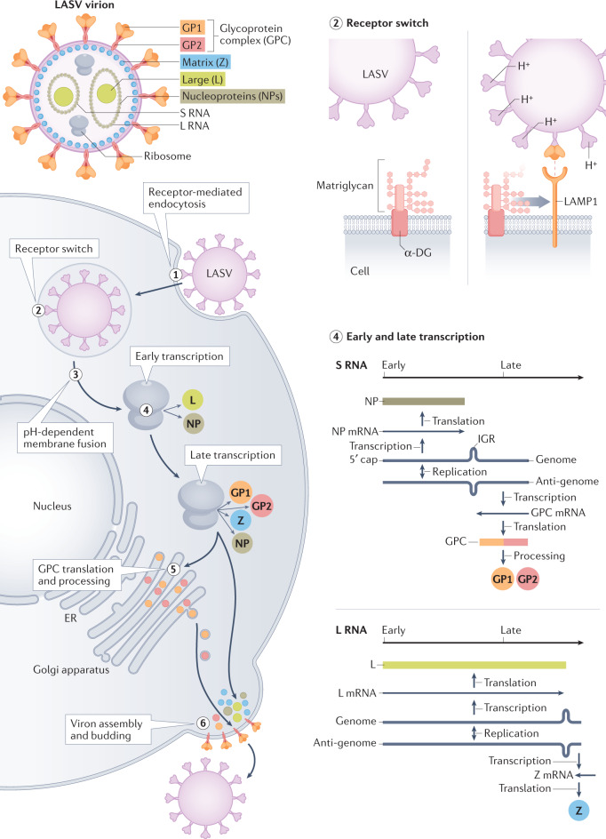
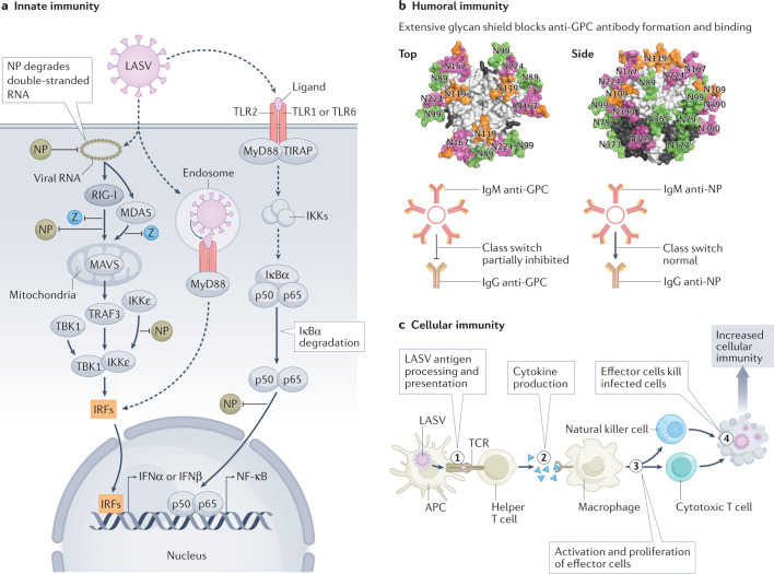
Lassa virus (LASV) is endemic in the rodent populations of Sierra Leone, Nigeria and other countries in West Africa. Spillover to humans occurs frequently and results in Lassa fever, a viral haemorrhagic fever (VHF) associated with a high case fatality rate. Despite advances, fundamental gaps in knowledge of the immunology, epidemiology, ecology and pathogenesis of Lassa fever persist. More frequent outbreaks, the potential for further geographic expansion of Mastomys natalensis and other rodent reservoirs, the ease of procurement and possible use and weaponization of LASV, the frequent importation of LASV to North America and Europe, and the emergence of novel LASV strains in densely populated West Africa have driven new initiatives to develop countermeasures for LASV. Although promising candidates are being evaluated, as yet there are no approved vaccines or therapeutics for human use. This Review discusses the virology of LASV, the clinical course of Lassa fever and the progress towards developing medical countermeasures.


Conflict of interest statement

R.F.G. is the co-founder of Zalgen Labs, a biotechnology company developing countermeasures for Lassa virus (LASV) and other emerging viruses.

Figures

Fig. 1
Fig. 1. Lassa fever endemic zone of West Africa.
The first described cases (squares) of Lassa fever were in the town of Lassa, Nigeria. The infected persons were transported to Jos. Seven lineages (I–VII) of Lassa virus (LASV) are present across West Africa. Important centres for Lassa fever research (circles) are located at the Kenema Government Hospital (KGH), the National Public Health Institute of Liberia (NPHIL), the Irrua Specialist Teaching Hospital (ISTH), Owo and Abakaliki. The African Center of Excellence for the Genetics of Infectious Diseases (ACEGID) is located at Redeemers University. The circles with Roman numerals I–VII represent the approximate ranges of the seven different LASV lineages.
Fig. 2
Fig. 2. Lassa virus transmission.
The major reservoir of Lassa virus (LASV) is Mastomys natalensis. LASV spreads among Mastomys via horizontal or vertical (congenital) routes. Other animal species can also be infected with LASV. Spillover of LASV occurs by exposure to excretions of Mastomys or intermediate hosts, or during preparation of infected animals for food. Human-to-human transmission can occur in the home or clinical setting.
Fig. 3
Fig. 3. Lassa virus structure, genome organization and replication strategy.
The Lassa virus (LASV) virion contains host ribosomes and two segments of single-stranded RNA that encode four proteins. Steps in LASV replication include: LASV enters cells via receptor-mediated endocytosis (step 1); LASV binds α-dystroglycan (α-DG) at the cell surface — as the endosomal pH drops, the LASV glycoprotein complex (GPC) undergoes a conformational shift that enables binding to lysosomal-associated membrane protein 1 (LAMP1) (step 2); LASV undergoes pH-dependent membrane fusion releasing the viral genome segments into the cytoplasm (step 3); both segments use an ambisense strategy — early transcription results in the synthesis of the Large (L) protein, an RNA-dependent RNA polymerase and the nucleoprotein (NP), and late transcription additionally involves synthesis of the GPC and the Zinc-binding (Z) protein (step 4); GPC translation and post-translational processing occur in the Golgi apparatus and result in association of GPC with the plasma membrane (step 5); and LASV acquires its membrane by budding at the cell surface (step 6). ER, endoplasmic reticulum; GP1, glycoprotein 1; IGR, inter-gene region; L RNA, large RNA; S RNA, small RNA.
Fig. 4
Fig. 4. Immune responses to Lassa virus.
a | Lassa virus (LASV) double-stranded RNA triggers the type I interferon pathway and involves retinoic acid-inducible gene I (RIG-I) and melanoma differentiation-associated protein 5 (MDA5). The exoribonuclease function of nucleoprotein (NP) degrades LASV double-stranded RNA, thereby blunting RIG-I and MDA5 activation. LASV Zinc-binding (Z) protein binds RIG-I and MDA5 and prevents mitochondrial antiviral signalling (MAVS) protein activation of tumour necrosis factor (TNF) receptor-associated factor 3 (TRAF3) and TANK-binding kinase 1 (TBK1), which are also involved in interferon induction via interferon-responsive factors (IRFs). NP binds RIG-I and IκB kinase-ε (IKKε) inhibiting activation of IRFs. NP blocks nuclear factor-κ light-chain enhancer of activated B cells (NF-κB) activation. The NF-κB pathway, which is involved in activating various aspects of innate immunity, is activated by recognition of LASV components via Toll-like receptors (TLRs) and involves Toll-interleukin 1 receptor (TIR) domain containing adaptor protein (TIRAP), myeloid differentiation primary response 88 (MyD88), various IKKs and IκBα. NP interferes with the p65 subunit of NF-κB via an unknown mechanism. In addition to MAVS activating IRFs (via TRAF3/TBK1), MAVS activates NF-κB (via TRAF6/IKK complex) independently of TLR signalling (not shown). Elements of panel a were inspired by ref.. b | LASV evades humoral immunity by elaborating a dense glycan shield on the glycoprotein complex (GPC) shown modelled onto trimeric LASV GPC [PDB:5VK2],. LASV also blocks or delays class switching from IgM that recognizes GPC to anti-GPC IgG. Class switching of anti-NP IgM to IgG is not affected. c | Antigen presenting cells (APCs), including dendritic cells and macrophages, phagocytose material from LASV-infected cells in local tissues, degrade it and display the resultant peptides (step 1). T cell proliferation requires recognition of the displayed peptides by the T cell receptor (TCR; co-receptor CD28 and co-stimulatory molecule B7 not shown) (step 2). Proliferation of effector cells is dependent on stimulation with cytokines. Direct killing of infected cells by natural killer cells or cytotoxic T cells involves recognition of viral peptide–major histocompatibility complex (MHC) class I complexes (step 3). Effector functions can also involve other immune cells (for example, antibody-dependent cellular cytotoxicity). Humans or experimental animals that fail to develop effective cellular immunity have a higher fatality rate than individuals who develop effective cellular immune responses (step 4).

References

    1. Andersen KG, et al. Clinical sequencing uncovers origins and evolution of Lassa virus. Cell. 2015;162:738–750. doi: 10.1016/j.cell.2015.07.020. - DOI - PMC - PubMed
    1. Pinneo L, Pinneo R. Mystery virus from Lassa. Am. J. Nurs. 1971;71:1352–1355. - PubMed
    1. Watts GM. Lily Lyman Pinneo. Lancet. 2013;380:1552. doi: 10.1016/S0140-6736(12)61871-6. - DOI
    1. Leifer E, Gocke DJ, Bourne H. Lassa fever, a new virus disease of man from West Africa. II. Report of a laboratory-acquired infection treated with plasma from a person recently recovered from the disease. Am. J. Trop. Med. Hyg. 1970;19:677–679. doi: 10.4269/ajtmh.1970.19.677. - DOI - PubMed
    1. Buckley SM, Casals J, Downs WG. Isolation and antigenic characterization of Lassa virus. Nature. 1970;227:174. doi: 10.1038/227174a0. - DOI - PubMed

Publication types