Clinical Trial
. 2022 Oct;28(10):2155-2161.
doi: 10.1038/s41591-022-01962-5. Epub 2022 Sep 12.

Neoadjuvant atezolizumab for resectable non-small cell lung cancer: an open-label, single-arm phase II trial

Collaborators, Affiliations
Clinical Trial

Neoadjuvant atezolizumab for resectable non-small cell lung cancer: an open-label, single-arm phase II trial

Jamie E Chaft et al. Nat Med. 2022 Oct.

Erratum in

Abstract

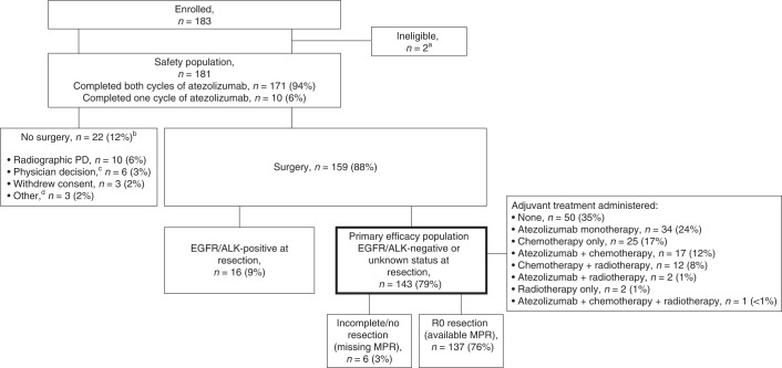
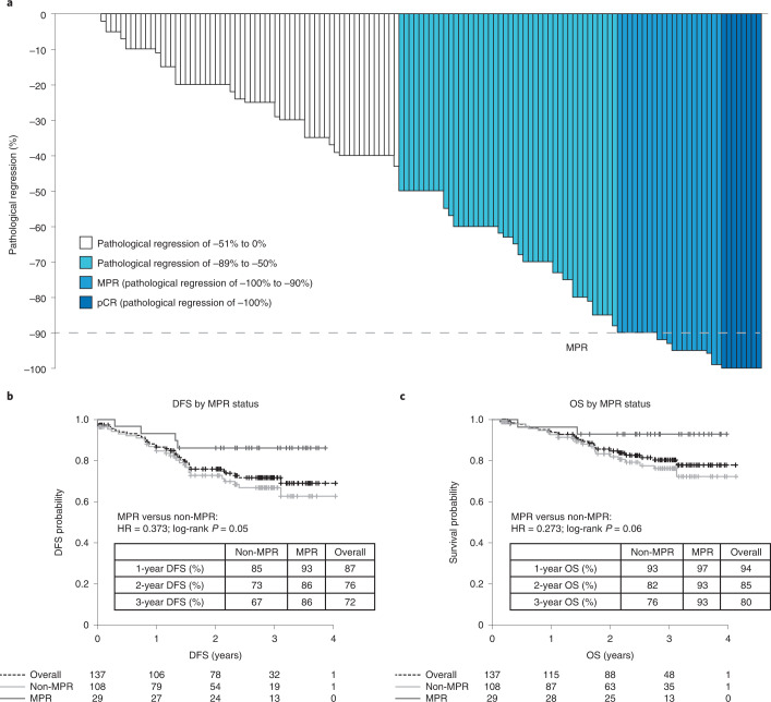
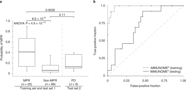
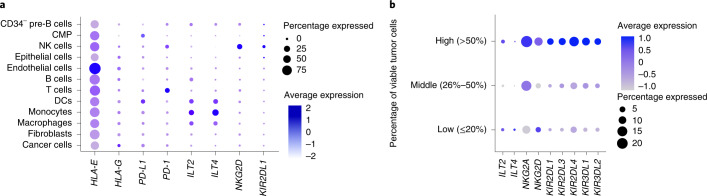
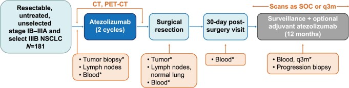
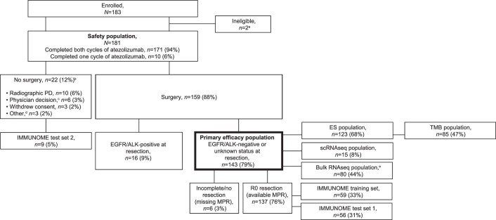
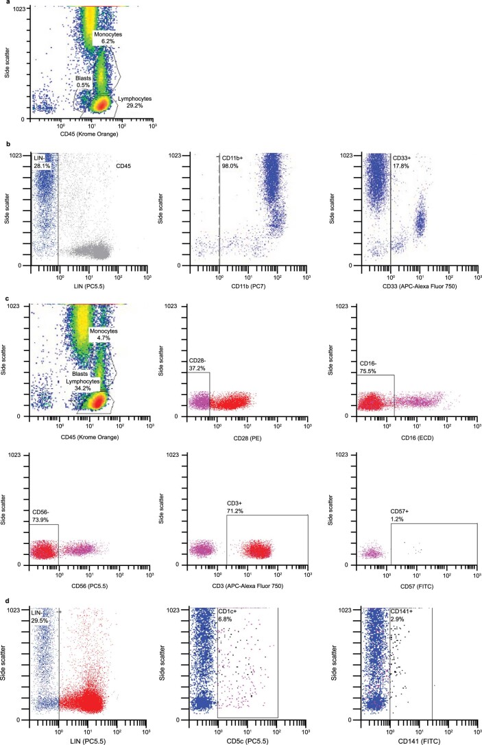
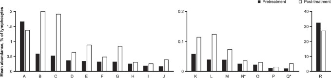
In an ongoing, open-label, single-arm phase II study ( NCT02927301 ), 181 patients with untreated, resectable, stage IB-IIIB non-small cell lung cancer received two doses of neoadjuvant atezolizumab monotherapy. The primary end point was major pathological response (MPR; ≤10% viable malignant cells) in resected tumors without EGFR or ALK alterations. Of the 143 patients in the primary end point analysis, the MPR was 20% (95% confidence interval, 14-28%). With a minimum duration of follow-up of 3 years, the 3-year survival rate of 80% was encouraging. The most common adverse events during the neoadjuvant phase were fatigue (39%, 71 of 181) and procedural pain (29%, 53 of 181), along with expected immune-related toxicities; there were no unexpected safety signals. In exploratory analyses, MPR was predicted using the pre-treatment peripheral blood immunophenotype based on 14 immune cell subsets. Immune cell subsets predictive of MPR in the peripheral blood were also identified in the tumor microenvironment and were associated with MPR. This study of neoadjuvant atezolizumab in a large cohort of patients with resectable non-small cell lung cancer was safe and met its primary end point of MPR ≥ 15%. Data from this single-arm, non-randomized trial suggest that profiles of innate immune cells in pre-treatment peripheral blood may predict pathological response after neoadjuvant atezolizumab, but additional studies are needed to determine whether these profiles can inform patient selection and new therapeutic approaches.


Conflict of interest statement

J.E.C. serves as an advisor to Genentech/Roche, AstraZeneca/MedImmune, Merck, Bristol Myers Squibb, Flame Biosciences, Janssen Oncology, Guardant Health, Regeneron/Sanofi and Novartis. She reports research funding from Genentech/Roche, Bristol Myers Squibb, AstraZeneca/MedImmune, Novartis and Merck. F.O. reports honoraria from Novartis, serves as an advisor to Epigenomics, Sanofi-Aventis and Decibio and has received travel, accommodations and expenses from Genentech/Roche and research funding (paid to her institution) from Genentech/Roche. M.G.K. reports speaking fees from AstraZeneca and Pfizer, consultant fees from Janssen and in-kind support for medical writing from Hoffman La Roche. P.A.B. is on advisory or data safety monitoring boards for Bristol Myers Squibb and Merck and reports membership on the Board of Directors for Verastem, with funding paid to his institution. I.I.W. reports grants and personal fees from Genentech/Roche, Bayer, Bristol Myers Squibb, AstraZeneca, Pfizer, HTG Molecular, GlaxoSmithKline, Guardant Health, Merck, Novartis, Sanofi and Amgen; personal fees from Asuragen, Flame, Daiichi Sankyo, Oncocyte, MSD and Platform Health; and grants from Adaptive, Adaptimmune, EMD Serono, Takeda, Karus, Johnson & Johnson, 4D, Iovance and Akoya, outside of the submitted work. D.J.K. serves as an advisor to AstraZeneca and Genentech/Roche and reports research funding from AADi, Genentech and Revolution Medicines. D.H.O. reports funding (paid to his institution) from Genentech, Merck, Pfizer, Palobiofarma and Bristol Myers Squibb. Y.T. reports no relationships to disclose. B.E.J. reports consultant fees from Checkpoint Therapeutics, Genentech, Hummingbird Diagnostics and Hengrui Therapeutics. J.M.L. reports grants, consulting fees and honoraria from AstraZeneca, Bristol Myers Squibb, Genentech and Novartis and leadership roles at AstraZeneca, Genentech and Novartis. G.L. reports research funding from Genentech, Innate Pharma, Novartis and Stemline Therapeutics. M.P. reports no relationships to disclose. M.S. reports no relationships to disclose. W.Y.B. reports no relationships to disclose. K.S. is an employee of Genentech and reports stock ownership with Roche. A.N. is an employee of Genentech and reports stock ownership with Roche. A.J. is an employee of Genentech and reports stock ownership with Roche. J.G. is an employee of Genentech and reports stock ownership with Roche. S.H. is an employee of Genentech and reports stock ownership with Roche. D.S. is an employee of Genentech and reports stock ownership with Roche. C.R. reports no relationships to disclose. E.T. reports honoraria from Intuitive Surgical. E.B.H. serves in a consulting or advisory role to Amgen, Ellipses Pharma, Janssen Oncology, Janssen Research & Development and Revolution Medicines; reports research funding (paid to his institution) from AstraZeneca, Genentech, Incyte, Janssen, Novartis, Revolution Medicines and Spectrum Pharmaceuticals; and reports patents, royalties or other intellectual property from Protein–Protein Interactions as Biomarkers Patent. C.J.M. reports no relationships to disclose. G.A.P. reports no relationships to disclose. S.N.W. reports research support grants from AbbVie, Ariad Pharmaceuticals, Genentech, Immunomedics, Millennium Pharmaceuticals, Roche, Astellas Pharma, Daiichi Sankyo, Cullinan Pearl, Verastem, GlaxoSmithKline/GSK, Janssen Research & Development, Elevation Oncology, Genentech, Loxo Oncology, Takeda Pharmaceuticals Company Limited and the SWOG Clinical Trials Partnership; has received honoraria from the American Society of Clinical Oncology; and serves as Chair of the Data Safety Monitoring Board for the Hoosier Cancer Research Network. V.W.R. is a member of the Data Safety and Monitoring Committee for the MARS2 trial (UK) and serves as Co-Chair of the National Cancer Institute Thoracic Staging Malignancy Committee. She reports funding (paid to her institution) from Genentech. D.P.C. serves in a consultant or advisory role to AbbVie, Agenus, AstraZeneca, Boehringer Ingelheim, Bristol Myers Squibb, EMD Serono, Genentech/Roche, Helsinn Healthcare, Incyte, Inivata, Inovio Pharmaceuticals, Janssen, Kyowa Hakko Kirin, Merck, Novartis, Pfizer, prIME Oncology and Takeda and research funding from Bristol Myers Squibb and Genentech.

Figures

Fig. 1
Fig. 1. Patient disposition.
Primary efficacy population is bolded. aTwo patients were determined to have hemangioma and solitary fibrous tumor at resection despite initial pathology consistent with NSCLC. bIncludes one EGFR-positive patient. cThe reasons were clinical progression (n = 3), physician did not want to delay patient surgery (n = 1), physician did not consider the patient a good surgical candidate (n = 1) and physician discontinued patient from the study because of an AE (n = 1). dOne patient was determined to have pre-existing congestive heart failure, one declined surgery and one was lost to follow-up.
Fig. 2
Fig. 2. Clinical outcomes in patients who had surgical resection and whose tumors did not have known EGFR or ALK alterations.
a, Pathological response (n = 143). Pathological regression is defined as percentage viable tumor cells – 100%. b, DFS by MPR status in patients with R0 resections (n = 137). c, OS by MPR status in patients with R0 resections (n = 137). HR, hazard ratio.
Fig. 3
Fig. 3. Performance of GAM–LASSO MPR predictive models.
a, Use of the GAM–LASSO model to predict MPR in test set 2, which consisted of patients within LCMC3 who were not included in either the training set (n = 57) or test set 1 (n = 54). MPR was not assessed in these nine patients because of no resection. The MPR and non-MPR cohorts derived from the merge of the model’s training set and test set 1. The maximum and minimum values of the boxes denote the IQR. The line within the IQR denotes the median. The extremities of the dashed lines represent the minimum and maximum values of the data, which are 1.5× below the first quartile and 1.5× above the third quartile. The parameters for null hypothesis testing via analysis of variance (ANOVA) were as follows: d.f. = 2, total sum of squares = 1.976, mean squares = 0.988, F-value = 32.799 and Pr(>F) = 4.914 × 10−12. The statistical details for the comparison of MPR and non-MPR were t = −5.47, d.f. = 27.02, two-sided P = 8.6 × 10−6 and 95% CI = −0.439 to −0.200. The statistical details for the comparison of MPR and PD were t = −3.18, d.f. = 28.45, two-sided P = 0.0035 and 95% CI = −0.383 to −0.083. The statistical details for the comparison of non-MPR and PD were t = −1.77, d.f. = 9.52, two-sided P = 0.11 and 95% CI = −0.195 to 0.023. No adjustment was made for multiplicity. b, ROC curves for the training set and test set 1. The dashed y = x line, which represents random assignment, is included for reference. aImmunophenotyping via flow cytometry. IQR, interquartile range; ROC, receiver operating characteristic.
Fig. 4
Fig. 4. scRNA-seq analysis of selected genes expressed in tumor tissue from 15 patients following treatment with neoadjuvant atezolizumab.
a, The expression of different NK cell surface receptors was determined by scRNA-seq. b, Tumor samples collected at resection were classified into three groups of five samples each on the basis of the percentage of viable tumor cells by pathological analysis: low (≤25% viable tumor cells), middle (26–50%) and high (>50%). Dot size represents the percentage of NK cells in the group expressing the gene. Color represents the scaled average normalized expression. NK cells were downsampled to have the same number of cells in each group. ILT2 is also known as LILRB1, ILT4 as LILRB2, KIR2DL1 as CD158a, NKG2D as CD314 and KLRK1 and PD-L1 as CD274. CD, cluster of differentiation; CMP, common myeloid progenitor; ILT, immunoglobulin-like transcript; KIR2DL1, killer cell immunoglobulin-like receptor DL1; LILRB, leukocyte immunoglobulin-like receptor subfamily B; NKG2, natural killer group protein 2.
Extended Data Fig. 1
Extended Data Fig. 1. Study design.
*Mandatory. CT, computed tomography; NSCLC, non-small cell lung cancer; PET, positron emission tomography; q3m, every 3 months; SOC, standard of care.
Extended Data Fig. 2
Extended Data Fig. 2. CONSORT diagram for the various biomarker analyses.
aTwo patients were determined to have hemangioma and solitary fibrous tumor at resection despite initial pathology consistent with NSCLC. bIncludes 1 EGFR-positive patient. cThe reasons were as follows: clinical progression (n = 3), physician did not want to delay patient surgery (n = 1), physician did not consider the patient a good surgical candidate (n = 1), and physician discontinued patient from the study because of an adverse event (n = 1). dOne patient was determined to have preexisting congestive heart failure, 1 declined surgery, and 1 was lost to follow-up. eEighty patients provided a total of 98 samples for the bulk RNA-seq analyses (54 at baseline and 44 at surgery). ALK, anaplastic lymphoma kinase; EGFR, epidermal growth factor receptor; ES, exome sequencing; MPR, major pathologic response; NSCLC, non-small cell lung cancer; PD, progressive disease; RECIST, Response Evaluation Criteria in Solid Tumors; RNA-seq, ribonucleic acid sequencing; scRNA-seq, single-cell ribonucleic acid sequencing; TMB, tumor mutational burden.
Extended Data Fig. 3
Extended Data Fig. 3. Relationship between MPR, histology, and TMB.
(a) Pathologic response by mutation status (n = 85). TMB showed a positive but not significant trend with pathologic response in non-squamous (R = 0.28; P = 0.05) and squamous (R = 0.23; P = 0.22) tumors. KEAP1 mutations were not significantly associated with pathologic response in either non-squamous (P = 0.46) or squamous tumors (P = 0.98). KRAS and STK11 mutations were found only in non-squamous tumors, where mutations in STK11 were found to significantly associate with pathologic response (P = 0.01), and mutations in KRAS were not (P = 0.87). (b) Pathologic response by the overlapping mutational status of KRAS and STK11 in patients with non-squamous NSCLC (n = 78). The maximum and minimum values of the boxes denote the IQR. The line within the IQR denotes the median. The extremities of the dashed lines represent the 5th to 95th percentiles. *TMB was only determined for the subset of patients with ES data from baseline and/or surgery with tumor purity ≥15%. P values for TMB in Panel a were determined via linear correlation test (Pearson). P values for mutation status in Panels a and b were determined via two-sided Wilcoxon rank sum test. ES, exome sequencing; IQR, interquartile range; KEAP1, Kelch-like ECH-associated protein 1; KRAS, Kirsten rat sarcoma viral oncogene homolog; MPR, major pathologic response; NS, non-significant; NSCLC, non-small cell lung cancer; STK11, serine/threonine kinase 11; TMB, tumor mutational burden.
Extended Data Fig. 4
Extended Data Fig. 4. DFS (a) and OS (b) by disease stage, use of adjuvant atezolizumab, and lymph node status (n = 143).
The MPR rate in patients who did and did not receive adjuvant atezolizumab was 41% (22/54) and 8% (7/89), respectively. DFS, disease-free survival; OS, overall survival.
Extended Data Fig. 5
Extended Data Fig. 5. Gating strategy for the IMMUNOME tubes described in Supplementary Table 5.
(a) Tubes 1–11 were gated on the basis of CD45 and side scatter. The immune cell populations of interest were those falling within the region ‘lymphocytes,’ which was defined by CD45bright, a low side scatter, and a lymphosum (CD3+ CD19+ CD56+ CD16+) totaling 95%–100%, with <5% myeloid contamination. Tube 1 (lymphosum) contained lymphosum markers, as well as myeloid markers (CD13/CD14). The gate established for Tube 1 was applied to Tubes 2–11. (b) Tube 12 (myeloid cells) used LIN (CD3+ CD19+ CD56+) as an exclusion gate. Plots were gated using three different strategies: LIN total, LIN CD11b+, and LIN CD33+. (c) Tube 13 (senescent cells) used both lymphocyte and sequential gating to find the immune cell population of interest. Lymphocyte gating isolated CD28 CD16 CD56 CD3+ cells, which were sequentially gated on CD57. Events falling with the CD57+ region were considered senescent cells. Senescent cells were further classified into subsets defined by positivity and negativity for CD4, CD8, KLRG1, and CD127. (d) Tube 14 (dendritic cells) used LIN as an exclusion gate. Plots were gated using three different strategies: LIN total, LIN CD1c+, and LIN CD141+. CD, cluster of differentiation; KLRG1, killer cell lectin-like receptor subfamily G member 1; LIN, lineage.
Extended Data Fig. 6
Extended Data Fig. 6. ROC curves for test set 1 (n = 54), which considered only peripheral blood IMMUNOME, as well as for test set 1 in combination with histology (non-squamous [n = 32] vs. squamous [n = 22]), nodal status (N1/N2 [n = 35] vs. N0 [n = 19]), PD-L1 expression (n = 54), sex (female [n = 26] vs. male [n = 28]), or smoking status/history (never [n = 6] vs. current/former [n = 48]).
The dashed y = x line, which represents random assignment, is included for reference. aImmunophenotyping via flow cytometry. PD-L1, programmed death-ligand 1; ROC, receiver operating characteristic.
Extended Data Fig. 7
Extended Data Fig. 7. Immune cell subsets with significant (two-sided p < 0.05) changes in abundance before (baseline) and after (at surgery) neoadjuvant treatment with atezolizumab. No adjustment was made for multiplicity.
A, CD45+ CD62L+ CD27+ CD56/16 CD45RO+ CCR7 CD45RA CD4 CD3+ CD8+ (p = 0.0081); B, CD45+ CD33+ HLA-DR+ CD124 CD14+ CD11b+ CD66b CD16 CD33+ CD15+ (p = 0.0042); C, CD45+ HLA-DR+ CD124 CD14+ CD11b+ CD66b CD16 CD33+ CD15+ (p = 0.0042); D, CD45+ CD94+ NKG2D+ CD3 CD56+ CD117 NKG2A+ CD127 CD161 CD16+ (p = 0.0036); E, CD45+ LIN+ HLA-DR+ CD33+ CD16+ CD11b+ CD15+ (p = 0.0092); F, CD45+ HLA-DR CD69+ CD19 CD56+ CD16 CD134 CD4 CD3+ CD8+ (p = 0.0079); G, CD45+ CD11b+ HLA-DR+ CD124 CD14+ CD11b+ CD66b CD16 CD33+ CD15+ (p = 0.0157); H, CD45+ γ/δ- α/β+ CD19 CD56+ CD16 CD13/14 CD4+ CD3+ CD8 (p = 0.0113); I, CD45+ HLA-DR CD69+ CD3+ KIR3DL1 KIR2DL2 NKG2A CD56+ KIR2DL1 CD16 (p < 0.0001); J, CD45+ CD107a/b- CD159c+ CD3 KIR3DL1 KIR2DL2 NKp80+ CD56+ KIR2DL1 CD16+ (p = 0.0025); K, CD45+ HLA-DR CD69+ CD19 CD56+ CD16 CD134 CD4 CD3+ CD8 (p = 0.0004); L, CD45+ CD16+ CD336 CD3 CD244+ CD335+ NKG2D+ CD56 CD161+ CD337 (p = 0.0002); M, CD45+ γ/δ- α/β+ CD19 CD56+ CD16 CD13/14 CD4 CD3+ CD8 (p = 0.0191); N, CD45+ HLA-DR CD69+ CD3+ KIR3DL1 KIR2DL2 NKG2A+ CD56+ KIR2DL1 CD16 (p = 0.0307); O, CD45+ CD62L CD27+ CD56/16+ CD45RO CCR7 CD45RA+ CD4 CD3+ CD8+ (p = 0.0400); P, CD45+ CD94 NKG2D CD3+ CD56+ CD117 NKG2A CD127+ CD161+ CD16+ (p = 0.0480); Q, CD45+ HLA-DR+ CD69+ CD3 KIR3DL1 KIR2DL2 NKG2A+ CD56 KIR2DL1 CD16+ (p = 0.0014); R, CD45+ LIN HLA-DR CD33 CD16+ CD11b+ CD15+ (p = 0.0030). *Pre-treatment predictor of MPR. LIN included CD19, CD3, and CD56. NKG2A is also known as CD159a, NKG2D as CD314 and KLRK1, KIR2DL1 as CD158a, KIR2DL2 as CD158b, CD335 as NKp46, and CD337 as NKp30. α/β, α/β chains of the T cell receptor; γ/δ, γ/δ chains of the T cell receptor; CCR7, C-C motif chemokine receptor 7; CD, cluster of differentiation; HLA, human leukocyte antigen; KIR, killer cell immunoglobulin-like receptor; KLR, killer cell lectin-like receptor; LIN, lineage; MPR, major pathologic response; NK, natural killer; NKG2, natural killer group protein 2.
Extended Data Fig. 8
Extended Data Fig. 8. Association between gene expression in NK cells and the percentage of viable tumor cells.
The expression of different NK cell surface receptors was determined by scRNA-seq. Tumor samples collected at resection were classified into 3 groups of 5 samples each based on the percentage of viable tumor cells by pathologic analysis: low (≤25% viable tumor cells), middle (26%–50%), and high (>50%). UMAP plots of NK cells show normalized expression of several receptors in the 3 groups of viable tumor cell numbers. NK cells were down-sampled to have the same number of cells in each group. ILT2 is also known as LILRB1, ILT4 as LILRB2, NKG2A as CD159a, NKG2D as CD314 and KLRK1, KIR2DL1 as CD158a, KIR2DL3 as CD158b, KIR2DL4 as CD158d, KIR3DL1 as CD158e1, and KIR3DL2 as CD158k. CD, cluster of differentiation; ILT, immunoglobulin-like transcript; KIR, killer cell immunoglobulin-like receptor; KLR, killer cell lectin-like receptor; LILRB, leukocyte immunoglobulin-like receptor subfamily B; max, maximum; min, minimum; NK, natural killer; NKG2, natural killer group protein 2; scRNA-seq, single-cell ribonucleic acid sequencing; UMAP, uniform manifold approximation and projection.
Extended Data Fig. 9
Extended Data Fig. 9. Association of (a) ILT2 and (b) PDL1 transcripts with the cell type enrichment scores of specific immune cell subsets derived from bulk RNA-seq at baseline and at surgery by tumor histology.
Relative immune cell abundance was estimated from RNA-seq data using the cell type enrichment analysis tool xCell and correlated with the abundance (log2 cpm) of ILT2 or PD-L1. Results are shown for all baseline and surgical samples with RNA-Seq, split by histology. A positive correlation coefficient (blue) indicates samples with an increased abundance of a given immune cell signature also having an increased abundance of either ILT2 or PD-L1. Asterisks indicate significant correlations (unadjusted Pearson correlation two-sided P < 0.05). ILT2 is also known as LILRB1 and PD-L1 as CD274. CD, cluster of differentiation; cpm, counts per million reads mapped; DC, dendritic cell; ILT2, immunoglobulin-like transcript 2; LILRB, leukocyte immunoglobulin-like receptor subfamily B; NK, natural killer; PD-L1, programmed death-ligand 1; RNA-seq, ribonucleic acid sequencing.
Extended Data Fig. 10
Extended Data Fig. 10. Association of ILT2 and PD-L1 transcripts with pathologic response at baseline and at surgery by tumor histology.
Expression values for each transcript are presented as log2 (cpm + 1). Two-sided P-values for Pearson (R) and Spearman (ρ [Rho]) correlation are shown. The grey band represents the 95% confidence interval for the linear regression line. No adjustment was made for multiplicity. Of the 54 patients at baseline with RNA-seq data, 52 also had pathologic response data available. ILT2 is also known as LILRB1 and PD-L1 as CD274. CD, cluster of differentiation; cpm, counts per million reads mapped; ILT2, immunoglobulin-like transcript 2; LILR1, leukocyte immunoglobulin-like receptor 1; MPR, major pathologic response; PD-L1, programmed death-ligand 1; RNA-seq, ribonucleic acid sequencing.

References

    1. Wakelee HA, et al. IMpower010: Primary results of a phase III global study of atezolizumab versus best supportive care after adjuvant chemotherapy in resected stage IB-IIIA non-small cell lung cancer (NSCLC) J. Clin. Oncol. 2021;39:8500. doi: 10.1200/JCO.2021.39.15_suppl.8500. - DOI
    1. Forde PM, et al. Neoadjuvant PD-1 blockade in resectable lung cancer. N. Engl. J. Med. 2018;378:1976–1986. doi: 10.1056/NEJMoa1716078. - DOI - PMC - PubMed
    1. Cascone T, et al. Neoadjuvant nivolumab or nivolumab plus ipilimumab in operable non-small cell lung cancer: the phase 2 randomized NEOSTAR trial. Nat. Med. 2021;27:504–514. doi: 10.1038/s41591-020-01224-2. - DOI - PMC - PubMed
    1. Herbst RS, et al. Predictive correlates of response to the anti-PD-L1 antibody MPDL3280A in cancer patients. Nature. 2014;515:563–567. doi: 10.1038/nature14011. - DOI - PMC - PubMed
    1. Scagliotti GV, et al. Randomized phase III study of surgery alone or surgery plus preoperative cisplatin and gemcitabine in stages IB to IIIA non-small-cell lung cancer. J. Clin. Oncol. 2012;30:172–178. doi: 10.1200/JCO.2010.33.7089. - DOI - PubMed

Publication types

MeSH terms

Associated data