. 2023 Apr 4;33(8):4262-4279.
doi: 10.1093/cercor/bhac341.

A neural stem cell paradigm of pediatric hydrocephalus

Affiliations

A neural stem cell paradigm of pediatric hydrocephalus

Phan Q Duy et al. Cereb Cortex. .

Abstract

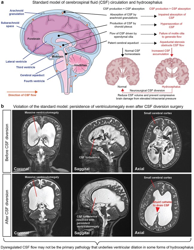
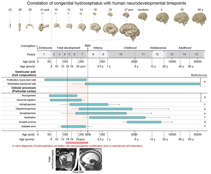
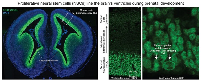
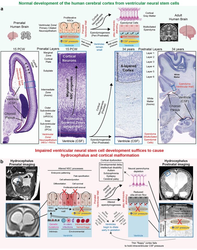
Pediatric hydrocephalus, the leading reason for brain surgery in children, is characterized by enlargement of the cerebral ventricles classically attributed to cerebrospinal fluid (CSF) overaccumulation. Neurosurgical shunting to reduce CSF volume is the default treatment that intends to reinstate normal CSF homeostasis, yet neurodevelopmental disability often persists in hydrocephalic children despite optimal surgical management. Here, we discuss recent human genetic and animal model studies that are shifting the view of pediatric hydrocephalus from an impaired fluid plumbing model to a new paradigm of dysregulated neural stem cell (NSC) fate. NSCs are neuroprogenitor cells that comprise the germinal neuroepithelium lining the prenatal brain ventricles. We propose that heterogenous defects in the development of these cells converge to disrupt cerebrocortical morphogenesis, leading to abnormal brain-CSF biomechanical interactions that facilitate passive pooling of CSF and secondary ventricular distention. A significant subset of pediatric hydrocephalus may thus in fact be due to a developmental brain malformation leading to secondary enlargement of the ventricles rather than a primary defect of CSF circulation. If hydrocephalus is indeed a neuroradiographic presentation of an inborn brain defect, it suggests the need to focus on optimizing neurodevelopment, rather than CSF diversion, as the primary treatment strategy for these children.

Keywords: cerebrospinal fluid; hydrocephalus; neural stem cell; neurodevelopmental disorders; neuroprogenitor.


Figures

Fig. 1
Fig. 1
Standard model of CSF circulation and hydrocephalus pathology. a) Standard model of CSF circulation in the adult human brain. Hydrocephalus is characterized by enlargement of the cerebral ventricles classically attributed to dysregulated CSF flow, leading to overaccumulation of CSF that distends the ventricles and raises the intracranial pressure. Neurosurgical CSF diversion that aims to reduce CSF volume and intracranial pressure is the primary treatment strategy for hydrocephalus. Image modified from Servier medical art (https://smart.servier.com) permitted by the creative commons attribution license (https://creativecommons.org/licenses/by/3.0/). b) Clinical case that demonstrates an example wherein the standard model of CSF circulation does not sufficiently explain the pathogenesis of hydrocephalus. Patient is a 3-year-old girl who presented with progressive macrocephaly. Neuroimaging prior to surgery (top panels) demonstrated extreme communicating ventriculomegaly with strikingly turbulent CSF flow and small cerebral cortex. A ventriculoperitoneal shunt was placed to divert CSF from the ventricles into the peritoneal cavity. Postoperative imaging showed complete resolution of CSF turbulence with persistence of ventriculomegaly. Although the patient’s progressive macrocephaly was arrested, she continued to exhibit mild neurocognitive impairments. Thus, reinstatement of CSF homeostasis may address some consequences of disease and prevent further deterioration but does not necessarily target the underlying developmental pathology. Images from patient were modified with permission from (Duy and Kahle 2021).
Fig. 2
Fig. 2
Timeline of key events in human cerebral cortex development and correlation to in utero diagnosis of hydrocephalus. Figure modified from Silbereis et al., Neuron, 2016 (Silbereis et al. 2016). Top and bottom panels provide a timeline of human cerebrocortical development (Kang et al. 2011) with age in postconceptional days (pcd), postconceptional weeks (pcw), and postnatal years (y). The schematic provides approximate timing and sequence of cellular processes in the ventricular wall and prefrontal cortex. Bars indicate the peak developmental period in which each feature is acquired; dotted lines indicate that feature acquisition occurs to lesser degrees at these ages, and arrows indicate that the feature is present thereafter throughout life. A human MRI showing in utero diagnosis of hydrocephalus is shown, corresponding to a period during which proliferative NSCs make up the ventricular wall well before appearance of multiciliated ependymal cells. Right column lists relevant references: a) Coletti et al. (2018); b) Bystron et al. (2006), Meyer (2007), Workman et al. (2013); c) Choi and Lapham (1978), deAzevedo et al. (2003), Kang et al. (2011); d) Kang et al. (2011), Yeung et al. (2014); e) Huttenlocher (1979), Kwan et al. (2012), Molliver et al. (1973), Petanjek et al. (2011); f) Miller et al. (2012), Yakovlev and Lecours (1967); g) Huttenlocher (1979), Petanjek et al. (2011); h) Kostovic and Rakic (1990).
Fig. 3
Fig. 3
NSCs line the cerebral ventricles before birth. Shown are fluorescent images of a coronal section from an embryonic day 15.5 wild-type mouse brain that was stained with DAPI and SOX2 (a marker of NSCs). SOX2+ NSCs line the entire cerebral ventricular system. Zoomed in images show neuroprogenitor cell division at the apical surface of the ventricular wall directly adjacent to CSF. NSCs are therefore the predominant cell types at the brain–CSF interface before birth. Images were modified from (Allocco et al. 2019) permitted by the creative commons attribution license (https://creativecommons.org/licenses/by/4.0/).
Fig. 4
Fig. 4
A NSC paradigm of human pediatric hydrocephalus. a) Normal development of the human ventricular system and cerebral cortex. During gestation, the ventricular wall is composed of a germinal neuroepithelium that has 2 crucial functions. First, as the epithelial barrier between fluid and brain parenchymal component, the germinal neuroepithelium has an important function in resisting the pressure generated by CSF in the ventricular lumen. Second, the germinal neuroepithelium contains proliferative NSCs that produce all neurons and macroglia of the cerebral cortex. After birth, this neuroepithelium is replaced by multiciliated ependymal cells that may contribute to local CSF flow driven by motile cilia, though in humans, the roles of ependymal ciliary motion in generating CSF circulation remain debated. MR image of control prenatal human brain from Gholipour et al. Scientific Reports, 2017 (Gholipour et al. 2017); MR image of control adult human brain from OpenNeuro dataset ds000221; images of histological sections from control prenatal and adult human brains from the BrainSpan atlas. NECs, neuroepithelial cells; RGCs, radial glia cells; IPCs, intermediate progenitor cells; oRGCs, outer radial glia cells. b) Pathophysiologic explanation of how altered ventricular NSCs during gestation lead to hydrocephalus and cortical maldevelopment. The developmental consequences of perturbed NSC fate are 2-fold. First, the germinal neuroepithelium of the developing brain ventricles weakens and thus fails to resist intraventricular CSF pressure, initiating ventricular dilation early in gestation. Second, abnormal development of the germinal neuroepithelium leads to abnormal cortical neurogenesis, leading to a thin and “floppy” cerebral cortex wall that also fails to hold the CSF pressure, leading to progression of ventriculomegaly. Impaired NSC fate can theoretically also result in abnormal ependymogenesis and failure of motile cilia-driven CSF flow, leading to secondary effects on CSF circulation. However, altered development of the ependyma is neither sufficient or required to cause hydrocephalus as discussed in the main text, thus the contribution of disrupted ependyma to the primary developmental pathogenesis of pediatric hydrocephalus remains questionable and should only be considered as a secondary consequence that may potentially worsen the phenotype but not absolutely required for initiation of the disease phenotype.

References

    1. Adle-Biassette H, Saugier-Veber P, Fallet-Bianco C, Delezoide A-LL, Razavi F, Drouot N, Bazin A, Beaufrère A-M, Bessières B, Blesson S, et al. Neuropathological review of 138 cases genetically tested for X-linked hydrocephalus: evidence for closely related clinical entities of unknown molecular bases. Acta Neuropathol. 2013:126(3):427–442. 10.1007/s00401-013-1146-1. - DOI - PubMed
    1. Alders GL, Minuzzi L, Sarin S, Frey BN, Hall GB, Samaan Z. Volumetric MRI analysis of a case of severe Ventriculomegaly. Front Hum Neurosci. 2018:12:495. 10.3389/fnhum.2018.00495. - DOI - PMC - PubMed
    1. Al-Dosari MS, Al-Owain M, Tulbah M, Kurdi W, Adly N, Al-Hemidan A, Masoodi TA, Albash B, Alkuraya FS. Mutation in MPDZ causes severe congenital hydrocephalus. J Med Genet. 2013:50(1):54–58. 10.1136/jmedgenet-2012-101294. - DOI - PubMed
    1. Allocco AA, Jin SC, Duy PQ, Furey CG, Zeng X, Dong W, Nelson-Williams C, Karimy JK, DeSpenza T, Hao LT, et al. Recessive inheritance of congenital hydrocephalus with other structural brain abnormalities caused by compound heterozygous mutations in ATP1A3. Front Cell Neurosci. 2019:13:425. 10.3389/fncel.2019.00425. - DOI - PMC - PubMed
    1. Alvin MD, Miller PE. Compensated hydrocephalus. Lancet (London, England). 2016:387(10036):2422. 10.1016/S0140-6736(16)00089-1. - DOI - PubMed

Publication types