

Polysaccharide Electrospun Nanofibers for Wound Healing Applications
- PMID: 36097445
- PMCID: PMC9464040
- DOI: 10.2147/IJN.S371900
Polysaccharide Electrospun Nanofibers for Wound Healing Applications
Abstract
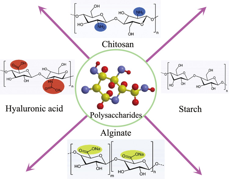
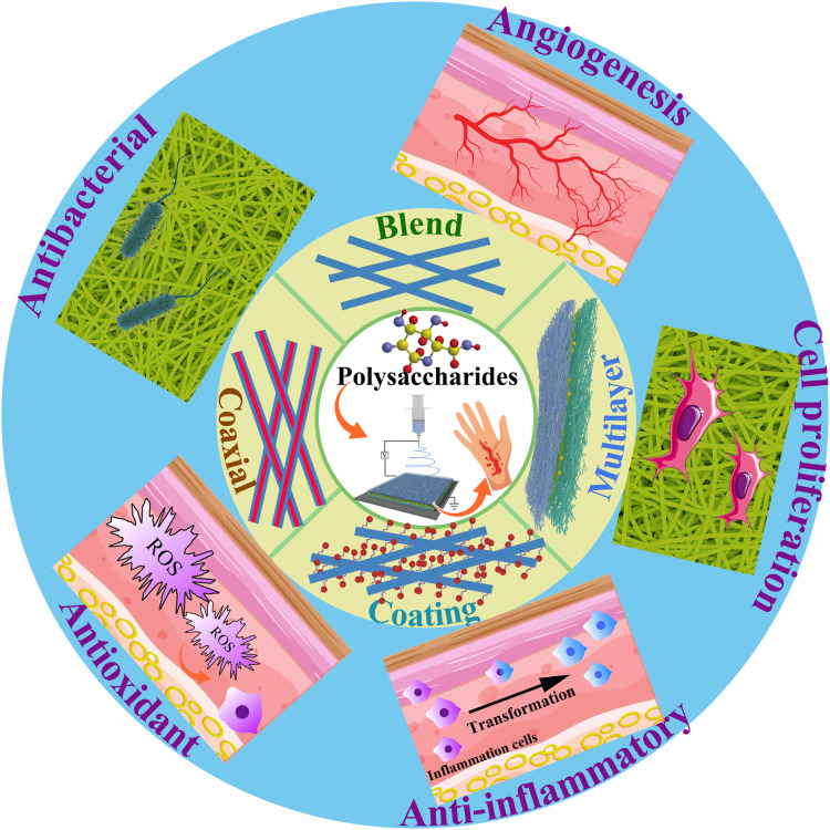
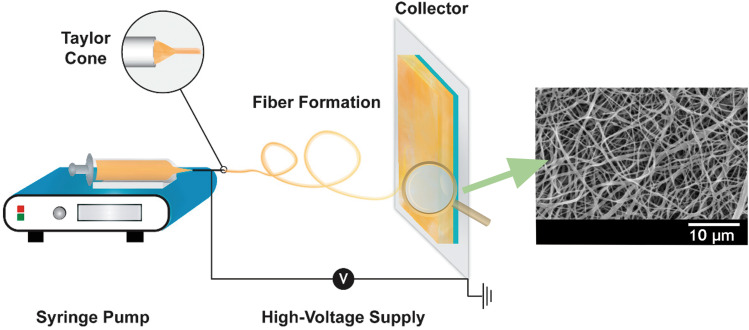
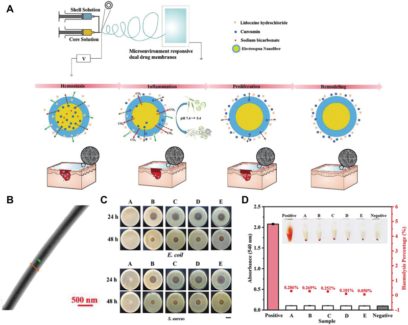
As a type of biological macromolecule, natural polysaccharides have been widely used in wound healing due to their low toxicity, good biocompatibility, degradability and reproducibility. Electrospinning is a versatile and simple technique for producing continuous nanoscale fibers from a variety of natural and synthetic polymers. The application of electrospun nanofibers as wound dressings has made great progress and they are considered one of the most effective wound dressings. This paper reviews the preparation of polysaccharide nanofibers by electrospinning and their application prospects in the field of wound healing. A variety of polysaccharide nanofibers, including chitosan, starch, alginate, and hyaluronic acid are introduced. The preparation strategy of polysaccharide electrospun nanofibers and their functions in promoting wound healing are summarized. In addition, the future prospects and challenges for the preparation of polysaccharide nanofibers by electrospinning are also discussed.
Keywords: electrospun nanofibers; function; polysaccharide; preparation strategy; wound healing.
© 2022 Tan et al.
Conflict of interest statement
The authors declare no conflicts of interest in relation to this work.
Figures
Similar articles
-
Hyaluronic acid and chitosan-based electrospun wound dressings: Problems and solutions.Int J Biol Macromol. 2022 May 1;206:74-91. doi: 10.1016/j.ijbiomac.2022.02.117. Epub 2022 Feb 24. Int J Biol Macromol. 2022. PMID: 35218807 Review.
-
Preparation of animal polysaccharides nanofibers by electrospinning and their potential biomedical applications.J Biomed Mater Res A. 2015 Feb;103(2):807-18. doi: 10.1002/jbm.a.35187. Epub 2014 Apr 28. J Biomed Mater Res A. 2015. PMID: 24733749 Review.
-
A review on polysaccharides mediated electrospun nanofibers for diabetic wound healing: Their current status with regulatory perspective.Int J Biol Macromol. 2023 Apr 15;234:123696. doi: 10.1016/j.ijbiomac.2023.123696. Epub 2023 Feb 16. Int J Biol Macromol. 2023. PMID: 36801273 Review.
-
Polysaccharides and proteins as natural polymers for electrospun wound dressings: A review of healing potential, challenges, and crosslinking strategies.Int J Biol Macromol. 2025 Aug;320(Pt 4):145960. doi: 10.1016/j.ijbiomac.2025.145960. Epub 2025 Jul 17. Int J Biol Macromol. 2025. PMID: 40683510 Review.
-
An Overview of Biopolymeric Electrospun Nanofibers Based on Polysaccharides for Wound Healing Management.Pharmaceutics. 2020 Oct 17;12(10):983. doi: 10.3390/pharmaceutics12100983. Pharmaceutics. 2020. PMID: 33080849 Free PMC article. Review.
Cited by
-
Alginate-Based Electrospun Nanofibers and the Enabled Drug Controlled Release Profiles: A Review.Biomolecules. 2024 Jul 3;14(7):789. doi: 10.3390/biom14070789. Biomolecules. 2024. PMID: 39062503 Free PMC article. Review.
-
Therapeutic applications of nanobiotechnology.J Nanobiotechnology. 2023 May 6;21(1):148. doi: 10.1186/s12951-023-01909-z. J Nanobiotechnology. 2023. PMID: 37149615 Free PMC article. Review.
-
Application of Deferoxamine in Tissue Regeneration Attributed to Promoted Angiogenesis.Molecules. 2024 Apr 29;29(9):2050. doi: 10.3390/molecules29092050. Molecules. 2024. PMID: 38731540 Free PMC article. Review.
-
Nanofiber Scaffolds as Drug Delivery Systems Promoting Wound Healing.Pharmaceutics. 2023 Jun 26;15(7):1829. doi: 10.3390/pharmaceutics15071829. Pharmaceutics. 2023. PMID: 37514015 Free PMC article. Review.
-
A critical overview of challenging roles of medicinal plants in improvement of wound healing technology.Daru. 2024 Jun;32(1):379-419. doi: 10.1007/s40199-023-00502-x. Epub 2024 Jan 15. Daru. 2024. PMID: 38225520 Free PMC article.
References
Publication types
MeSH terms
Substances
LinkOut - more resources
Full Text Sources
Research Materials
