

Pulsed-field ablation: Computational modeling of electric fields for lesion depth analysis
- PMID: 36097449
- PMCID: PMC9463712
- DOI: 10.1016/j.hroo.2022.05.009
Pulsed-field ablation: Computational modeling of electric fields for lesion depth analysis
Abstract
Background: Pulsed-field ablation (PFA) is an emerging and promising nonthermal technology for cardiac ablation. The effective applied voltage to achieve adequate irreversible myocardial injury is not well studied. The pulsed-field strength remains independent of tissue contact; therefore, PFA is assumed to be an ablation technology, not mandating the need for tissue contact.
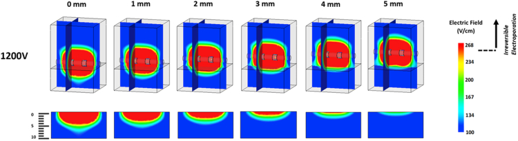
Objective: Determine the effect of applied voltage and distance to surface on depth of myocardial injury using PFA.
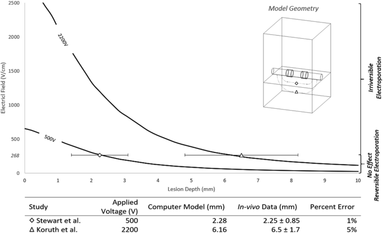
Methods: A computational model was developed and validated based on extracted data from in vivo studies to examine the effect of different applied voltages and the impact of distance between the catheter and endocardial surface on the depth of irreversible myocardial injury using PFA.
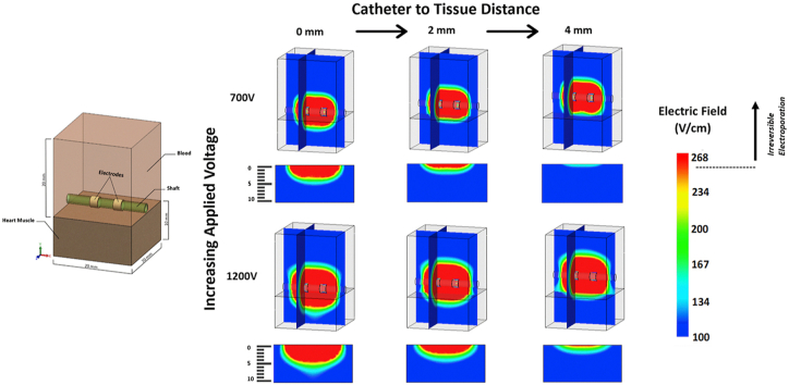
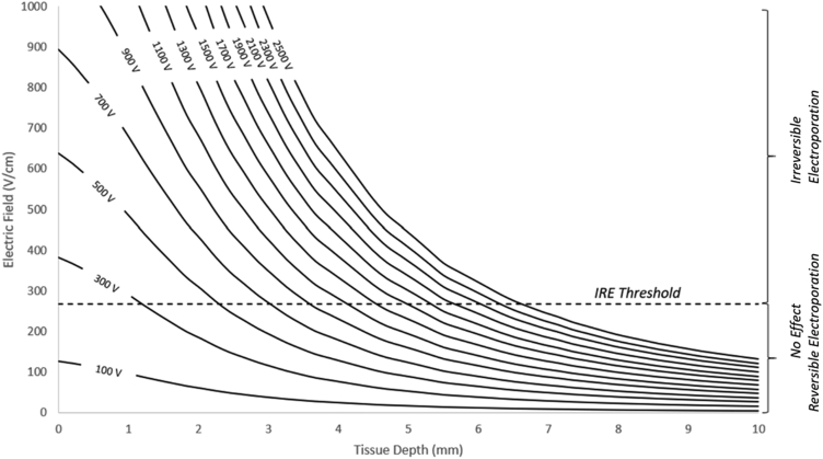
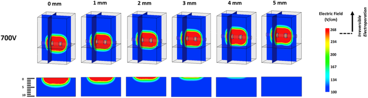
Results: The depth of lesions created by PFA are dose-dependent, and there is a direct correlation between applied PFA voltages and depth of irreversible myocardial injury. The minimum applied voltage of PFA required to create a lesion deeper than 1 mm is 300 volts. The catheter-tissue contact plays a pivotal role in determining lesion depth. With optimal catheter contact in the absence of trabeculation, the minimal applied energy required to achieve a 3-mm-deep lesion is 700 volts. A minor increase in the catheter-tissue distance of 1-2 mm doubles the minimum required applied voltage, increasing it to 1500 volts.
Conclusion: PFA is an important new technology that is proposed to be more efficacious and safer than currently used thermal ablation. Here we demonstrate the impact of dose dependence and the need for maintaining tissue contact during ablation.
Keywords: Catheter ablation; Irreversible electroporation; PFA doses; Pulsed-field ablation; Tissue contact.
Crown Copyright © 2022 Published by Elsevier Inc. on behalf of Heart Rhythm Society.
Figures

References
-
- Gonzalez R., Scheinman M., Margaretten W., Rubinstein M. Closed-chest electrode-catheter technique for His bundle ablation in dogs. Am J Physiol. 1981;241:H283–H287. - PubMed
-
- Gallagher J.J., Svenson R.H., Kasell J.H., et al. Catheter technique for closed-chest ablation of the atrioventricular conduction system. N Engl J Med. 1982;306:194–200. - PubMed
-
- Scheinman M.M., Morady F., Hess D.S., Gonzalez R. Catheter-induced ablation of the atrioventricular junction to control refractory supraventricular arrhythmias. JAMA. 1982;248:851–855. - PubMed
-
- Tovar O., Tung L. Electroporation of cardiac cell membranes with monophasic or biphasic rectangular pulses. Pacing Clin Electrophysiol. 1991;14:1887–1892. - PubMed
-
- Reddy V.Y., Koruth J., Jais P., et al. Ablation of atrial fibrillation with pulsed electric fields: an ultra-rapid, tissue-selective modality for cardiac ablation. JACC Clin Electrophysiol. 2018;4:987–995. - PubMed
LinkOut - more resources
Full Text Sources
Other Literature Sources
