Review
. 2022 Aug 30:16:100412.
doi: 10.1016/j.mtbio.2022.100412. eCollection 2022 Dec.

Scaffolds in the microbial resistant era: Fabrication, materials, properties and tissue engineering applications

Affiliations
Review

Scaffolds in the microbial resistant era: Fabrication, materials, properties and tissue engineering applications

Ángel Serrano-Aroca et al. Mater Today Bio. .

Abstract

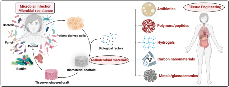
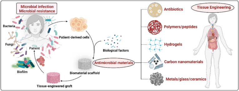
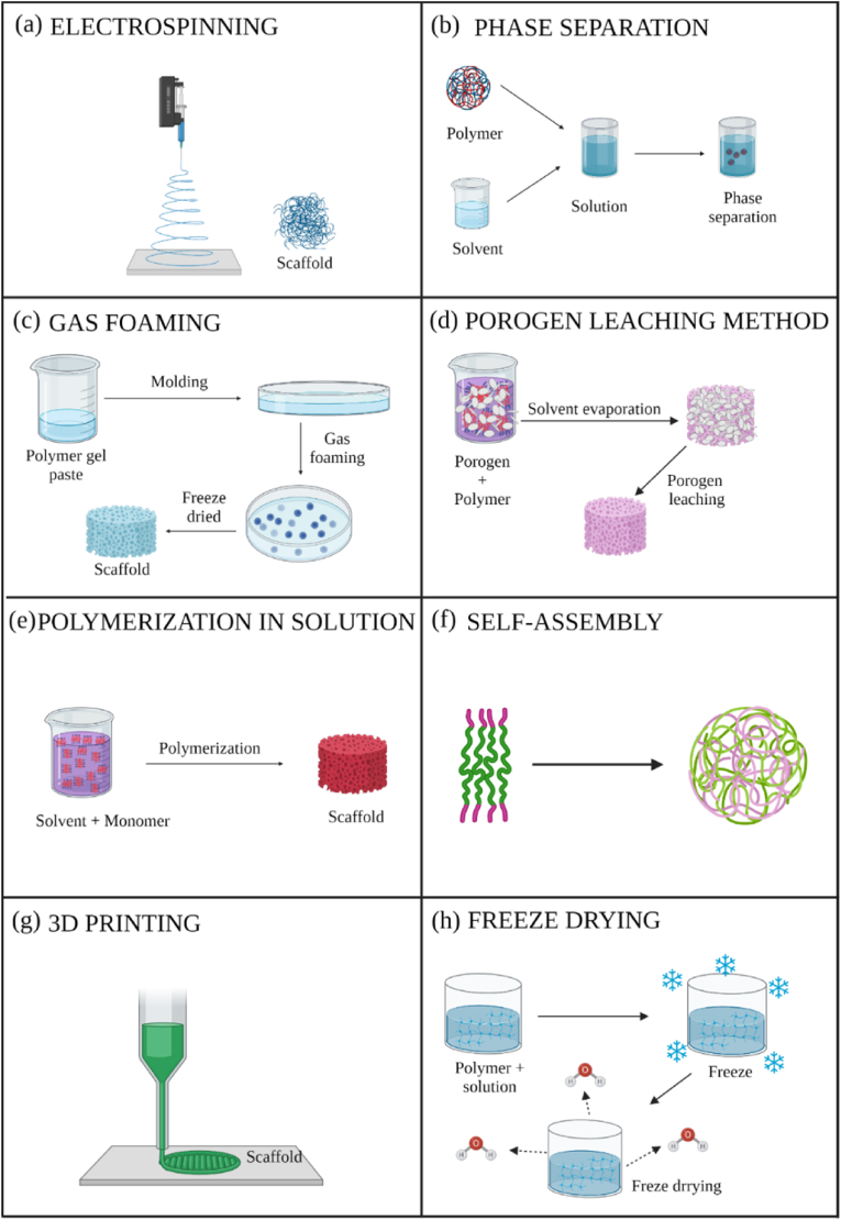
Due to microbial infections dramatically affect cell survival and increase the risk of implant failure, scaffolds produced with antimicrobial materials are now much more likely to be successful. Multidrug-resistant infections without suitable prevention strategies are increasing at an alarming rate. The ability of cells to organize, develop, differentiate, produce a functioning extracellular matrix (ECM) and create new functional tissue can all be controlled by careful control of the extracellular microenvironment. This review covers the present state of advanced strategies to develop scaffolds with antimicrobial properties for bone, oral tissue, skin, muscle, nerve, trachea, cardiac and other tissue engineering applications. The review focuses on the development of antimicrobial scaffolds against bacteria and fungi using a wide range of materials, including polymers, biopolymers, glass, ceramics and antimicrobials agents such as antibiotics, antiseptics, antimicrobial polymers, peptides, metals, carbon nanomaterials, combinatorial strategies, and includes discussions on the antimicrobial mechanisms involved in these antimicrobial approaches. The toxicological aspects of these advanced scaffolds are also analyzed to ensure future technological transfer to clinics. The main antimicrobial methods of characterizing scaffolds' antimicrobial and antibiofilm properties are described. The production methods of these porous supports, such as electrospinning, phase separation, gas foaming, the porogen method, polymerization in solution, fiber mesh coating, self-assembly, membrane lamination, freeze drying, 3D printing and bioprinting, among others, are also included in this article. These important advances in antimicrobial materials-based scaffolds for regenerative medicine offer many new promising avenues to the material design and tissue-engineering communities.

Keywords: Antimicrobial activity; Biomaterials; Fabrication; Scaffolds; Tissue engineering.


Conflict of interest statement

The authors declare that they have no known competing financial interests or personal relationships that could have appeared to influence the work reported in this paper.

Figures

Image 1
Graphical abstract
Fig. 1
Fig. 1
Antimicrobial scaffolds to prevent microbial infections in tissue engineering applications. Created with Biorender by Ángel Serrano-Aroca.
Fig. 2
Fig. 2
Production methods for antimicrobial scaffolds: (a) electrospinning; (b) phase separation; (c) gas foaming; (d) porogen leaching method; (e) polymerization in solution; (f) self-assembly; (g) 3D printing; (h) freeze drying.
Fig. 3
Fig. 3
Scanning electron microscope images of chitosan and PVA blended electrospun fibers at a magnification of x10000. Chitosan and PVA were dissolved in formic acid at 7% w/w and in distilled water at 9% w/w, respectively. The two solutions were mixed and electrospun in the indicated chitosan: PVA specified volume ratios of 50:50 (A), 30:70 (B) and 0:100 (C). Electrospun fibers made of a mixture of chitosan dissolved in formic acid (or 0.2 ​M acetic acid) at 2% w/w and mixed with a solution of 9% w/w PVA in a volume ratio of 50:50 (D). Adapted with permission from Ref. [69]. Copyright 2004 Elsevier.
Fig. 4
Fig. 4
Fused deposition modeling 3D-printing scaffolds for bone tissue regeneration: morphology and surface microstructure. Scaffold images of PCL (A), PCL/PDA (B), PCL/AgNPs (C), PCL/PDA/AgNPs (D). Scanning electron microscopy photographs of PCL (E, I), PCL/PDA (F, J), PCL/AgNPs (G, L), PCL/PDA/AgNPs (H, M) scaffolds. Reprinted with permission from Ref. [63]. Copyright 2019 Elsevier.
Fig. 5
Fig. 5
Main bioprinting technological methods: laser-induced forward transfer (a), inkjet printing (b) and robotic dispensing (c). Adapted with permission from Ref. [66]. Copyright 2013 John Wiley and Sons.
Fig. 6
Fig. 6
Tissue engineering application fields for antibacterial, antifungal and antibiofilm scaffolds. Created with Biorender by Ángel Serrano-Aroca.
Fig. 7
Fig. 7
(a) PCL scaffold (top view); (b) details and pore size; (c) scaffold with conductive TrGO particles (top view); (d) detailed image of a scaffold pore. Reprinted with permission under a Creative Commons CC BY 4.0 License from Ref. [145]. Copyright 2020 MDPI.
Fig. 8
Fig. 8
View of sintered robocast biphasic calcium phosphate scaffolds for bone tissue engineering. Optical (b) and scanning electron microscope view from the top (a) and cross-section (c) of the ceramic scaffold. Reprinted with permission from Ref. [185]. Copyright 2019 Elsevier.
Fig. 9
Fig. 9
(A) Uncrosslinked scaffold after freeze-drying, the printed struts shrunk sharply and showed one-level macroporous structures. (B) Crosslinked scaffold after freeze drying showed multi-level porous structures. Reprinted with permission from Ref. [196]. Copyright 2020 Elsevier.
Fig. 10
Fig. 10
Scaffolds composed of copper-loaded-zeolitic-imidazolate-frameworks (ZIF-8) and PLGA (PLGA/Cu(I)@ZIF-8): (a) Transmission electron microscope (TEM) image of Cu(I)@ZIF-8 nanoparticles; (b) Particle size distribution of Cu(I)@ZIF-8 nanoparticles; (c) digital image; (d, e) TEM images of PLGA/Cu(I)@ZIF-8 scaffolds; (f) Load–displacement curve of PLGA and PLGA/Cu(I)@ZIF-8 scaffolds. Reprinted with permission under a Creative Commons CC BY 4.0 License from Ref. [194]. Copyright 2020 Springer Nature.
Fig. 11
Fig. 11
Scanning electron microscope (SEM) images of unmodified and Cipro-modified (2 or 5% w/w) HPPS, obtained by using 5 or 10% w/w of PLA and a SEM image of ciprofloxacin used for the scaffolds modification Reprinted with permission under a Creative Commons CC BY 4.0 License from Ref. [275]. Copyright 2020 MDPI.
Fig. 12
Fig. 12
Scanning electron microscope images of Ag-MBGN after soaking in SBF for 14 days at different magnifications. Reprinted with permission under a Creative Commons CC BY License from Ref. [283]. Copyright 2019 Elsevier.
Fig. 13
Fig. 13
Hydrogel morphologies after swelling in phosphate buffered saline. Scale bar was 500 ​μm. Reprinted with permission from Ref. [354]. Copyright 2015 Elsevier.
Fig. 14
Fig. 14
Scanning electron microscope images of scaffolds of different magnifications: (a) and (a′) Collagen, (b) and (b′) Collagen-rGO-200, (c) and (c′) Collagen-rGO-400, (d) and (d′) Collagen-rGO-600 and (e) and (e′) Collagen-rGO-800. Reprinted with permission from Ref. [374]. Copyright 2019 Elsevier.
Fig. 15
Fig. 15
Preparation scheme of the multifunctional auricle scaffold by 3D printing and subsequent activation by polydopamine (pDA) and coated layer-by-layer with EPL and FIB. The pDA-EFE auricle scaffold obtained showed bioactive, antibacterial, angiogenesi enhancing, and tissue ingrowth-promoting properties. Reprinted with permission from Ref. [378]. Copyright 2022 Elsevier.
Fig. 16
Fig. 16
Normalized width of the antimicrobial “halo” of a scaffold calculated by the inhibition zone (diz) and the scaffold diameter (d). Reprinted by kind permission of ref. [410]. Copyright 2018 MyJoVE Corporation.
Fig. 17
Fig. 17
Schematic representation of a CDC Biofilm Reactor used to study biofilm formation on scaffold prepared with in the form of disks. Bioreactor fabricated by BioSurface Technologies Corporation (http://biofilms.biz/). Reprinted with permission under a Creative Commons CC BY 4.0 License from Ref. [416]. Copyright 2020 MDPI.
Fig. 18
Fig. 18
Schematic illustration of antimicrobial mechanism in: (a) 3D-printed biocompatible scaffolds based on calcium-deficient hydroxyapatite (CDHA) with gold nanoparticles. Reprinted with permission from Ref. [204]. Copyright 2019 Elsevier; (b) gelatin-based and Zn2+-incorporated composite hydrogel (Gel@Zn) for bacterial elimination to promote infected wound healing. Reprinted with permission from Ref. [292]. Copyright 2022 Elsevier.

References

    1. Ambekar R.S., Kandasubramanian B. Progress in the advancement of porous biopolymer scaffold: tissue engineering application. Ind. Eng. Chem. Res. 2019;58:6163–6194. doi: 10.1021/acs.iecr.8b05334. - DOI
    1. DE Boer J.B. third ed. Academic Press; Oxford, United Kingdom: 2022. TISSUE ENGINEERING.
    1. Ratner B.D., Hoffman A.S., Schoen F.J., Lemons J.E. Academic Press; Toronto, Canada: 2012. Biomaterials Science: an Introduction to Materials in Medicine.
    1. Ahmadian E., Dizaj S.M., Eftekhari A., Dalir E., Vahedi P., Hasanzadeh A., Samiei M. The potential applications of hyaluronic acid hydrogels in biomedicine. Drug Res. 2020;70:6–11. doi: 10.1055/a-0991-7585. - DOI - PubMed
    1. Moreno-Manzano V., Zaytseva-Zotova D., López-Mocholí E., Briz-Redón Á., Strand B.L., Serrano-Aroca Á. Injectable gel form of a decellularized bladder induces adipose-derived stem cell differentiation into smooth muscle cells in vitro. Int. J. Mol. Sci. 2020;21 doi: 10.3390/IJMS21228608. 8608. 21 (2020) 8608. - DOI - PMC - PubMed

LinkOut - more resources