Arterial spin labeling magnetic resonance evaluates changes of cerebral blood flow in patients with mild traumatic brain injury
- PMID: 36097769
- PMCID: PMC10950119
- DOI: 10.11817/j.issn.1672-7347.2022.210754
Arterial spin labeling magnetic resonance evaluates changes of cerebral blood flow in patients with mild traumatic brain injury
Abstract
Objectives: The patients with mild traumatic brain injury (mTBI) accounts for more than 80% of the patients with brain injury. Most patients with mTBI have no abnormalities in CT examination. Therefore, most patients choose to self-care and recover rather than seeking medical treatment. In fact, mTBI may result in persistent cognitive decline and neurobehavioral dysfunction. In addition, changes occurred in neurochemistry, metabolism, and cells after injury may cause changes in cerebral blood flow (CBF), which is one of the causes of secondary injury and slow brain repair. This study aims to evaluate the changes of CBF with the progression of the disease in patients with mTBI based on arterial spin labeling (ASL) magnetic resonance imaging technology.
Methods: In the outpatient or emergency department of the Second Affiliated Hospital of Wenzhou Medical University, 43 mTBI patients were collected as an mTBI group, and 43 normal subjects with age, gender, and education level matching served as a control group. They all received clinical neuropsychology and cognitive function evaluation and magnetic resonance imaging. In the mTBI group, 22 subjects were followed up at acute phase, 1 month, 3 months, and 12 months. Based on the control group, the abnormal regions of CBF in the whole brain of mTBI patients were analyzed. The abnormal regions were taken as the regions of interest (ROI). The correlation of the values of the CBF in ROIs with clinical indications, cognitive function, and the changes of CBF in ROI at each time point during the follow-up were analyzed.
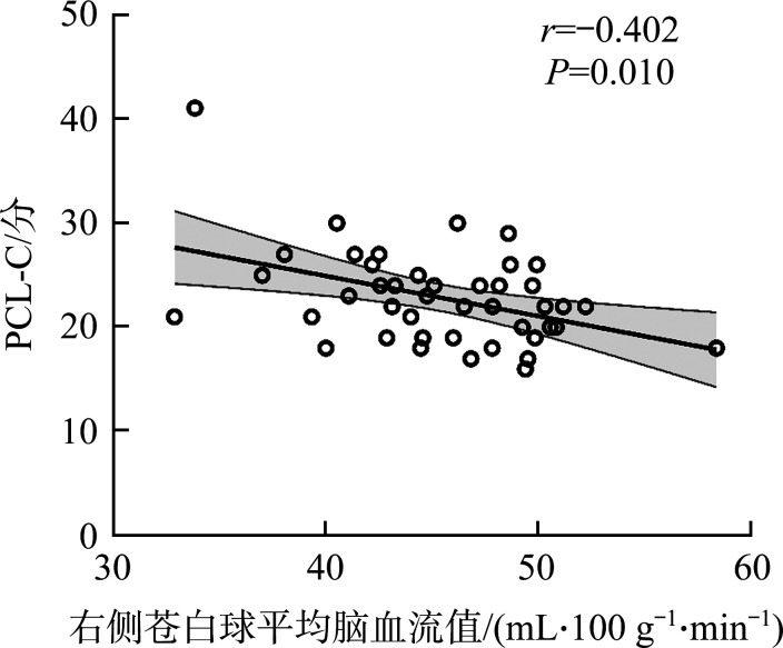
Results: Compared with the control group, the CBF in the bilateral dorsolateral superior frontal gyrus and auxiliary motor areas in the cortical region, as well as the right putamen, caudate nucleus, globus pallidus, and parahippocampus in the subcutaneous regions in the acute phase of the mTBI group were significantly increased (all P<0.01, TFCE-FWE correction). The analysis results of correlation of CBF with neuropsychology and cognitive domain showed that in the mTBI group, whole brain (r=0.528, P<0.001), right caudate nucleus (r=0.512, P<0.001), putamen (r=0.486, P<0.001), and globus pallidus (r=0.426, P=0.006) values of the were positively correlated with Backward Digit Span Test (BDST) score (reflectting working memory ability), and the right globus pallidus CBF was negatively correlated with the Post-Traumatic Stress Disorder Cheeklist-CivilianVersion (PCL-C) score (r=-0.402, P=0.010). Moreover, the follow-up study showed that abnormal CBF in these areas had not been restored. The correlation of CBF was negatively correlated with PCL-C and BDST at 1 months, 3 months, and 12 months (all P>0.05).
Conclusions: The elevated CBF value is one of the stress characteristics of brain injury in the mTBI patients at the acute phase. There is abnormal elevation of CBF values in multiple cortex or subcortical areas. Multi-time point studies show that there is no obvious change of CBF in abnormal areas, suggesting that potential clinical treatment is urgently needed for the mTBI patients.
目的: 轻度脑外伤患者占脑外伤患者的80%以上。多数轻度脑外伤患者在CT检查上未见异常,因此绝大部分患者选择自我调养、恢复而非就医。事实上,轻度脑外伤发生后可能会导致持续性的认知功能下降和神经行为功能障碍,神经化学、代谢和细胞的改变可能会引发脑血流的改变,而脑血流的改变被认为是继发性损伤和脑修复缓慢的原因之一。本研究旨在利用动脉自旋标记(arterial spin labeling,ASL)磁共振成像技术评估轻度脑外伤患者脑血流值随病程进展的变化情况。方法: 采集在温州医科大学附属第二医院门诊或急诊就医的43例轻度脑外伤患者的临床信息,同时纳入年龄、性别和受教育程度匹配的43例健康受试者作为对照组,所有受试者均接受脑部磁共振成像扫描,并进行临床神经心理学评估和认知功能评估。对其中22名轻度脑外伤受试者在急性期、第1个月、第3个月和第12个月进行随访。以对照组为基准,分析轻度脑外伤患者的异常脑血流区域,并以异常区域作为后续的感兴趣区(region of interest,ROI)。分析各ROI中的脑血流值与临床指征、认知功能的相关性以及轻度脑外伤患者在后续随访的各时间点ROI中脑血流值的变化。结果: 与对照组比较,轻度脑外伤组急性期在皮质区域的双侧背外侧额上回和双侧辅助运动区以及在皮下核团的右侧壳核、尾状核、苍白球、旁海马区域的脑血流值显著升高(均P<0.01,TFCE-FWE校正)。脑血流值与神经心理学评分以及各认知域认知量表评分的相关性分析结果显示:轻度脑外伤组全脑(r=0.528,P<0.001)、右侧尾状核(r=0.512,P<0.001)、壳核(r=0.486,P<0.001)和苍白球(r=0.426,P=0.006)脑血流值与评估工作记忆能力的倒背数字广度测验(Backward Digit Span Task,BDST)评分呈正相关,而右侧苍白球脑血流值与创伤后应激障碍评分(Post-Traumatic Stress Disorder Cheeklist-Civilian Version,PCL-C)呈负相关(r=-0.402,P=0.010)。在随访期间,这些区域的异常脑血流改变均未恢复,而在第1个月,3个月和12个月时脑血流值与PCL-C和BDST之间无相关性(均P>0.05)。结论: 脑血流值代谢升高是急性期轻度脑外伤患者脑损伤的应激特征之一;其多个皮层或皮层下区域的脑血流值均存在异常的升高,且这些异常区域的脑血流值在后续随访期间均无明显改变,提示轻度脑外伤患者亟需采取潜在的临床治疗手段。.
Keywords: arterial spin labeling; cerebral blood flow; magnetic resonance imaging; mild traumatic brain injury.
Conflict of interest statement
作者声称无任何利益冲突。
Figures


Similar articles
-
Cerebral Blood Flow and Its Connectivity Deficits in Mild Traumatic Brain Injury at the Acute Stage.Neural Plast. 2020 Jul 1;2020:2174371. doi: 10.1155/2020/2174371. eCollection 2020. Neural Plast. 2020. PMID: 32684919 Free PMC article.
-
Arterial Spin Labeling Perfusion Study in the Patients with Subacute Mild Traumatic Brain Injury.PLoS One. 2016 Feb 12;11(2):e0149109. doi: 10.1371/journal.pone.0149109. eCollection 2016. PLoS One. 2016. PMID: 26871696 Free PMC article.
-
Chronic cerebral blood flow alterations in traumatic brain injury and sports-related concussions.Brain Inj. 2022 Jul 3;36(8):948-960. doi: 10.1080/02699052.2022.2109746. Epub 2022 Aug 10. Brain Inj. 2022. PMID: 35950271
-
A Systematic Review of ASL Perfusion MRI in Mild TBI.Neuropsychol Rev. 2023 Mar;33(1):160-191. doi: 10.1007/s11065-020-09451-7. Epub 2020 Aug 18. Neuropsychol Rev. 2023. PMID: 32808244 Free PMC article.
-
Neuroimaging in Pediatric Patients with Mild Traumatic Brain Injury: Relating the Current 2018 Centers for Disease Control Guideline and the Potential of Advanced Neuroimaging Modalities for Research and Clinical Biomarker Development.J Neurotrauma. 2021 Jan 1;38(1):44-52. doi: 10.1089/neu.2020.7100. Epub 2020 Oct 21. J Neurotrauma. 2021. PMID: 32640874 Free PMC article. Review.
Cited by
-
Concussion and the Autonomic, Immune, and Endocrine Systems: An Introduction to the Field and a Treatment Framework for Persisting Symptoms.J Pers Med. 2025 Jan 17;15(1):33. doi: 10.3390/jpm15010033. J Pers Med. 2025. PMID: 39852225 Free PMC article. Review.
-
Abnormal volumetric brain morphometry and cerebral blood flow in adolescents with depression.World J Psychiatry. 2023 Jun 19;13(6):386-396. doi: 10.5498/wjp.v13.i6.386. eCollection 2023 Jun 19. World J Psychiatry. 2023. PMID: 37383288 Free PMC article.
-
Updated Review of Neurologic Concussion Biomarkers for Time-sensitive Point-of-care Testing.J Emerg Trauma Shock. 2025 Apr-Jun;18(2):74-89. doi: 10.4103/jets.jets_76_24. Epub 2025 Jun 19. J Emerg Trauma Shock. 2025. PMID: 40666393 Free PMC article. Review.
References
MeSH terms
Substances
Grants and funding
LinkOut - more resources
Full Text Sources
Medical