. 2023 Feb;93(2):336-347.
doi: 10.1002/ana.26509. Epub 2022 Oct 8.

Neuroprotection of Low-Frequency Repetitive Transcranial Magnetic Stimulation after Ischemic Stroke in Rats

Affiliations

Neuroprotection of Low-Frequency Repetitive Transcranial Magnetic Stimulation after Ischemic Stroke in Rats

Cathrin M Buetefisch et al. Ann Neurol. 2023 Feb.

Abstract

Objective: Stroke is a leading cause of human death and disability. Effective early treatments with reasonable therapeutic windows remain critically important to improve the outcomes of stroke. Transcranial magnetic stimulation (TMS) is an established noninvasive technique that has been applied clinically and in animal research for multiple brain disorders, but few studies have examined acute neuroprotection against ischemic stroke. The present investigation tested the novel approach of low-frequency repetitive TMS (rTMS) as an acute treatment after ischemic stroke.

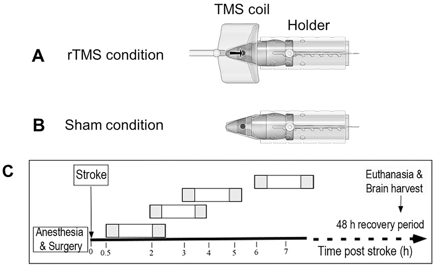
Methods: Adult male rats received focal ischemic surgery through occlusion of the right middle cerebral artery for 60 minutes. The rats received either rTMS or sham treatment with 1.5-, 3-, 4-, or 7-hour delay after the onset of stroke. Low-frequency and low-intensity rTMS was applied to the rat brain for two 30-minute episodes separated by a 1-hour interval.

Results: Three days after stroke, compared to stroke controls, rats receiving rTMS treatment with a 1.5-hour delay showed a 35% reduction of infarct volume. Protective effects were also seen with 3- or 4-hour-delayed treatments by rTMS, shown as reduced infarct volume and cell death. rTMS treatment upregulated the antiapoptotic factor Bcl-2 and downregulated the proapoptotic caspase-3 cleavage, expressions of Bax and matrix metallopeptidase-9. In sensorimotor functional assessments 3 to 21 days after stroke, rats receiving rTMS treatment with a 1.5- or 3-hour delay showed significantly better performance compared to stroke controls.

Interpretation: These results support the inference that low-frequency rTMS may be feasible as a neuroprotective acute treatment after ischemic stroke. ANN NEUROL 2023;93:336-347.


Conflict of interest statement

Potential Conflicts of Interest

Nothing to report.

Figures

FIGURE 1:
FIGURE 1:
Transcranial magnetic stimulation (TMS) device and experimental design. (A, B) The illustration shows the rat holder and the location of the TMS coil in reference to the animal’s head. The funnel that accommodated the rat’s head contained a window covering the surface of the head that was covered with mesh. The rodent holder was fixated on the surface of the table and, for the repetitive TMS (rTMS) group, the coil was fixated against the skull over the sensorimotor cortex. For the sham rTMS group, the sham animal was placed next to an animal receiving real rTMS treatment, so that the former was exposed to the sound of the discharging coil without receiving magnetic stimulation. (C) The timeline of the experimental design. Stroke animals were subjected to the focal ischemic surgery of 60-minute middle cerebral artery occlusion under isoflurane anesthesia. At the end of the surgery, animals were allowed to recover from anesthesia. At 1.5, 3, 4, or 7 hours after the onset of the ischemic insult, awake animals were stimulated twice for 30 minutes (dark squares in rectangle) with a 1-hour intersession interval and then returned to their home cage for full recovery. Animals were sacrificed 72 hours after stroke for infarct measurement, cell death assays, and Western blot analysis.
FIGURE 2:
FIGURE 2:
Acute repetitive transcranial magnetic stimulation (rTMS) treatment reduced infarct volume after ischemic stroke. rTMS treatment was initiated at 1.5, 3, 4, or 7 hours after the onset of the ischemic insult. (A) Triphenyltetrazolium chloride staining 3 days after stroke was performed in brain sections to reveal the infarction volume (white color) in the right cortex. Representative images show brain sections from a sham rTMS rat and an rTMS-treated rat 1.5 hours after stroke. The bar graph on the right shows the quantified data of significantly reduced infarct volume in the rTMS-treated group (~35% reduction; n = 17 per group, Student t test, *p < 0.05 vs sham rTMS). (B–D) rTMS treatment was applied with 3-, 4-, or 7-h delay after stroke. Both 3- and 4-hour-delayed treatments still showed significant neuroprotection in reducing the infarct volume (n = 10, 8 for 3-hour-delayed group; 10, 10 for 4-hour-delayed group; and 10, 10 for 7-hour-delayed group, respectively; Student t test, *p < 0.05 vs sham rTMS). The protective effect was not significant when rTMS was applied 7 hours after the onset of stroke.
FIGURE 3:
FIGURE 3:
Repetitive transcranial magnetic stimulation (rTMS) acute treatments prevented neuronal cell death after ischemic stroke. Cellular immunohistochemical assays were performed on brain sections 3 days after stroke. (A) In the peri-infarct cortex of sham-treated rats, terminal deoxynucleotide transferase–mediated deoxyuridine triphosphate nick-end labeling (TUNEL) staining (green) demonstrated massive cell death, whereas stroke rats receiving rTMS treatment 1.5 hours after the onset of stroke exhibited fewer TUNEL-positive cells. (B, C) Quantified bar graphs summarize TUNEL-positive total cells (B) and TUNEL/NeuN double positive neurons (C) in the peri-infarct region (n = 10 for sham and rTMS groups, respectively; *p < 0.05 vs sham, **p < 0.01 vs sham). (D) Representative images of NeuN (red) and TUNEL (green) staining in the peri-infarct region. rTMS was applied 3 hours after stroke. (E, F) The bar graphs summarize cell counting data of the experiments with 3-hour-delayed treatment of rTMS. Both the total TUNEL-positive cells and the TUNEL-positive neurons were significantly reduced in stroke rats receiving rTMS (Student t test, F = 8.96 and F = 6.718, respectively; *p < 0.05 vs controls, n = 6 in stroke + sham rTMS and 7 in stroke + rTMS group). (G) Quantified data of TUNEL/NeuN double positive cells after rTMS treatment with 4-hour delay (Student t test, F = 21.37, **p < 0.01 vs sham rTMS, n = 5 per group).
FIGURE 4:
FIGURE 4:
Effect of repetitive transcranial magnetic stimulation (rTMS) on protein regulation in the peri-infarct cortex. Western blotting was applied to measure pro- and antiapoptotic proteins 3 days after stroke in the peri-infarct cortex. (A) Representative gel showing the protein levels of different genes with 3-hour-delayed treatment of rTMS. (B) Summarized data from A, showing upregulation of Bcl-2 and downregulations of Bax, matrix metallopeptidase-9 (MMP-9), and caspase-3 by rTMS treatment (1-way analysis of variance [ANOVA], *p < 0.05 between stroke + sham rTMS [sham] and control, **p < 0.05 between sham and rTMS, n = 4–5 per group). (C) Representative gel showing gene expression in the peri-infarct region with 4-hour-delayed rTMS treatment. (D) Summarized data show a significant increase in Bcl-2 expression and decreased levels of Bax and caspase-3. The expression level of MMP-9 was not significantly changed under this condition (1-way ANOVA, *p < 0.05 vs control, **p < 0.05 vs sham rTMS, n = 4–5 per group).
FIGURE 5:
FIGURE 5:
Functional benefits of repetitive transcranial magnetic stimulation (rTMS) treatments. Sensorimotor functional activities were tested to evaluate functional recovery after ischemic stroke using the adhesive dot removal test before and 3 days poststroke, and the corner test at 14 and 21 days poststroke. (A) The time durations for the animal to contact and remove the adhesive dot were measured and compared between sham and 1.5-hour-delayed rTMS groups. The ischemic insult to the right sensorimotor cortex significantly prolonged the time needed for the animal to feel and contact the adhesive dot on the affected left paw, whereas stroke rats in the rTMS group showed significantly faster time to contact and remove the (2-way analysis of variance [ANOVA] followed by Bonferroni correction, *p < 0.01 between control and stroke + sham [sham] groups, **p < 0.01 between sham and rTMS groups, n = 13 in control and rTMS groups, respectively). (B) The adhesive removal test showed significantly improved performance (faster time) in stroke rats that received rTMS with 3-hour delay after stroke tested 3 days after stroke (2-way ANOVA followed by Bonferroni correction, *p < 0.001 vs before stroke, **p < 0.001 vs control, n = 12 in sham and n = 11 in rTMS group). (C) Stroke rats that received the rTMS treatment with 7-hour delay showed no difference from sham (2-way ANOVA, n = 11 for each group). (D) Sensorimotor function associated with whisker activities was assessed using the corner test at 14 and 21 days after stroke. Normal animals make equal left (L) and right (R) turns in this test (ratio = 1), whereas stroke animals show biased turn direction due to the ischemic injury to one side of the sensorimotor cortex. Stroke animals that received the rTMS treatment with 3-hour delay showed significantly improved turns at these two time points (2-way ANOVA, *p < 0.05 for sham vs control, **p < 0.05 for rTMS vs sham, n = 11 for each group).

References

    1. Go AS, Mozaffarian D, Roger VL, et al. Heart disease and stroke statistics—2014 update: a report from the American Heart Association. Circulation 2014;129:e28–e292. - PMC - PubMed
    1. Hacke W, Kaste M, Bluhmki E, et al. Thrombolysis with alteplase 3 to 4.5 hours after acute ischemic stroke. N Engl J Med 2008;359:1317–1329. - PubMed
    1. Haile WB, Wu J, Echeverry R, et al. Tissue-type plasminogen activator has a neuroprotective effect in the ischemic brain mediated by neuronal TNF-alpha. J Cereb Blood Flow Metab 2012;32:57–69. - PMC - PubMed
    1. Lansberg MG, Schrooten M, Bluhmki E, et al. Treatment time-specific number needed to treat estimates for tissue plasminogen activator therapy in acute stroke based on shifts over the entire range of the modified Rankin Scale. Stroke 2009;40:2079–2084. - PMC - PubMed
    1. Stankowski JN, Gupta R. Therapeutic targets for neuroprotection in acute ischemic stroke: lost in translation? Antioxid Redox Signal 2011;14:1841–1851. - PMC - PubMed

Publication types