. 2022 Sep 30;131(8):687-700.
doi: 10.1161/CIRCRESAHA.122.320916. Epub 2022 Sep 13.

Extracellular Vesicles Regulate Sympatho-Excitation by Nrf2 in Heart Failure

Affiliations

Extracellular Vesicles Regulate Sympatho-Excitation by Nrf2 in Heart Failure

Changhai Tian et al. Circ Res. .

Abstract

Background: Chronic heart failure (CHF) is associated with redox imbalance. Downregulation of Nrf2 (nuclear factor [erythroid-derived 2]-like 2) plays important roles in disrupting myocardial redox homeostasis and mediating sympathetic nerve activity in the setting of CHF. However, it is unclear if circulating extracellular vesicles (EVs) elicit sympathetic excitation in CHF by disrupting central redox homeostasis. We tested the hypothesis that cardiac-derived EVs circulate to the presympathetic rostral ventrolateral medulla and contribute to oxidative stress and sympathetic excitation via EV-enriched microRNA-mediated Nrf2 downregulation.

Methods: Data were collected on rats with CHF post-myocardial infarction (MI) and on human subjects with ischemic CHF. EVs were isolated from tissue and plasma, and we determined the miRNAs cargo that related to targeting Nrf2 translation. We tracked the distribution of cardiac-derived EVs using in vitro labeled circulating EVs and cardiac-specific membrane GFP+ transgenic mice. Finally, we tested the impact of exogenously loading of antagomirs to specific Nrf2-related miRNAs on CHF-EV-induced pathophysiological phenotypes in normal rats (eg, sympathetic and cardiac function).

Results: Nrf2 downregulation in CHF rats was associated with an upregulation of Nrf2-targeting miRNAs, which were abundant in cardiac-derived and circulating EVs from rats and humans. EVs isolated from the brain of CHF rats were also enriched with Nrf2-targeting miRNAs and cardiac-specific miRNAs. Cardiac-derived EVs were taken up by neurons in the rostral ventrolateral medulla. The administration of cardiac-derived and circulating EVs from CHF rats into the rostral ventrolateral medulla of normal rats evoked an increase in renal sympathetic nerve activity and plasma norepinephrine compared with Sham-operated rats, which were attenuated by exogenously preloading CHF-EVs with antagomirs to Nrf2-targeting miRNAs.

Conclusions: Cardiac microRNA-enriched EVs from animals with CHF can mediate crosstalk between the heart and the brain in the regulation of sympathetic outflow by targeting the Nrf2/antioxidant signaling pathway. This new endocrine signaling pathway regulating sympathetic outflow in CHF may be exploited for novel therapeutics.

Keywords: antioxidant; extracellular vesicles; heart failure; oxidative stress; sympathetic nervous system.


Conflict of interest statement

DISCLOSURES

The authors declare no competing interests.

Figures

Figure 1.
Figure 1.. Nrf2 is dysregulated in the RVLM of animals with CHF.
Western blots showing Nrf2 expression (as indicated by the arrow) in the RVLM of Sham and CHF rats at 6 weeks post-MI or Sham surgery (A). Mean data and individual values quantifying Nrf2 expression in Sham and CHF RVLM punches (B); mean and individual values of miRNAs targeting 3’-UTR of Nrf2 mRNA in the RVLM: miR-27a, miR-28a and miR-34a (C) (n=4, ± SEM); qRT-PCR results show Nrf2 downstream targets, including heme oxygenase 1 (HO-1) mRNA level (D) and catalase mRNA level (E) in RVLM; Representative sections of immunostaining with 8-OHdG antibody show DNA/RNA oxidation (F) in the RVLM of Sham (top panel) and CHF (bottom panel) rats, respectively. Summary of the percentages of (G) 8-OHdG-positive cells in the RVLM of Sham (n=4) and CHF groups (n=4, ± SEM). *denotes p=0.0286 and all p values were derived using the Mann-Whitney test (nonparametric test). Scale bar is 20 μm.
Figure 2.
Figure 2.. Cardiac-derived EVs are abundant with miRNAs targeting Nrf2 translation in CHF.
TEM image shows typical morphology of cardiac EVs isolated from the LV of the rat heart (A); EV size was determined by measuring individual EV diameter (nm), and statistical size distribution of LV EVs (B); Representative western blots for CD81, CD9, HSC70 and TSG101 in LV EVs (C); mean and individual data showing miRNAs targeting 3’-UTR of Nrf2 mRNA (miR-27a (D), miR-28a (E) and miR-34a (F)), and cardiac-specific miRNAs (miR-1 (G), miR-499 (H) and miR-208 (I)) were analyzed by real-time qRT-PCR with specific primers, U6 snRNA was used as an internal control (n=6, ± SEM). Shapiro-Wilk and Kolmogorov-Smirnov test were used to evaluate normality, and p values were derived using an unpaired t-test with Welch’s correction.
Figure 3.
Figure 3.. Plasma EVs of CHF rats are abundant with Nrf2 targeting miRNAs.
EVs isolated from rat plasma by differential centrifugation were subjected to TEM (A), Nanosight analysis (B) and western blot with CD63, CD9, HSC70 and TSG101 antibodies (C); qRT-PCR results show miR-27a (D), miR-28a (E) and miR-34a (F) levels in EVs (Sham: n=6; CHF: n=7; ±SEM). Cel-mir-39 was used as a spike-in control. Shapiro-Wilk and Kolmogorov-Smirnov test were used to evaluate normality. P values were derived using an unpaired t-test with Welch’s correction.
Figure 4.
Figure 4.. Circulating EVs are taken up by neurons in the RVLM.
Circulating EVs from rat plasma were labeled with PKH26 Red Fluorescent Cell Linker and administrated to mice by IP injection. Four hours post injection, mice were perfused with ice-cold PBS and 4% PFA, and the brainstem was sectioned in the area shown in the midsagittal section in (A), and stained with anti-MAP2 antibody and nuclear stained with DAPI. The dashed red rectangular area incorporating the RVLM was screened under confocal microscopy (B). Scale bar in B is 20 μm; EV distribution in neurons in the RVLM are shown at high magnification (C). Scale bar is 10 μm.
Figure 5.
Figure 5.. Cardiac-derived EVs abundant with miRNAs targeting Nrf2 in CHF are distributed to the brainstem.
The EVs isolated from rat brain by employing enzyme-based methods and differential centrifugation and subjected to TEM (A); EV size was determined by measuring individual EV diameter (nm), and mean values for average size (B) and Western blot analysis with CD63, CD9, TSG101 and HSC70 antibodies (C). qRT-PCR results show the relative expression of miRNAs targeting Nrf2 mRNA (D, Sham: n=5; CHF: n=7; ±SEM); Cardiac-specific miRNAs in the EVs isolated from brain (E, Sham: n=5; CHF: n=6; ±SEM), D), p values were derived by the Mann-Whitney test (nonparametric test) and adjusted using the Bonferroni correction.
Figure 6.
Figure 6.. HF-derived EVs contribute to Nrf2 reduction and oxidative stress in the RVLM resulting in sympatho-excitation.
Schematic diagram for plasma EV isolation, labeling with PKH26, transfection with miRNA negative control and inhibitors, and stereotaxic bilateral microinjection into the RVLM of normal rats (A); Three days post-injection, rats were euthanized and perfused with PBS and 4% PFA. The brain stem was sectioned and subjected to immunostaining with Nrf2 and MAP2 (B), and βIII-tubulin and 4-HNE antibodies (C), respectively. Representative images were used to demonstrate neurons with and without PKH26+ EVs in the RVLM. Arrows indicate MAP2+/Nrf2+/PKH26+ neurons (B), and β III-Tubulin+/4-HNE+/PKH26+ neurons (C). The nuclei were stained with DAPI. Scale bar is 20 μm; plasma NE concentration in 3 groups of rats is shown in (D) (n=5, ± SEM). Statistical analysis was performed using Kruskal-Wallis test and p values were derived by the Dunn’s multiple comparisons test.
Figure 7.
Figure 7.. CHF-derived EVs microinjected into the RVLM elicit sympatho-excitation, and impair cardiac function.
Representative tracings showing baseline renal sympathetic nerve activity and induced arterial baroreflex sensitivity in anesthetized rats in response to bilateral microinjection of circulating Sham-EVs/NC, CHF-EVs/NC and CHF-EVs/antagomirs, respectively (A); Mean and individual data showing mean arterial pressure (B, MAP), heart rate (C), and Left ventricular end-diastolic pressure (D, LVEDP); dp/dtmax/min (G); Baseline RSNA (E), and baroreflex sensitivity (F, H). Shapiro-Wilk and Kolmogorov-Smirnov tests were used for normal distribution, and p values were derived from unpaired t with Welch’s correction (F, left panel, n=5–6, ± SEM), and the Dunn’s multiple comparisons test (nonparametric test) (F, right panel; E and H).
Figure 8.
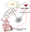
Figure 8.. A schematic overview of a heart-brain communication pathway that modulates central Nrf2 and oxidative stress in pre-sympathetic neurons.
Cardiac-derived EVs contribute to oxidative stress in the RVLM via EV-miRNA-mediated Nrf2 downregulation, resulting in sympathetic excitation and potentially contributing to further cardiac dysfunction. EV loading with antagomirs targeting Nrf2-related miRNAs into CHF-derived EVs attenuates these pathophysiological phenotypes of CHF.

References

    1. Tian C, Gao L, Zhang A, Hackfort BT, Zucker IH. Therapeutic Effects of Nrf2 Activation by Bardoxolone Methyl in Chronic Heart Failure. J Pharmacol Exp Ther. 2019;371:642–651. doi: 10.1124/jpet.119.261792 - DOI - PMC - PubMed
    1. Farías JG, Molina VM, Carrasco RA, Zepeda AB, Figueroa E, Letelier P, Castillo RL. Antioxidant Therapeutic Strategies for Cardiovascular Conditions Associated with Oxidative Stress. Nutrients. 2017;9. doi: 10.3390/nu9090966 - DOI - PMC - PubMed
    1. Bhimaraj A, Tang WH. Role of oxidative stress in disease progression in Stage B, a pre-cursor of heart failure. Heart Fail Clin. 2012;8:101–111. doi: 10.1016/j.hfc.2011.08.003 - DOI - PMC - PubMed
    1. Li J, Ichikawa T, Villacorta L, Janicki JS, Brower GL, Yamamoto M, Cui T. Nrf2 protects against maladaptive cardiac responses to hemodynamic stress. Arterioscler Thromb Vasc Biol. 2009;29:1843–1850. doi: 10.1161/atvbaha.109.189480 - DOI - PubMed
    1. Gao L, Zimmerman MC, Biswal S, Zucker IH. Selective Nrf2 Gene Deletion in the Rostral Ventrolateral Medulla Evokes Hypertension and Sympathoexcitation in Mice. Hypertension. 2017;69:1198–1206. doi: 10.1161/HYPERTENSIONAHA.117.09123 - DOI - PMC - PubMed

Publication types