. 2022 Sep 28;96(18):e0071622.
doi: 10.1128/jvi.00716-22. Epub 2022 Sep 13.

A Glu-Glu-Tyr Sequence in the Cytoplasmic Tail of the M2 Protein Renders Influenza A Virus Susceptible to Restriction of the Hemagglutinin-M2 Association in Primary Human Macrophages

Affiliations

A Glu-Glu-Tyr Sequence in the Cytoplasmic Tail of the M2 Protein Renders Influenza A Virus Susceptible to Restriction of the Hemagglutinin-M2 Association in Primary Human Macrophages

Sukhmani Bedi et al. J Virol. .

Abstract

Influenza A virus (IAV) assembly at the plasma membrane is orchestrated by at least five viral components, including hemagglutinin (HA), neuraminidase (NA), matrix (M1), the ion channel M2, and viral ribonucleoprotein (vRNP) complexes, although particle formation is observed with expression of only HA and/or NA. While these five viral components are expressed efficiently in primary human monocyte-derived macrophages (MDMs) upon IAV infection, this cell type does not support efficient HA-M2 association and IAV particle assembly at the plasma membrane. Both defects are specific to MDMs and can be reversed upon disruption of F-actin. However, the relationship between the two defects is unclear. Here, we examined whether M2 contributes to particle assembly in MDMs and if so, which region of M2 determines the susceptibility to the MDM-specific and actin-dependent suppression. An analysis using correlative fluorescence and scanning electron microscopy showed that an M2-deficient virus failed to form budding structures at the cell surface even after F-actin was disrupted, indicating that M2 is essential for virus particle formation at the MDM surface. Notably, proximity ligation analysis revealed that a single amino acid substitution in a Glu-Glu-Tyr sequence (residues 74 to 76) in the M2 cytoplasmic tail allowed the HA-M2 association to occur efficiently even in MDMs with intact actin cytoskeleton. This phenotype did not correlate with known phenotypes of the M2 substitution mutants regarding M1 interaction or vRNP packaging in epithelial cells. Overall, our study identified M2 as a target of MDM-specific restriction of IAV assembly, which requires the Glu-Glu-Tyr sequence in the cytoplasmic tail. IMPORTANCE Human MDMs represent a cell type that is nonpermissive to particle formation of influenza A virus (IAV). We previously showed that close proximity association between viral HA and M2 proteins is blocked in MDMs. However, whether MDMs express a restriction factor against IAV assembly or whether they lack a dependency factor promoting assembly remained unknown. In the current study, we determined that the M2 protein is necessary for particle formation in MDMs but is also a molecular target of the MDM-specific suppression of assembly. Substitutions in the M2 cytoplasmic tail alleviated the block in both the HA-M2 association and particle production in MDMs. These findings suggest that MDMs express dependency factors necessary for assembly but also express a factor(s) that inhibits HA-M2 association and particle formation. High conservation of the M2 sequence rendering the susceptibility to the assembly block highlights the potential for M2 as a target of antiviral strategies.

Keywords: M2; actin cytoskeleton; influenza A virus; macrophage; restriction factor; virus assembly.


Conflict of interest statement

The authors declare no conflict of interest.

Figures
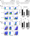
FIG 1
FIG 1
Generation and characterization of ΔM2 WSN virus. (A) The strategy used for generation of the WSN M segment that fails to encode the M2 protein (WSN M SS) is depicted. (B to D) dTHP1 cells (B) and MDMs (C) were infected with WT or ΔM2 virus at an MOI of 0.1 for 18 h. Infected cells were analyzed for cell surface expression of HA and M2 and intracellular expression of M1 by flow cytometry. Representative flow plots for mock-infected and virus-infected cells are shown. Percentages of cells positive for viral proteins (boxed) are shown. Relative MFIs for signal of indicated proteins for positive cell populations (gated in B and C) are shown. Data are means ± standard deviations and are from at least three independent experiments. *, P < 0.05; ns, nonsignificant.
FIG 2
FIG 2
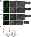
F-actin disruption restores virus bud formation in MDM in an M2-dependent manner. MDMs were infected with WSN at an MOI of 0.1 for 14 h. Infected cells were treated with vehicle (DMSO) or 5 μM Lat B for 4 h, before fixation and immunostaining with anti-HA. After identification of HA-positive cells by confocal fluorescence microscopy, cells were processed for SEM. The same cells were identified based on grid positions and analyzed by SEM. (A) Representative SEM images for WSN-infected HA-positive cells. Fluorescence images corresponding to the SEM images are also included. Boxed areas for SEM images are magnified and shown on the right of original images. Alphabetic labels are used to distinguish between the individual boxed areas. (B) The numbers of ~100-nm buds identified in SEM images were counted within the same-sized area (100 μm2) in each cell. Data are shown for 10 to 20 cells from two independent experiments. Error bars represent standard errors of means. ***, P < 0.005; ns, nonsignificant.
FIG 3
FIG 3
dTHP1 cells support IAV particle assembly in an M2-dependent manner. dTHP1 cells were infected with WT or ΔM2 virus for 18 h. Infected cells were fixed and immunostained with anti-HA antibody. After identification of HA-positive cells by confocal fluorescence microscopy, cells were processed for SEM. The same cells were identified based on grid positions and analyzed by SEM. (A) Representative SEM images for WSN-infected HA-positive cells. Fluorescence images corresponding to the SEM images are also included. Boxed areas for SEM images are magnified and shown on the right of original images. Alphabetic labels are used to distinguish between the individual boxed areas. (B) The numbers of ~100-nm buds identified in SEM images were counted within the same-sized area (100 μm2) in each cell. (C) Fluorescence intensity (FI) of surface HA expression was determined for each cell analyzed in panel B. Data are shown for 10 to 20 cells from two independent experiments. Error bars represent standard errors of means. **, P < 0.01; ns, nonsignificant.
FIG 4
FIG 4
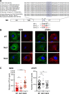
Alanine substitutions at amino acid positions 74 to 76 of the cytoplasmic tail of M2 circumvent the inhibition of HA-M2 association in MDMs. (A) Schematic showing conservation of M2 residues 71 to 76 (shaded), including the M2 Glu-Glu-Tyr sequence, across the representative IAV subtypes including the pandemic 1918 (H1N1), recently circulating human strains, and avian strains. The mutations introduced in the cytoplasmic tail of M2 are also shown. Amino acid residues 71 to 73 or 74 to 76 were replaced with alanine to generate Mut1 and Mut2 M2 mutants, respectively. (B and C) dTHP1 cells and MDMs were infected with WT WSN or M2 mutant viruses at an MOI of 0.1 for 18 h. Cells were fixed and examined by PLA using goat anti-HA and mouse anti-M2 antibodies. To identify infected cells, total surface M2 was also detected using a fluorescently tagged anti-mouse IgG secondary antibody (shown in green). Nuclei were stained with 4′,6-diamidino-2-phenylindole (DAPI; blue). (B) Representative maximum intensity projection images, which were reconstructed from z-stacks corresponding to the focal planes ranging from the middle plane of the nucleus to the bottom of the cells. (C) Number of PLA spots for each cell. Data are shown for experiments with MDMs from three independent donors, and 8 to 10 cells were analyzed per experiment. Error bars represent standard errors of means. ****, P < 0.0001; ns, nonsignificant.
FIG 5
FIG 5
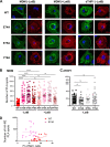
Amino acids at positions 74, 75, and 76 of the M2 protein are individually important for suppression of HA-M2 association in MDMs. dTHP1 cells and MDMs were infected with WT WSN or M2 mutant viruses at an MOI of 0.1 for 14 h. Infected cells were treated with vehicle (DMSO) or 5 μM Lat B for 4 h, before fixation. Fixed cells were examined by PLA using goat anti-HA and mouse anti-M2 antibodies. In addition, total surface M2 was also detected using a fluorescently tagged anti-mouse IgG secondary antibody (shown in green). Nuclei were stained with DAPI (blue). (A) Representative maximum intensity projection images, which were reconstructed from z-stacks corresponding to the focal planes ranging from the middle plane of the nucleus to the bottom of the cells. (B and C) Numbers of PLA spots were determined for each cell. (B) Data are shown for three independent experiments with MDMs, and 8 to 10 cells were analyzed per experiment. (C) Data are shown for three independent experiments with dTHP1 cells, and 8 to 10 cells were analyzed per experiment. Error bars represent standard errors of means. ****, P < 0.0001; ns, nonsignificant. (D) Increased HA-M2 association in MDMs expressing E74A M2 proteins does not correlate with the surface expression level of M2. MDMs were infected with WT WSN or E74A M2 mutant viruses at an MOI of 0.1 for 14 h and processed for PLA and M2 detection as described for panel A. Scatterplots between fluorescence intensity (FI) for cell surface M2 signals and number of HA-M2 PLA plots are shown for MDMs infected with WT and E74A mutant viruses. The best-fit line was determined for each set of x-y data points using linear regression analyses. Data were collected from MDMs of two different donors.
FIG 6
FIG 6
The E74A and E75A but not Y76A substitutions in the M2 cytoplasmic tail enhance virus particle release from MDM. vRNA release efficiency (ratio of virus-associated vRNA to the cell-associated vRNA) was measured in MDM and dTHP1 cultures. MDMs were infected with WT WSN or M2 mutant viruses at an MOI of 0.1. PB2 vRNA copy number was measured in MDM and dTHP1 culture supernatants and cell lysates at 14 hpi. A significant increase in vRNA release efficiency was observed with the E74A and E75A M2 mutants. Data were collected from MDMs of six different donors. Error bars represent standard deviations. *, P < 0.05; **, P < 0.005, ***, P < 0.001; ns, nonsignificant.

Similar articles

Cited by

References

    1. Nayak DP, Balogun RA, Yamada H, Zhou ZH, Barman S. 2009. Influenza virus morphogenesis and budding. Virus Res 143:147–161. 10.1016/j.virusres.2009.05.010. - DOI - PMC - PubMed
    1. Rossman JS, Lamb RA. 2011. Influenza virus assembly and budding. Virology 411:229–236. 10.1016/j.virol.2010.12.003. - DOI - PMC - PubMed
    1. Dou D, Revol R, Ostbye H, Wang H, Daniels R. 2018. Influenza A virus cell entry, replication, virion assembly and movement. Front Immunol 9:1581. 10.3389/fimmu.2018.01581. - DOI - PMC - PubMed
    1. Zhang J, Pekosz A, Lamb RA. 2000. Influenza virus assembly and lipid raft microdomains: a role for the cytoplasmic tails of the spike glycoproteins. J Virol 74:4634–4644. 10.1128/jvi.74.10.4634-4644.2000. - DOI - PMC - PubMed
    1. Leser GP, Lamb RA. 2005. Influenza virus assembly and budding in raft-derived microdomains: a quantitative analysis of the surface distribution of HA, NA and M2 proteins. Virology 342:215–227. 10.1016/j.virol.2005.09.049. - DOI - PubMed

Publication types

LinkOut - more resources