Review
. 2022 Nov;305(2):265-276.
doi: 10.1148/radiol.212808. Epub 2022 Sep 13.

Pulse-Echo Quantitative US Biomarkers for Liver Steatosis: Toward Technical Standardization

Affiliations
Review

Pulse-Echo Quantitative US Biomarkers for Liver Steatosis: Toward Technical Standardization

David T Fetzer et al. Radiology. 2022 Nov.

Abstract

Excessive liver fat (steatosis) is now the most common cause of chronic liver disease worldwide and is an independent risk factor for cirrhosis and associated complications. Accurate and clinically useful diagnosis, risk stratification, prognostication, and therapy monitoring require accurate and reliable biomarker measurement at acceptable cost. This article describes a joint effort by the American Institute of Ultrasound in Medicine (AIUM) and the RSNA Quantitative Imaging Biomarkers Alliance (QIBA) to develop standards for clinical and technical validation of quantitative biomarkers for liver steatosis. The AIUM Liver Fat Quantification Task Force provides clinical guidance, while the RSNA QIBA Pulse-Echo Quantitative Ultrasound Biomarker Committee develops methods to measure biomarkers and reduce biomarker variability. In this article, the authors present the clinical need for quantitative imaging biomarkers of liver steatosis, review the current state of various imaging modalities, and describe the technical state of the art for three key liver steatosis pulse-echo quantitative US biomarkers: attenuation coefficient, backscatter coefficient, and speed of sound. Lastly, a perspective on current challenges and recommendations for clinical translation for each biomarker is offered.


Conflict of interest statement

Disclosures of conflicts of interest: D.T.F. Institutional research agreements with and material support from GE Healthcare, Philips Healthcare, and Siemens Healthineers; advisory board consulting fees from GE Healthcare and Philips Healthcare. I.M.R.M. Research agreements with and equipment loans from Siemens Healthineers and General Electric; consultant, Siemens Healthineers; patents planned, issued, or pending; co-chair, American Institute of Ultrasound in Medicine (AIUM)–RSNA Quantitative Imaging Biomarkers Alliance (QIBA) Pulse-Echo Quantitative Ultrasound (PEQUS) Committee; member, Imaging Metrology and Standards Subcommittee and Ultrasound Subcommittee of the American Association of Physicists in Medicine. M.W. Meeting attendance support from GE Healthcare; co-chair, RSNA QIBA PEQUS Committee. M.L.R. New ultrasound equipment evaluation and lecture payment, Philips Medical; lecture payment, World Federation of Ultrasound in Medicine and Biology; associate editor, Radiology; subspecialty editor, Journal of Ultrasound in Medicine; editorial board, Ultrasound Quarterly. A.O. No relevant relationships. K.A.W. Royalties from CIRS; patents planned, issued, or pending; research agreement with Siemens Healthineers; chair, AIUM Bioeffects Committee and AAPM Task Group 333 MR-guided Focused Ultrasound Quality Assurance; member, AAPM TG353-Pulsed Doppler and Color Flow Ultrasound System Performance Assessment using Flow Phantoms, AIUM Technical Standards Committee, AIUM QIBA PEQUS Biomarker Committee, AIUM QIBA Shear Wave Speed Biomarker Committee, and AIUM QIBA Coordinating Committee; editorial board, The Journal of the Acoustical Society of America and Ultrasonic Imaging. J.O. No relevant relationships. T.A.S. Internal startup funding and participation on a data safety monitoring board, Kettering University; consultant, Riverside Research. J.B.F. Board member and meeting attendance support, AIUM and International Contrast Ultrasound Society; grant or equipment support from Philips Healthcare and GE Healthcare. T.J.H. Grants or contracts from Siemens Healthineers, General Electric, Philips, Samsung, Clarius, Echosens, and Mindray; chair, RSNA QIBA; equipment loans and technical support, Siemens Healthineers. A.E.S. Institutional payments, Allergan, Analogic Corporation, Boehringer Ingelheim, Department of Defense, Echosens, Fujifilm Healthcare, Foundation for the National Institutes of Health (NIH), Genentech, General Electric, Gilead, Intercept, Medimmune, NIH, Novo Nordisk, Partners Healthcare, Philips, Supersonic Imagine, Takeda, Toshiba Medical Systems, and Siemens; royalties or licenses, Elsevier and Katharos Labs; consulting fees, Astra Zeneca, Bracco Diagnostics, Bristol-Myers Squibb, General Electric, Gerson Lehman Group, Guidepoint Global Advisors, Supersonic Imagine, Novartis, Pfizer, Philips, Parexel Informatics, and WorldCare Clinical; legal consulting, Matis Baum and O'Connor; board of governors, AIUM; stockholder, Ochre Bio, Rhino Healthtech, Katharos Labs, Avira, Autonomous, Quantix Bio, Evidence Based Psychology, and Klea.

Figures

None
Graphical abstract
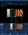
Image shows the controlled attenuation parameter (CAP), which is a
nonimaging sonographic method to estimate steatosis available on a
vibration-controlled transient elastography point-of-care device (FibroScan;
Echosens).
Figure 1:
Image shows the controlled attenuation parameter (CAP), which is a nonimaging sonographic method to estimate steatosis available on a vibration-controlled transient elastography point-of-care device (FibroScan; Echosens).
Diagrams depict the physics principles behind three PEQUS
biomarkers–attenuation coefficient (AC), backscatter coefficient, and
speed of sound (SOS)–for (left) healthy liver and (right) steatotic
liver. The color bar legend at the top right indicates ultrasound pulse
amplitude as it travels through tissue and is attenuated to a great extent in
steatotic liver (AC of 1.0 dB/cm-MHz). The inserts show the liver microstructure
that produces backscattered echo, which is greater in steatotic liver due to
hepatocytes filled and expanded by lipid vacuoles. The vertical array of numbers
on the left side of each diagram indicates the propagation depth and time of
ultrasound pulse arrival based on the SOS reported at the top of the
array.
Figure 2:
Diagrams depict the physics principles behind three PEQUS biomarkers–attenuation coefficient (AC), backscatter coefficient, and speed of sound (SOS)–for (left) healthy liver and (right) steatotic liver. The color bar legend at the top right indicates ultrasound pulse amplitude as it travels through tissue and is attenuated to a great extent in steatotic liver (AC of 1.0 dB/cm-MHz). The inserts show the liver microstructure that produces backscattered echo, which is greater in steatotic liver due to hepatocytes filled and expanded by lipid vacuoles. The vertical array of numbers on the left side of each diagram indicates the propagation depth and time of ultrasound pulse arrival based on the SOS reported at the top of the array.
 US images show various commercial implementations of the attenuation
coefficient, including (A) ATI (Canon Medical Systems), (B) ATT (FujiFilm
Medical Systems), (C) UGAP (GE Healthcare), (D) Atten (Philips Healthcare), and
(E) TAI (Samsung Medison). Images A, C, and D were acquired at the Massachusetts
General Hospital Center for Ultrasound Research & Translation laboratory
as a component of the Non-Invasive Biomarkers of Metabolic Liver Disease
(NIMBLE) study, which is supported by the Foundation for the National Institutes
of Health and the private sector.
Figure 3:
US images show various commercial implementations of the attenuation coefficient, including (A) ATI (Canon Medical Systems), (B) ATT (FujiFilm Medical Systems), (C) UGAP (GE Healthcare), (D) Atten (Philips Healthcare), and (E) TAI (Samsung Medison). Images A, C, and D were acquired at the Massachusetts General Hospital Center for Ultrasound Research & Translation laboratory as a component of the Non-Invasive Biomarkers of Metabolic Liver Disease (NIMBLE) study, which is supported by the Foundation for the National Institutes of Health and the private sector.
US images show commercial implementations of the attenuation coefficient
and speed of sound, including (A) Att and SSp (Hologic Supersonic Imagine) and
(B) ACS and SOS (E-Scopics).
Figure 4:
US images show commercial implementations of the attenuation coefficient and speed of sound, including (A) Att and SSp (Hologic Supersonic Imagine) and (B) ACS and SOS (E-Scopics).
US image shows sound speed index (SSI) quantification by Mindray, a
commercial implementation of the speed of sound.
Figure 5:
US image shows sound speed index (SSI) quantification by Mindray, a commercial implementation of the speed of sound.
US image shows a fat fraction tool (Siemens Healthineers) derived from
pulse-echo quantitative US features. The image was acquired at the Massachusetts
General Hospital Center for Ultrasound Research & Translation laboratory
as a component of the Non-Invasive Biomarkers of Metabolic Liver Disease
(NIMBLE) study, which is supported by the Foundation for the National Institutes
of Health and the private sector.
Figure 6:
US image shows a fat fraction tool (Siemens Healthineers) derived from pulse-echo quantitative US features. The image was acquired at the Massachusetts General Hospital Center for Ultrasound Research & Translation laboratory as a component of the Non-Invasive Biomarkers of Metabolic Liver Disease (NIMBLE) study, which is supported by the Foundation for the National Institutes of Health and the private sector.
Diagram depicts the strategy for the phantom-based groundwork study
planned by the American Institute of Ultrasound in Medicine–RSNA
Quantitative Imaging Biomarkers Alliance Pulse-Echo Quantitative Ultrasound
Biomarker Committee. The study will include the participation of 10 US vendors
and 24 clinical, academic, and government institutions for an expected total of
34 sites. At least three sites will perform measurements on at least one
specified imaging system from each vendor. Each site will perform measurements
on the four phantoms with two appraisers, and each appraiser will perform 10
repetitions at three different depths.
Figure 7:
Diagram depicts the strategy for the phantom-based groundwork study planned by the American Institute of Ultrasound in Medicine–RSNA Quantitative Imaging Biomarkers Alliance Pulse-Echo Quantitative Ultrasound Biomarker Committee. The study will include the participation of 10 US vendors and 24 clinical, academic, and government institutions for an expected total of 34 sites. At least three sites will perform measurements on at least one specified imaging system from each vendor. Each site will perform measurements on the four phantoms with two appraisers, and each appraiser will perform 10 repetitions at three different depths.

References

    1. Younossi ZM , Koenig AB , Abdelatif D , Fazel Y , Henry L , Wymer M . Global epidemiology of nonalcoholic fatty liver disease-Meta-analytic assessment of prevalence, incidence, and outcomes . Hepatology 2016. ; 64 ( 1 ): 73 – 84 . - PubMed
    1. Eslam M , Sanyal AJ , George J ; International Consensus Panel. MAFLD: A consensus-driven proposed nomenclature for metabolic associated fatty liver disease . Gastroenterology 2020. ; 158 ( 7 ): 1999 – 2014.e1 . - PubMed
    1. Chalasani N , Younossi Z , Lavine JE , et al. . The diagnosis and management of nonalcoholic fatty liver disease: Practice guidance from the American Association for the Study of Liver Diseases . Hepatology 2018. ; 67 ( 1 ): 328 – 357 . - PubMed
    1. Hamer OW , Aguirre DA , Casola G , Lavine JE , Woenckhaus M , Sirlin CB . Fatty liver: imaging patterns and pitfalls . RadioGraphics 2006. ; 26 ( 6 ): 1637 – 1653 . - PubMed
    1. Brunt EM , Janney CG , Di Bisceglie AM , Neuschwander-Tetri BA , Bacon BR . Nonalcoholic steatohepatitis: a proposal for grading and staging the histological lesions . Am J Gastroenterol 1999. ; 94 ( 9 ): 2467 – 2474 . - PubMed

Publication types