

Atypical follicular hyperplasia with light chain-restricted germinal centers after COVID-19 booster: a diagnostic pitfall
- PMID: 36098816
- PMCID: PMC9469053
- DOI: 10.1007/s00428-022-03400-w
Atypical follicular hyperplasia with light chain-restricted germinal centers after COVID-19 booster: a diagnostic pitfall
Abstract
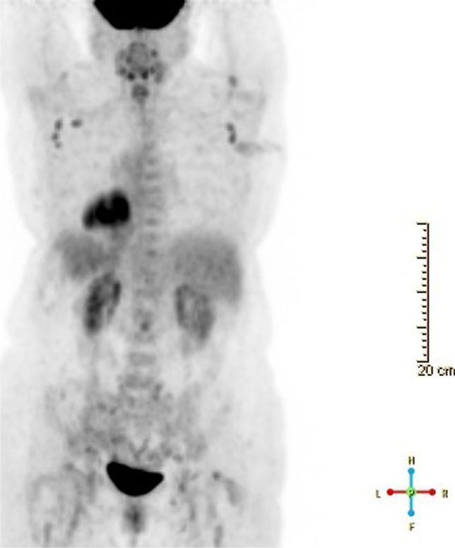
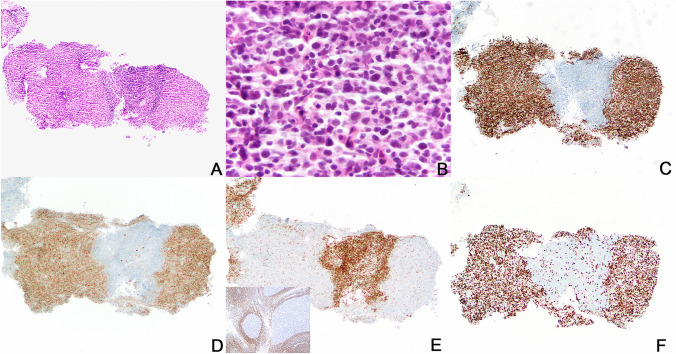
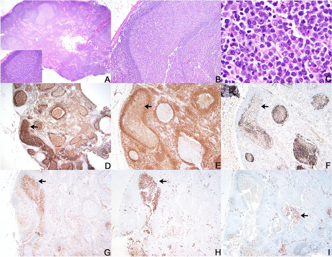
There has been a surge in COVID-19 vaccine-associated lymphadenopathy (LAD), including after the booster dose of vaccine. This can create diagnostic dilemmas in oncology patients as the relatively sudden LAD can mimic metastasis or cancer recurrence, at a risk of leading to additional but unnecessary anti-neoplastic therapy. Here we report the histopathologic features in a case of persistent LAD occurring in a patient with history of breast invasive ductal carcinoma which followed a COVID-19 vaccine booster. A needle core and then excisional biopsy showed atypical follicular hyperplasia with features that histologically and phenotypically could mimic follicular lymphoma, but the findings were ultimately interpreted to be reactive in nature and related temporally to COVID-19 vaccine. To our knowledge, this is the first case of an atypical lymphoproliferative lesion with features potentially mimicking lymphoma associated with COVID-19 vaccine.
Keywords: COVID-19; COVID-19 vaccine; COVID-19-related lymphadenopathy; Mimicker of lymphoma.
© 2022. The Author(s), under exclusive licence to Springer-Verlag GmbH Germany, part of Springer Nature.
Conflict of interest statement
The authors declare no competing interests.
Figures
Similar articles
-
COVID-19 post-vaccination lymphadenopathy: Report of cytological findings from fine needle aspiration biopsy.Diagn Cytopathol. 2021 Dec;49(12):E467-E470. doi: 10.1002/dc.24863. Epub 2021 Aug 25. Diagn Cytopathol. 2021. PMID: 34432391 Free PMC article.
-
The life and death of the germinal center.Ann Diagn Pathol. 2020 Feb;44:151421. doi: 10.1016/j.anndiagpath.2019.151421. Epub 2019 Nov 13. Ann Diagn Pathol. 2020. PMID: 31751845 Review.
-
Lobular carcinoma in situ diagnosed by core needle biopsy: when should it be excised?Mod Pathol. 2003 Feb;16(2):120-9. doi: 10.1097/01.MP.0000051930.68104.92. Mod Pathol. 2003. PMID: 12591964
-
Excisional biopsy should be performed if lobular carcinoma in situ is seen on needle core biopsy.Arch Pathol Lab Med. 2002 Jun;126(6):697-701. doi: 10.5858/2002-126-0697-EBSBPI. Arch Pathol Lab Med. 2002. PMID: 12033958
-
Reactive Lymphadenopathy in the Pediatric Population with a Focus on Potential Mimics of Lymphoma.Semin Diagn Pathol. 2023 Nov;40(6):371-378. doi: 10.1053/j.semdp.2023.05.004. Epub 2023 Jun 1. Semin Diagn Pathol. 2023. PMID: 37295994 Review.
Cited by
-
COVID-19 vaccine-associated lymphadenopathy: a review.Infez Med. 2024 Jun 1;32(2):119-130. doi: 10.53854/liim-3202-1. eCollection 2024. Infez Med. 2024. PMID: 38827838 Free PMC article. Review.
-
Case Report: Gene expression profiling of COVID-19 vaccination-related lymphadenopathies reveals evidence of a dominantly extrafollicular immune response.Front Immunol. 2023 Nov 14;14:1285168. doi: 10.3389/fimmu.2023.1285168. eCollection 2023. Front Immunol. 2023. PMID: 38035070 Free PMC article.
-
Atypical lymphoid proliferations associated with therapeutic intervention: a report of the 2024 EA4HP/SH lymphoma workshop.Virchows Arch. 2025 Aug 13. doi: 10.1007/s00428-025-04197-0. Online ahead of print. Virchows Arch. 2025. PMID: 40801926 Review.
-
Indolent cutaneous lymphoma with gamma/delta expression after COVID-19 vaccination.JAAD Case Rep. 2023 Feb;32:74-76. doi: 10.1016/j.jdcr.2022.12.001. Epub 2022 Dec 13. JAAD Case Rep. 2023. PMID: 36530557 Free PMC article. No abstract available.
-
Highly atypical lymphoid proliferation after COVID-19 vaccine, with spontaneous regression.J Hematop. 2024 Sep;17(3):171-174. doi: 10.1007/s12308-024-00587-6. Epub 2024 May 22. J Hematop. 2024. PMID: 38776051 No abstract available.
References
-
- WHO Coronavirus (2022) (COVID-19) Dashboard. https://covid19.who.int/. Accessed 25 July 2022
-
- Our World in Data. https://ourworldindata.org. Accessed 25 July 2022
Publication types
MeSH terms
Substances
Supplementary concepts
LinkOut - more resources
Full Text Sources
Medical
