. 2022 Oct 28;479(20):2195-2216.
doi: 10.1042/BCJ20220401.

Co-ordinated control of the ADP-heptose/ALPK1 signalling network by the E3 ligases TRAF6, TRAF2/c-IAP1 and LUBAC

Affiliations

Co-ordinated control of the ADP-heptose/ALPK1 signalling network by the E3 ligases TRAF6, TRAF2/c-IAP1 and LUBAC

Tom Snelling et al. Biochem J. .

Abstract

ADP-heptose activates the protein kinase ALPK1 triggering TIFA phosphorylation at Thr9, the recruitment of TRAF6 and the subsequent production of inflammatory mediators. Here, we demonstrate that ADP-heptose also stimulates the formation of Lys63- and Met1-linked ubiquitin chains to activate the TAK1 and canonical IKK complexes, respectively. We further show that the E3 ligases TRAF6 and c-IAP1 operate redundantly to generate the Lys63-linked ubiquitin chains required for pathway activation, which we demonstrate are attached to TRAF6, TRAF2 and c-IAP1, and that c-IAP1 is recruited to TIFA by TRAF2. ADP-heptose also induces activation of the kinase TBK1 by a TAK1-independent mechanism, which require TRAF2 and TRAF6. We establish that ALPK1 phosphorylates TIFA directly at Thr177 as well as Thr9 in vitro. Thr177 is located within the TRAF6-binding motif and its mutation to Asp prevents TRAF6 but not TRAF2 binding, indicating a role in restricting ADP-heptose signalling. We conclude that ADP-heptose signalling is controlled by the combined actions of TRAF2/c-IAP1 and TRAF6.

Keywords: ADP-heptose; ALPK1; TAK1; TBK1; TIFA; TRAF.


Conflict of interest statement

The authors declare that there are no competing interests associated with the manuscript.

Figures

Figure 1.
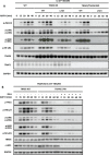
Figure 1.. ADP-heptose induces the ALPK1- and TIFA-dependent activation of MAP kinases and the canonical IKK complex.
(A,B) IL-1R* HEK293 cells (A) or BMDM from wild-type (WT) mice (B) were stimulated for the times indicated with ADP-heptose (ADP-H) or IL-1β (A) and ADP-H or R848 (B). (C) Parental, ALPK1 KO and TIFA KO HEK293 cells were stimulated for the times indicated with ADP-H, (D) ALPK1 KO HEK293 cells were transfected with empty vector (-), FLAG-ALPK1 (WT) or FLAG-ALPK1[K1067M] (K1067M) and 48 h later were stimulated for 20 min with ADP-H. (AD) Cell extracts were analysed directly by SDS–PAGE and immunoblotting was performed using antibodies that recognise phosphorylated (p) forms of the indicated proteins, or with antibodies recognising all forms of TIFA, ALPK1, IκBα and GAPDH. In (C,D), ALPK1 was detected after its immunoprecipitation from the extracts with an ALPK1 antibody (see Methods). The molecular mass markers needed to gauge the size of each band are shown in Supplementary Figure S7.
Figure 2.
Figure 2.. TAK1 and its protein kinase activity are required for ADP-heptose to activate MAP kinases and the canonical IKK complex.
(A) The parental and two independently isolated clones of TAK1 KO IL-1R* HEK293 cells were stimulated for the times indicated with ADP-heptose (ADP-H). (B) As in (A), except that WT TAK1 or the kinase-inactive TAK1[D175A] mutant were stably expressed in TAK1 KO cells under a doxycycline-inducible promoter and expression induced for 16 h with 50 ng/ml doxycycline, prior to stimulation with ADP-H for the times indicated. (C,D) IL-1R* HEK293 cells were treated for 1 h with 2 µM (5Z)-7-oxozeaenol (C) or 5 µM NG25 (D) and then stimulated with ADP-H for the times indicated. (A–C) Cell extracts were subjected to SDS–PAGE and immunoblotted with the antibodies indicated. The molecular mass markers needed to gauge the size of each band are shown in Supplementary Figure S7.
Figure 3.
Figure 3.. TRAF6 is required for ADP-heptose signalling in HEK293 cells and primary macrophages, but its E3 ligase activity is not.
(A) Parental and two independently isolated clones of TRAF6 KO IL-1R* HEK293 cells were stimulated for the times indicated with ADP-heptose (ADP-H). (B), As in (A), except that WT TRAF6, TRAF6[L74H] or TRAF6[120–522] were stably expressed in TRAF6 KO IL-1R* HEK293 cells under a constitutive promoter and stimulated for 20 min with ADP-H or IL-1β. (C) Foetal liver macrophages from wild-type (WT) and TRAF6 KO embryos were stimulated for the times indicated with ADP-H. (D,E) BMDM from WT and TRAF6[L74H] mice were stimulated for the times indicated with ADP-H (D) or R848 (E). (AE) Cell extracts were subjected SDS–PAGE and immunoblotted using the indicated antibodies. An asterisk indicates protein(s) recognised non-specifically by an antibody. The molecular mass markers needed to gauge the size of each band are shown in Supplementary Figure S7.
Figure 4.
Figure 4.. Proteins enriched in TRAF6 immunoprecipitates in ADP-heptose-stimulated HEK293 cells.
(A) IL-1R* HEK293 cells were stimulated for 20 min with ADP-heptose (ADP-H) and proteins in cell extracts immunoprecipitated with FLAG-TRAF6 were analysed by TMT mass spectrometry. The figure shows a volcano plot identifying the proteins where enrichment attained statistical significance and where the fold increase was >1.5-fold. (B) HEK293 cells were stimulated for 15 min with ADP-H or IL-1β and the proteins shown that were captured from the cell extracts on Halo-NEMO beads were identified by SDS–PAGE followed by immunoblotting with the antibodies indicated. (C) WT BMDM were stimulated for 15 min with ADP-H or R848 and analysed with the same antibodies used in (B), except for c-IAP1, where a mouse-specific antibody was used (see Methods). Ubiquitylated forms of proteins are indicated by (Ub)n.
Figure 5.
Figure 5.. The TRAF6 and TRAF2/c-IAP1 operate redundantly to generate the ubiquitin chains required for ADP-heptose signalling.
(A) Parental, TRAF6 KO or TRAF2/6 double KO IL-1R* HEK293 cells in which WT TRAF6 or TRAF6[L74H] had been stably re-expressed under a constitutive promoter were stimulated for the times indicated with ADP-H. (B) TRAF6 KO IL-1R* HEK293 cells in which WT TRAF6 or TRAF6[L74H] had been stably re-expressed as in (A) were treated for 5 h with 5 µM BV-6 prior to stimulation for the indicated times with ADP-H. (A,B) The cell extracts were subjected to SDS–PAGE and immunoblotting was performed with the antibodies indicated. The molecular mass markers needed to gauge the size of each band are shown in Supplementary Figure S7.
Figure 6.
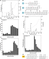
Figure 6.. ALPK1 phosphorylates TIFA directly at Thr9 in vitro.
(A) TIFA KO HEK293 cells were re-transfected with 500 ng of DNA plasmid encoding FLAG-TIFA or the indicated FLAG-TIFA mutants. After 24 h, the cells were stimulated for 20 min with ADP-heptose (ADP-H) and cell extracts subjected to SDS-PAGE and immunoblotting with the antibodies indicated. (B) Cell extracts (1.0 mg protein) from (A) were immunoprecipitated with anti-FLAG, then subjected to SDS-PAGE and immunoblotting. (C-E) ALPK1 KO HEK293 cells were re-transfected with 2.5 mg plasmid DNA encoding empty vector (-), FLAG-ALPK1 and FLAG-ALPK1[K1067M] as indicated. After 48 h, ALPK1 was immunoprecipitated from the cell extracts with FLAG antibody and phosphorylation performed for 20 min with 8 µM GST-TIFA or GST-TIFA mutants, 10 mM magnesium acetate and 0.1 mM [γ-32P]ATP (1000 cpm/pmol) with or without or without 100 nM ADP-H. The 32P-radioactivity incorporated was visualised by autoradiography (C, D) and quantitated by Cerenkov counting (E). In (D, E), phosphorylation of WT TIFA was compared with TIFA[T9A] in three experiments, but phosphorylation of WT TIFA with TIFA[T9S] was compared once. (F) FLAG-ALPK1 was immunoprecipitated from the cell extracts as in (C-E) and phosphorylation performed for 20 min with 8 µM of TIFA and 0.1 mM unlabelled ATP with or without 100 nM ADP-H. The reactions were stopped by denaturation in SDS, separated by SDS-PAGE and the band corresponding to TIFA was excised, digested for 24 h with chymotrypsin and the resulting peptides identified by mass spectrometry. The phospho-peptides identified are shown along with their b- and y- ions. Where localisation confidence was > 80%, the phosphorylated residue has been indicated in red.
Figure 7.
Figure 7.. ALPK1 phosphorylates TIFA directly at Thr177 and Thr9.
(A) ALPK1 KO HEK293 cells were re-transfected with plasmid DNA encoding FLAG-ALPK1 and FLAG-ALPK1 immunoprecipitated and used to phosphorylate TIFA as in Fig 6F except using [g-32P]ATP. The 32P-labelled TIFA was excised, digested for 24 h with chymotrypsin and the digest separated by HPLC with on-line radioactivity detection (see Methods). The solid line indicates the 32P-radioactivity and the broken line the acetonitrile gradient. (B, C) The peptides corresponding to C2 (B) and C4 (C) in (A) were subjected to solid phase sequencing and 32P-radioactivity released at each cycle of Edman degradation quantitated by Cerenkov counting. (D) Peptides C1, C2 and C4 were subjected to mass spectrometry and the phospho-peptides detected are indicated along with their respective b- and y- ions. The site of phosphorylation is shown where localisation confidence was >90%. (E) As in (A) except that TIFA[T9A] was the substrate. (F) Peptide T177 from E was subjected to solid phase sequencing as in (B, C). (G) Peptide T177 was subjected to mass spectrometry and the mass fragmentation pattern is shown.
Figure 8.
Figure 8.. Mutations that disrupt the TRAF6-binding site of TIFA do not affect the interaction of TIFA with TRAF2.
(A) TIFA KO HEK293 cells were re-transfected with 500 ng of plasmid DNA encoding empty vector (-), FLAG-TIFA or FLAG-TIFA[E178A]. Twenty-four hours later the cells were stimulated for 20 min with ADP-H and the extracts subjected to SDS–PAGE and immunoblotting with the antibodies indicated. (B) Cell extracts from (A) were immunoprecipitated with anti-FLAG and the immunoprecipitates analysed as in (A). (C) As in (A), except that plasmid DNA encoding empty vector, FLAG-TIFA, FLAG-TIFA[T177A] or FLAG-TIFA[T177D] was transfected. (D) As in (B) but using extracts from (C). The molecular mass markers needed to gauge the size of each band are shown in Supplementary Figure S7.
Figure 9.
Figure 9.. Schematic of the ADP-heptose signalling pathway in HEK293 cells.
ADP-heptose enters the cytosol through an unidentified transporter where it binds to an allosteric site in ALPK1 inducing activation and enabling it to phosphorylate TIFA at Thr9 and Thr177. The phosphorylated Thr9 interacts with the FHA domain of another TIFA molecule, leading to TIFA polymerisation in a head-to-tail fashion. This permits TRAF6 to interact with the TRAF6-binding motif of TIFA leading to the oligomerisation of TRAF6 and activation of its E3 ligase activity. The polymerised TIFA also recruits TRAF2 to an unknown site. The E3 ligase c-IAP1, which forms a complex with TRAF2, combines with TRAF6 to generate Lys63-linked ubiquitin oligomers that become attached covalently to TRAF6, TRAF2 and c-IAP1 and interact with the TAB2 or TAB3 components of TAK1 complexes, inducing the autoactivation of TAK1. ADP-heptose also stimulates the formation of Met1-linked ubiquitin chains that interact with NEMO to facilitate activation of the canonical IKK complex by TAK1. ADP-heptose additionally stimulates the activation of TBK1, which can be mediated by either TRAF6 or TRAF2 via an unknown pathway that does not require TAK1. TBK1 is likely to function as a negative feedback regulator of the pathway to prevent the overproduction of inflammatory mediators. The phosphorylation of TIFA at Thr177 also appears to be a feedback control device to suppress the TRAF6-dependent arm of the pathway. TBK1 may function at least in part, to restrict the activation of the canonical IKK complex. By analogy with IL-1 and TLR signalling, the K63-Ub and M1-Ub chains are depicted as being linked to one another, although this has not yet been established for the ADP-H signalling pathway. The degradation of TIFA (not shown) may also contribute to the feedback control of ADP-heptose signalling.

References

    1. Medzhitov, R. (2007) Recognition of microorganisms and activation of the immune response. Nature 449, 819–826 10.1038/nature06246 - DOI - PubMed
    1. Stein, S.C., Faber, E., Bats, S.H., Murillo, T., Speidel, Y., Coombs, N.et al. (2017) Helicobacter pylori modulates host cell responses by CagT4SS-dependent translocation of an intermediate metabolite of LPS inner core heptose biosynthesis. PLoS Pathog. 13, e1006514 10.1371/journal.ppat.1006514 - DOI - PMC - PubMed
    1. Schäffer, C. and Messner, P. (2004) Surface-layer glycoproteins: an example for the diversity of bacterial glycosylation with promising impacts on nanobiotechnology. Glycobiology 14, 31R–42R 10.1093/glycob/cwh064 - DOI - PubMed
    1. Tang, W., Guo, Z., Cao, Z., Wang, M., Li, P., Meng, X., et al. (2018) D-Sedoheptulose-7-phosphate is a common precursor for the heptoses of septacidin and hygromycin B. Proc. Natl Acad. Sci. U.S.A. 115, 2818–2823 10.1073/pnas.1711665115 - DOI - PMC - PubMed
    1. Zhou, P., She, Y., Dong, N., Li, P., He, H., Borio, A., et al. (2018) Alpha-kinase 1 is a cytosolic innate immune receptor for bacterial ADP-heptose. Nature 561, 122–126 10.1038/s41586-018-0433-3 - DOI - PubMed

Publication types

MeSH terms