. 2022 Oct 4;61(19):2106-2117.
doi: 10.1021/acs.biochem.2c00354. Epub 2022 Sep 13.

Effect of Phosphate and Ferritin Subunit Composition on the Kinetics, Structure, and Reactivity of the Iron Core in Human Homo- and Heteropolymer Ferritins

Affiliations

Effect of Phosphate and Ferritin Subunit Composition on the Kinetics, Structure, and Reactivity of the Iron Core in Human Homo- and Heteropolymer Ferritins

Aliaksandra A Reutovich et al. Biochemistry. .

Abstract

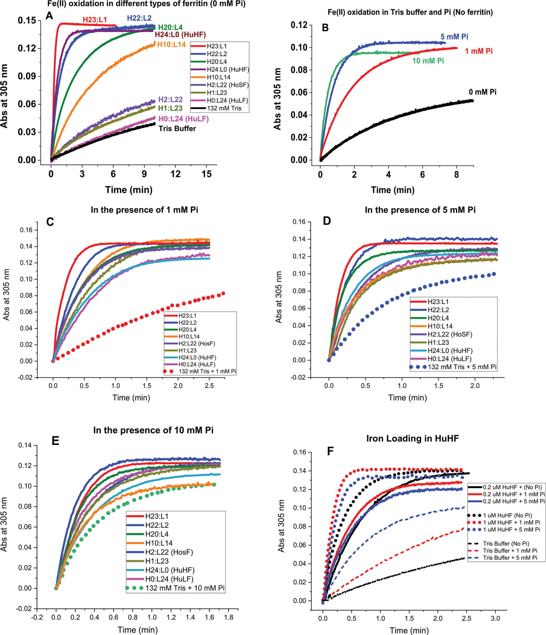
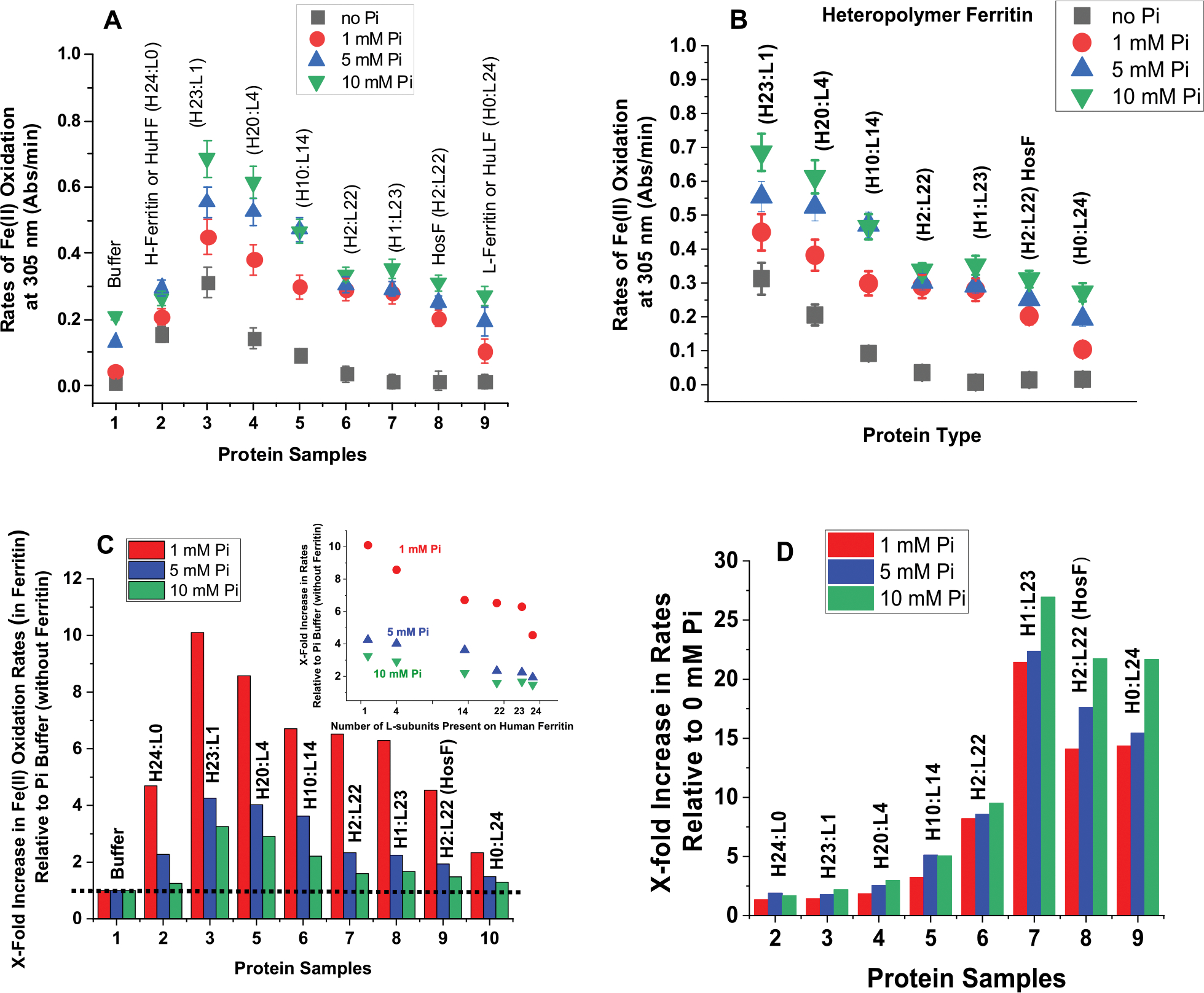
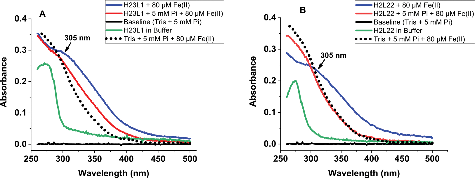
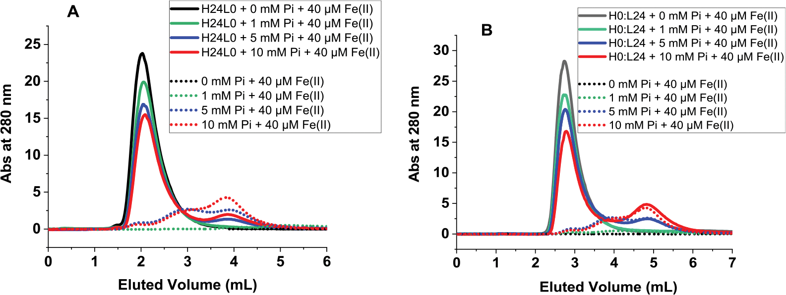
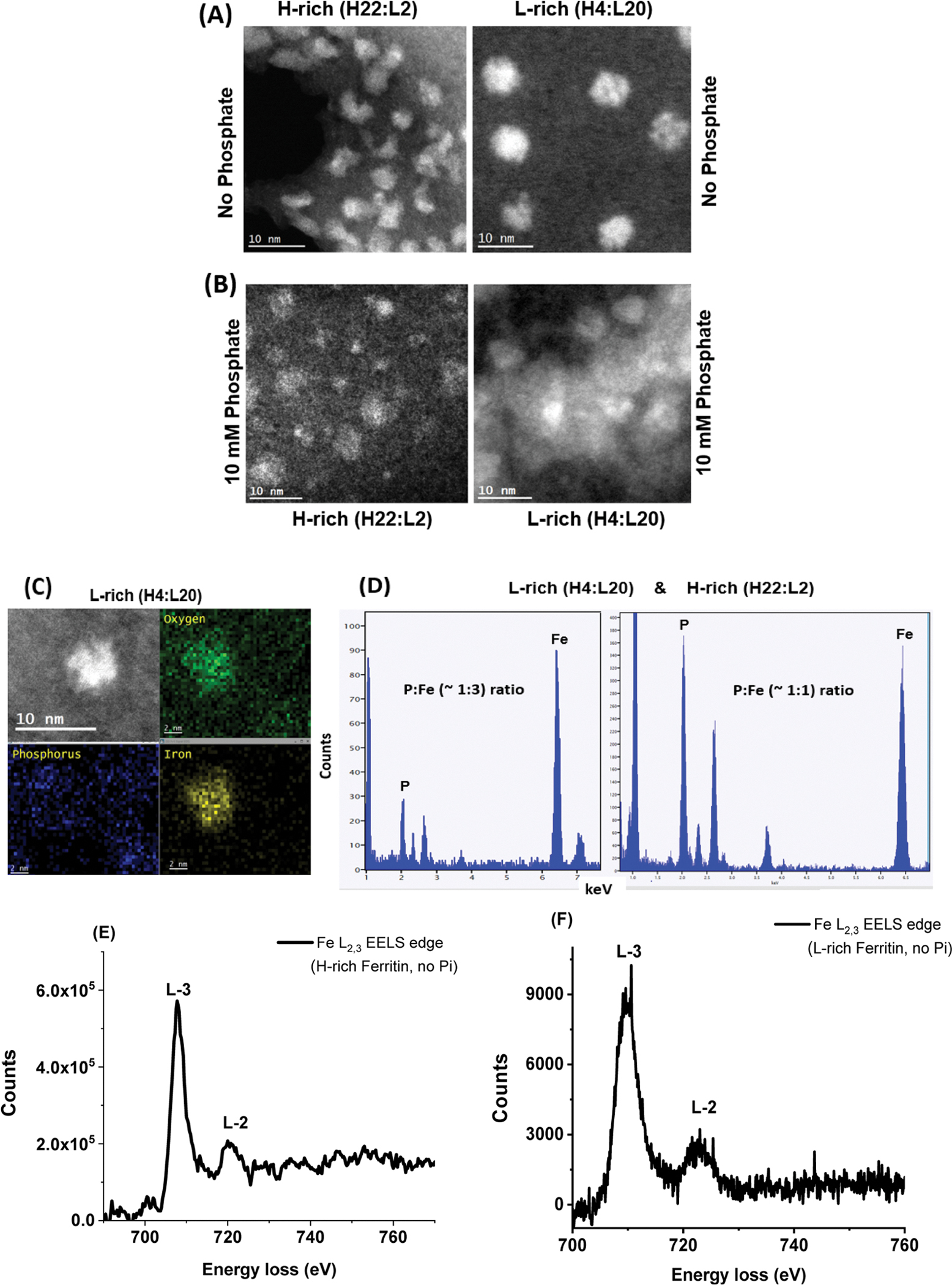
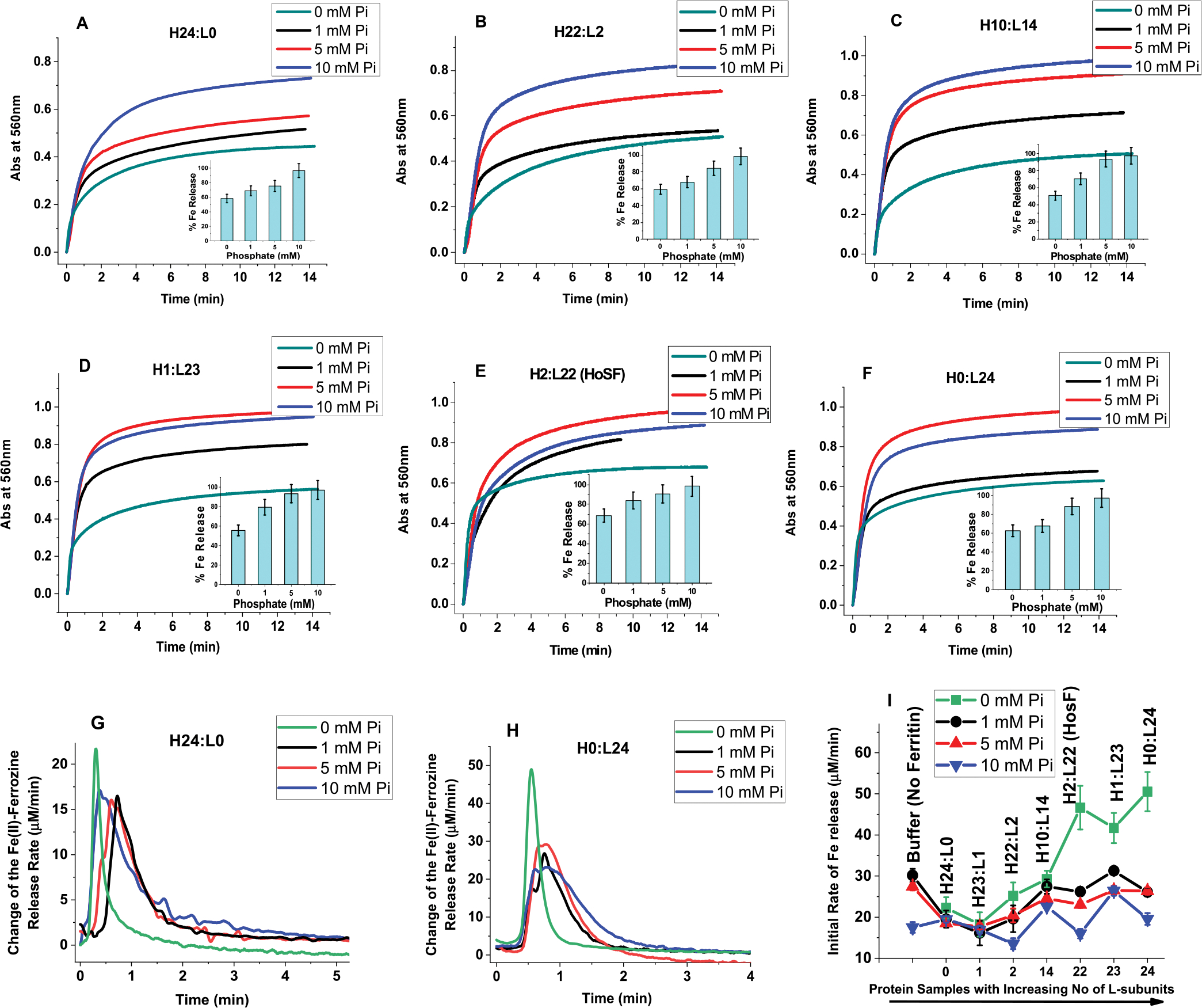
Ferritins are highly conserved supramolecular protein nanostructures that play a key role in iron homeostasis. Thousands of iron atoms can be stored inside their hollow cavity as a hydrated ferric oxyhydroxide mineral. Although phosphate associates with the ferritin iron nanoparticles, the effect of physiological concentrations on the kinetics, structure, and reactivity of ferritin iron cores has not yet been explored. Here, the iron loading and mobilization kinetics were studied in the presence of 1-10 mM phosphate using homopolymer and heteropolymer ferritins having different H to L subunit ratios. In the absence of ferritin, phosphate enhances the rate of ferrous ion oxidation and forms large and soluble polymeric Fe(III)-phosphate species. In the presence of phosphate, Fe(II) oxidation and core formation in ferritin is significantly accelerated with oxidation rates several-fold higher than with phosphate alone. High-angle annular dark-field scanning transmission electron microscopy measurements revealed a strong phosphate effect on both the size and morphology of the iron mineral in H-rich (but not L-rich) ferritins. While iron nanoparticles in L-rich ferritins have spherical shape in the absence and presence of phosphate, iron nanoparticles in H-rich ferritins change from irregular shapes in the absence of phosphate to spherical particles in the presence of phosphate with larger size distribution and smaller particle size. In the presence of phosphate, the kinetics of iron-reductive mobilization from ferritin releases twice as much iron than in its absence. Altogether, our results demonstrate an important role for phosphate, and the ferritin H and L subunit composition toward the kinetics of iron oxidation and removal from ferritin, as well as the structure and reactivity of the iron mineral, and may have an important implication on ferritin iron management in vivo.


Conflict of interest statement

The authors declare no competing financial interest.

Figures

Figure 1.
Figure 1.
Iron oxidation kinetics at 305 nm in the presence and absence of phosphate using light absorption spectroscopy. Conditions (A–E): 132 mM Tris buffer with 0, 1, 5, or 10 mM sodium phosphate; 0.2 μM ferritin; 40 μM FeSO4; pH 7.4; and 25.0 °C. Panel F displays the iron oxidation kinetics of recombinant human H-ferritin at 0.2 and 1 μM ferritin in 1 and 5 mM phosphate compared to Tris buffer. Panel F: effect of ferritin concentration on iron loading into human H-ferritin. We note that the number of H and L subunits in any heteropolymer ferritin is likely an average value representing a heterogeneous mixture of heteropolymers whose H and L subunit composition could vary between 5 and 8%.
Figure 2.
Figure 2.
(A,B) Plots of Fe(II) oxidation rates as a function of ferritin type. (C) Normalized rates of Fe(II) oxidation in different types of ferritin in 1 mM (red), 5 mM (blue), and 10 mM (green) phosphate buffer relative to those in the absence of ferritin. (D) Normalized Fe(II) oxidation rates in ferritin in 1 mM (red), 5 mM (blue), and 10 mM (green) phosphate buffer relative to those in the absence of phosphate. The inset of (C) shows a linear decrease in Fe(II) oxidation rates as a function of the number of L-subunits present in human ferritin. The dotted line in (C) illustrates the Fe(II) oxidation rates in phosphate and represents our baseline. The experimental conditions are the same as those of Figure 1.
Figure 3.
Figure 3.
Absorbance scans of (A) H-rich (H23:L1) and (B) L-rich (H2:L22) ferritins in the presence (or absence) of iron and 5 mM phosphate. The spectrum of the Fe(III)–phosphate complex (as control) is shown as a dotted line. Conditions: 0.2 μM ferritin, 132 mM Tris buffer pH 7.4, and 80 μM Fe(II). Spectra were collected after 2 h of adding iron.
Figure 4.
Figure 4.
Size exclusion chromatography of iron-loaded ferritin in the presence of phosphate (straight lines) and of Fe(III)–phosphate complexes (dotted lines) used as controls. Conditions: (A) 0.2 μM H-ferritin and (B) 0.2 μM L-ferritin, 40 μM Fe(II) (or 200 Fe/shell), in 50 mM Mops, 100 mM NaCl, pH 7.4, and 0, 1, 5, or 10 mM phosphate.
Figure 5.
Figure 5.
(A,B) STEM images of iron oxide nanoparticles formed within H-rich and L-rich ferritin in the absence (A) and presence (B) of 10 mM phosphate, at 10 nm magnification. (C,D) EDX spectra showing the estimated ratio of P/Fe in L-rich and H-rich ferritins. (E,F) STEM-EELS analysis shows the X-ray absorption spectra at the L3 and L2 edges of iron in H-rich and L-rich samples containing 1000 Fe atoms/core in the absence of phosphate.
Figure 6.
Figure 6.
Effect of phosphate on the reductive mobilization of iron from human ferritin. (A–F) Kinetics of iron release from different ferritins with the insets representing percent of iron released as a function of phosphate concentration. (G,H) Change of the Fe(II)-ferrozine release rate vs time shown only for H- and L-homopolymers (H24:L0 and H0:L24) to illustrate the major differences between H and H-rich ferritins and L or L-rich ferritins. (I) Initial rate of iron release (calculated for the first 30 s of reaction) as a function of increasing ferritin L-subunit content. Conditions: 0.067 μM ferritin, 132 mM Tris pH 7.4 at 25.0 °C, 5 mM FMN, 0.5 mM ferrozine, 5 mM NADH, 13.4 μM Fe(II) freshly added (or 200 Fe/shell), and 0–10 mM phosphate as indicated on each panel. The ferritins samples used in these experiments are those of Figure 1, after 3-fold dilution, and thus, the total amount of iron present in each sample is 26.8 μM (i.e., freshly added 13.4 μM Fe(II) + an average of 13.4 μM Fe(III) already present in the purified ferritin; see M&M for more details). The control experiment was performed under the exact same conditions but in the absence of ferritin using 26.8 μM Fe(II) to account for the total amount of Fe(II) present in ferritin. In all cases, the iron mobilization kinetics were performed with freshly prepared samples, and no differences were observed when the kinetics are performed (or repeated) using 1 or 2 week-old sample storage in the fridge at 4 °C.

Similar articles

Cited by

References

    1. Bou-Abdallah F The Iron Redox and Hydrolysis Chemistry of the Ferritins. Biochim. Biophys. Acta 2010, 1800, 719–731. - PubMed
    1. Ford GC; Harrison PM; Rice DW; Smith JM; Treffry A; White JL; Yariv J Ferritin: Design and Formation of an Iron-Storage Molecule. Philos. Trans. R. Soc. London, Ser. B 1984, 304, 551–565. - PubMed
    1. Harrison PM; Andrews SC; Artymiuk PJ; Ford GC; Guest JR; Hirzmann J; Lawson DM; Livingstone JC; Smith JMA; Treffry A; Yewdall SJ Probing Structure-Function Relations In Ferritin And Bacterioferritin. Adv. Inorg. Chem. 1991, 36, 449–486.
    1. Melman A; Bou-Abdallah F Iron Mineralization and Core Dissociation in Mammalian Homopolymeric H-Ferritin: Current Understanding and Future Perspectives. Biochim. Biophys. Acta 2020, 1864, 129700. - PubMed
    1. Mancias JD; Pontano Vaites L; Nissim S; Biancur DE; Kim AJ; Wang X; Liu Y; Goessling W; Kimmelman AC; Harper JW Ferritinophagy via NCOA4 Is Required for Erythropoiesis and Is Regulated by Iron Dependent HERC2-Mediated Proteolysis. ELife 2015, 4, No. e10308. - PMC - PubMed

Publication types