. 2022 Oct 10;7(19):e148309.
doi: 10.1172/jci.insight.148309.

Secondary bile acids mediate high-fat diet-induced upregulation of R-spondin 3 and intestinal epithelial proliferation

Secondary bile acids mediate high-fat diet-induced upregulation of R-spondin 3 and intestinal epithelial proliferation

Ji-Yao Li et al. JCI Insight. .

Retraction in

Abstract

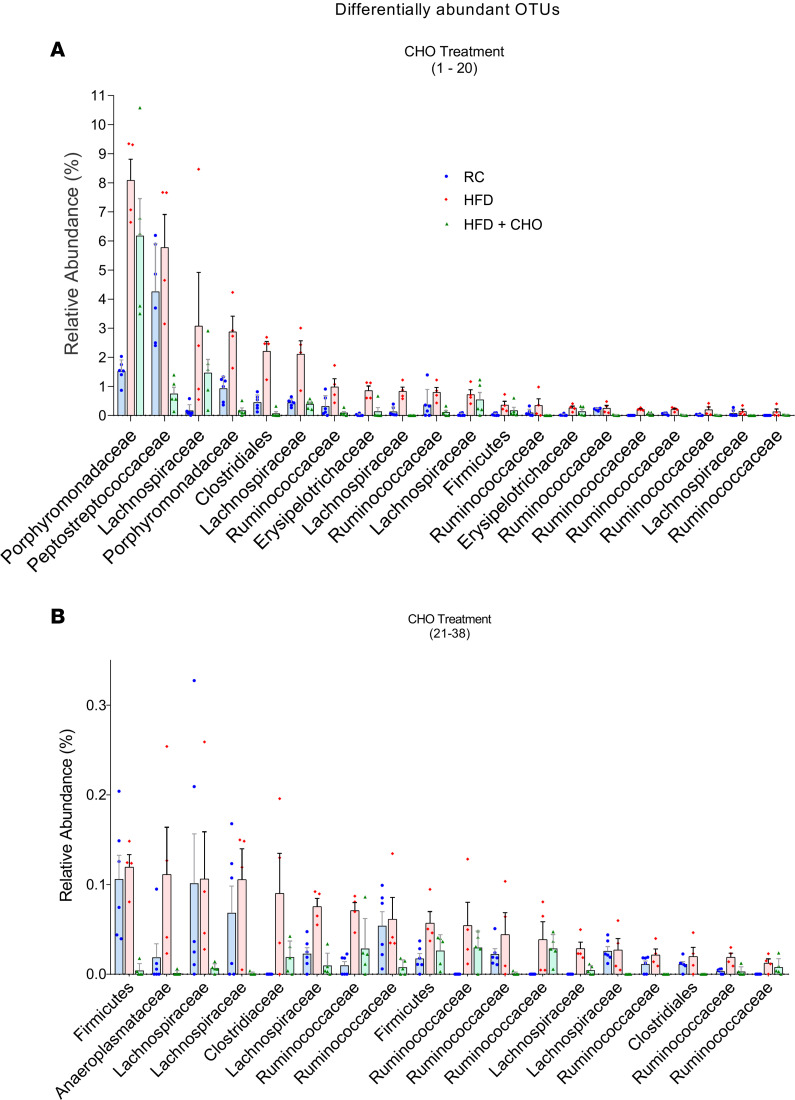
A high-fat diet (HFD) contributes to the increased incidence of colorectal cancer, but the mechanisms are unclear. We found that R-spondin 3 (Rspo3), a ligand for leucine-rich, repeat-containing GPCR 4 and 5 (LGR4 and LGR5), was the main subtype of R-spondins and was produced by myofibroblasts beneath the crypts in the intestine. HFD upregulated colonic Rspo3, LGR4, LGR5, and β-catenin gene expression in specific pathogen-free rodents, but not in germ-free mice, and the upregulations were prevented by the bile acid (BA) binder cholestyramine or antibiotic treatment, indicating mediation by both BA and gut microbiota. Cholestyramine or antibiotic treatments prevented HFD-induced enrichment of members of the Lachnospiraceae and Rumincoccaceae, which can transform primary BA into secondary BA. Oral administration of deoxycholic acid (DCA), or inoculation of a combination of the BA deconjugator Lactobacillus plantarum and 7α-dehydroxylase-containing Clostridium scindens with an HFD to germ-free mice increased serum DCA and colonic Rspo3 mRNA levels, indicating that formation of secondary BA by gut microbiota is responsible for HFD-induced upregulation of Rspo3. In primary myofibroblasts, DCA increased Rspo3 mRNA via TGR5. Finally, we showed that cholestyramine or conditional deletion of Rspo3 prevented HFD- or DCA-induced intestinal proliferation. We conclude that secondary BA is responsible for HFD-induced upregulation of Rspo3, which, in turn, mediates HFD-induced intestinal epithelial proliferation.

Keywords: Colorectal cancer; Gastroenterology; Mouse stem cells.


Conflict of interest statement

Conflict of interest: The authors have declared that no conflict of interest exists.

Figures

Figure 1
Figure 1. Rspo proteins were detected in intestine and intestinal myofibroblasts by RT-PCR and ISH.
RT-PCR of Rspo proteins 1 through 4 in the intestine of mouse (A), rat (B), and human colon and organoids (C). (D) Microphotograph of Rspo3 ISH in rat ileum. Clusters of silver grains representing Rspo3 mRNA are located beneath the crypts (left). Negative control was performed using Rspo3 sense probe (right). Double ISH shows that Rspo3 (silver grains) colocalized with α-SMA (purple), a marker for myofibroblasts, which was located beneath the crypts in (E) rat ileum and (F) colon. (G) Rspo3 colocalized with vimentin, a marker for fibroblasts, in rat colonic lamina propria. (H) Rspo3 colocalized with α-SMA underneath the crypts in mouse colon. Colocalization was defined as a cluster of silver grains overlapped with purple color. n = 3.
Figure 2
Figure 2. An HFD promotes upregulation of Rspo3, LGR4, LGR5, and β-catenin gene expression.
(A) An HFD increased gene expression of Rspo3, LGR4, LGR5, and β-catenin in mouse colon, as determined by qPCR. Gene expression was normalized to the expression level of GAPDH. n = 5 or 6. (B) In mouse colon, Rspo3 gene expression was also upregulated by diets high in protein and red meat but not by a high-carbohydrate diet. n = 6. (C) In rat colon, the expression of Rspo3, LGR4, LGR5, and β-catenin were upregulated by an HFD but reversed by concurrent feeding with the BA binder, cholestyramine (6%), with HFD. n = 5 to 10. (D) In germ-free mice, an HFD did not increase gene expression of Rspo3, LGR4, LGR5, or β-catenin in the colon. n = 5. (E) Conventionalization of germ-free mice with regular mouse fecal microbiota increased Rspo3 mRNA in the colon. n = 5. (F) Broad-spectrum antibiotics (ampicillin, 1 g/L; vancomycin, 500 mg/L; neomycin sulfate, 1 g/L; and metronidazole, 1 g/L in drinking water) reversed the effects of an HFD on Rspo3 in rat colon. n = 5. For statistical analysis, a 2-tailed unpaired Student’s t test (A, B, and D) or 1-way ANOVA with Bonferroni post hoc analysis (C, E, and F) was used. *P < 0.05, **P < 0.01. In all plots, data are shown as the mean ± SEM. Abx, antibiotics; Carb, carbohydrate; RC, regular chow.
Figure 3
Figure 3. The BA binder, cholestyramine (6% mixed in food), treatment prevented HFD-induced dysbiosis in rats.
Differentially abundant OTUs (A, 1 to 20; B, 21 to 38) that were significantly enriched by HFD and reversed by cholestyramine treatment. Linear discriminant analysis effect size was used to determine which OTUs were differentially abundant (P < 0.05). n = 4 to 6.
Figure 4
Figure 4. Antibiotics treatment prevented HFD-induced dysbiosis.
Differentially abundant OTUs (A, 1 to 20; B, 21 to 40) that were significantly enriched by HFD and reversed by Abx treatment. For the Abx treatment, rats were given ampicillin (1 g/L), vancomycin (500 mg/L), neomycin sulfate (1 g/L), and metronidazole (1 g/L) in drinking water while eating an HFD. Linear discriminant analysis effect size was used to determine which OTUs were differentially abundant (P < 0.05). n = 5. RC, regular chow.
Figure 5
Figure 5. Secondary BAs increased Rspo3 gene expression in myofibroblasts via TGR5.
(A) Primary myofibroblasts from rat colon stain positive for α-SMA and vimentin, and negative for desmin. (B) Myofibroblasts stain positive for α-SMA, vimentin, and Rspo3. (C) RT-PCR of Rspo3, BA membrane receptor TGR5, and nuclear receptor FXR-α and pregnane X (PRX) in primary myofibroblasts. (D) Secondary BA DCA, but not primary BA CA, increased Rspo3 gene expression in myofibroblasts. n = 6. (E) TGR5-specific agonist oleanolic acid (1 μM/L) increased Rspo3 gene expression, but the nuclear receptor–specific agonist GW4064 (1 μM/L) had no effect (left). Knockdown of TGR5 abolished the effect of DCA on Rspo3 expression (right). n = 5. (F) qPCR showed that TGR5 was decreased by 60% after knockdown. n = 5. One-way ANOVA with Bonferroni post hoc analysis (D and E) or 2-tailed unpaired Student’s t test (F) was used. *P < 0.05, **P < 0.01.
Figure 6
Figure 6. Cholestyramine treatment prevented HFD-induced BrdU incorporation into intestinal crypts.
BrdU IHC of mouse colon (A) and ileum (B) showing BrdU incorporation into crypts in the group of regular chow, HFD, or HFD plus cholestyramine. Data are summarized in bar graphs. One-way ANOVA with Bonferroni post hoc analysis was used to compare groups. *P < 0.05. n = 5.
Figure 7
Figure 7. Rspo3 mediates HFD-induced intestinal proliferation.
(A) Immunofluorescence of Ki67, a marker for proliferation, in mouse colon in the group fed regular chow (RC), HFD, or HFD plus cholestyramine. Data are summarized in bar graphs. n = 4. (B) Immunofluorescence of CK20, a marker of differentiation, in mouse colon in the same groups of mice. n = 4. (C) BrdU IHC in mouse colon in the groups of WT, RC; WT, HFD; and Rspo3 KO, HFD mice. Data are summarized in bar graphs. n = 3. (D) Immunofluorescence of Ki67 in the colon of the same groups of mice. *P < 0.05. n = 3. One-way ANOVA with Bonferroni post hoc analysis was used to compare groups.
Figure 8
Figure 8. Rspo3 mediates DCA-induced intestinal proliferation.
BrdU immunofluorescence of mouse colon (A) and ileum (B) in the group of WT, WT plus DCA (0.15% in drinking water), or Rspo3 KO plus DCA. Data are summarized in bar graphs. One-way ANOVA with Bonferroni post hoc analysis was used to compare groups. **P < 0.01. n = 6.

Similar articles

Cited by

References

    1. Okabayashi K, et al. Body mass index category as a risk factor for colorectal adenomas: a systematic review and meta-analysis. Am J Gastroenterol. 2012;107(8):1175–1185. doi: 10.1038/ajg.2012.180. - DOI - PubMed
    1. Ma Y, et al. Obesity and risk of colorectal cancer: a systematic review of prospective studies. PLoS One. 2013;8(1):e53916. doi: 10.1371/journal.pone.0053916. - DOI - PMC - PubMed
    1. Calle EE, Kaaks R. Overweight, obesity and cancer: epidemiological evidence and proposed mechanisms. Nat Rev Cancer. 2004;4(8):579–591. doi: 10.1038/nrc1408. - DOI - PubMed
    1. Arnold M, et al. Global patterns and trends in colorectal cancer incidence and mortality. Gut. 2017;66(4):683–691. doi: 10.1136/gutjnl-2015-310912. - DOI - PubMed
    1. Chan AT, Giovannucci EL. Primary prevention of colorectal cancer. Gastroenterology. 2010;138(6):2029–2043.e10. doi: 10.1053/j.gastro.2010.01.057. - DOI - PMC - PubMed

Publication types

LinkOut - more resources