. 2022 Sep-Oct;36(5):2149-2165.
doi: 10.21873/invivo.12941.

In Vivo Analysis of the Immune Response to Strontium- and Copper-doped Bioglass

Affiliations

In Vivo Analysis of the Immune Response to Strontium- and Copper-doped Bioglass

Mike Barbeck et al. In Vivo. 2022 Sep-Oct.

Abstract

Background: Bioglass is a highly adoptable bone substitute material which can be combined with so-called therapeutic ions. However, knowledge is poor regarding the influence of therapeutic ions on immune reactions and associated bone healing. Thus, the aim of this work was to investigate the influence of strontium- and copper-doped bioglass on the induction of M1 and M2 macrophages, as well as vascularization.

Materials and methods: Two types of alkali glass were produced based on ICIE16 bioglass via the melt-quench method with the addition of 5 wt% copper or strontium (ICIE16-Cu and ICIE16-Sr). Pure ICIE16 and 45S5 bioglass were used as control materials. The ion release and chemical composition of the bioglass were investigated, and an in vivo experiment was subcutaneously performed on Sprague-Dawley rats.

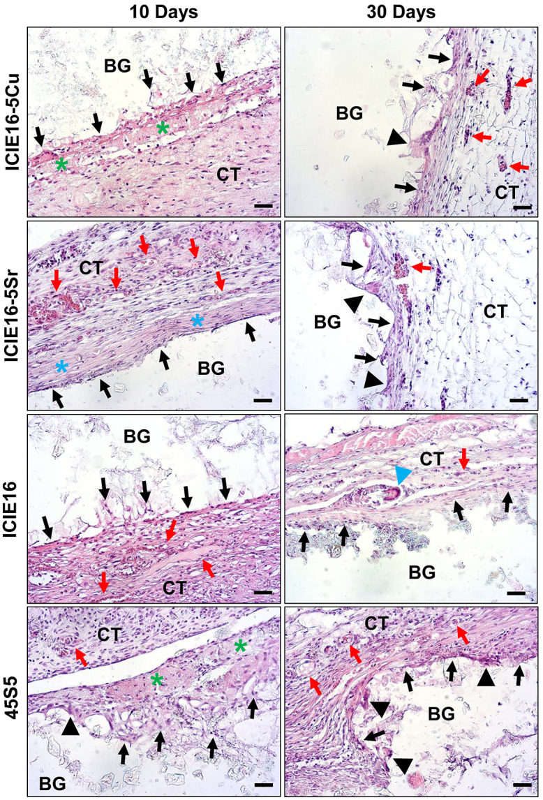
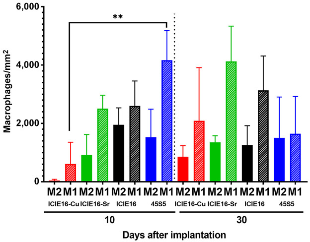
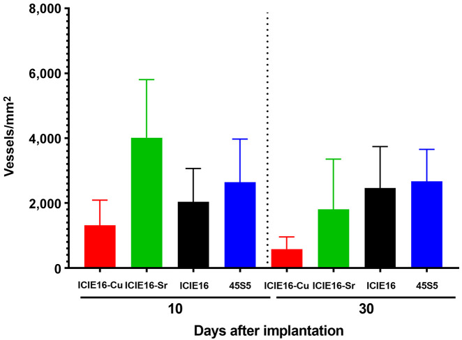
Results: Scanning electron microscopy revealed significant differences in the surface morphology of the bioglass materials. Energy dispersive X-ray spectroscopy confirmed the efficiency of the doping process by showing the ion-release kinetics. ICIE16-Cu exhibited a higher ion release than ICIE16-Sr. ICIE16-Cu induced low immune cell migration and triggered not only a low number of M1 and M2 macrophages but also of blood vessels. ICIE16-Sr induced higher numbers of M1 macrophages after 30 days. Both bioglass types induced numbers of M2 macrophages comparable with those found in the control groups.

Conclusion: Bioglass doping with copper and strontium did not significantly influence the foreign body response nor vascularization of the implantation bed in vivo. However, all the studied bioglass materials seemed to be biocompatible.

Keywords: 45S5; Bioglass; DIN EN ISO 10993-6; ICIE16; bone tissue regeneration; copper doping; hydroxyapatite deposition; ion release; macrophages; strontium doping; vascularization.


Conflict of interest statement

The Authors declare no conflicts of interest.

Figures

Figure 1
Figure 1. Exemplary scanning electron microscopy images of the surface morphology in 45S5 (A), ICIE16 (B), ICIE16-Sr (C) and ICIE16-Cu (D) bioglass (200× magnification; scale bars=10 µm).
Figure 2
Figure 2. Energy-dispersive X-ray spectroscopy spectra of the 45S5 (A), ICIE16 (B), ICIE16-Cu (C) and ICIE16-Sr (D) bioglass materials.
Figure 3
Figure 3. Release of Cu ions from ion-doped ICIE16-Cu and Sr ions from ICIE-16-Sr bioglass materials after immersion in simulated body fluid (SBF) for 3, 7, and 14 days. Significantly different at p<0.001: ***between metals; ###between bioglass samples of the same type.
Figure 4
Figure 4. Exemplary histological images showing the tissue reactions to four bioglass (BG) types within subcutaneous connective tissue (CT) at 10 days (left) and 30 days (right) after implantation. Black arrows: Mononuclear cells at the material–tissue interface; black arrowheads: multinucleated cells at the material surface; red arrows: blood vessels; green stars: fibrin network; blue arrowhead: multinucleated giant cell associated with a fragment of ICIE16 bioglass (hematoxylin and eosin staining, 20× magnification; scale bars=20 µm).
Figure 5
Figure 5. Exemplary histological images showing CD163-positive M2 macrophages (red staining) within the subcutaneous connective tissue (CT) at 10 days (left) and 30 days (right) after implantation of the four bioglass (BG) types. Stars: Cell walls at the material surface that were free of M2 macrophages (CD163 immunostaining, 20× magnification; scale bars=20 µm).
Figure 6
Figure 6. Exemplary histological images showing CD11c-positive M1 macrophages (red staining) within the subcutaneous connective tissue (CT) at 10 days (left) and 30 days (right) after implantation of the four bioglass (BG) types (CD11c immunostaining, 20× magnification; scale bars=20 µm).
Figure 7
Figure 7. Exemplary histological images showing blood vessels (red staining) within the subcutaneous connective tissue (CT) at 10 days (left) and 30 days (right) after implantation of the four bioglass (BG) types. (CD31 immunostaining; 200×, scale bars=20 µm).
Figure 8
Figure 8. Results of the histomorphometrical analysis of CD11c-positive M1 and CD163-positive M2 macrophages (cells/mm2). ***Significantly different at p<0.001 (n=4).
Figure 9
Figure 9. Results of the histomorphometrical analysis of vessel density (vessels/mm2) at 10 and 30 days after implantation of four bioglass types (n=4).

References

    1. Perić Kačarević Ž, Rider P, Alkildani S, Retnasingh S, Pejakić M, Schnettler R, Gosau M, Smeets R, Jung O, Barbeck M. An introduction to bone tissue engineering. Int J Artif Organs. 2020;43(2):69–86. doi: 10.1177/0391398819876286. - DOI - PubMed
    1. Ribas R, Schatkoski V, Montanheiro T, De menezes B, Stegemann C, Leite D, Thim G. Current advances in bone tissue engineering concerning ceramic and bioglass scaffolds: A review. Ceramics International. 2019;45(17):21051–21061. doi: 10.1016/j.ceramint.2019.07.096. - DOI
    1. Hench LL. The story of Bioglass. J Mater Sci Mater Med. 2006;17(11):967–978. doi: 10.1007/s10856-006-0432-z. - DOI - PubMed
    1. Chacko NL, Abraham S, Rao HN, Sridhar N, Moon N, Barde DH. A Clinical and radiographic evaluation of periodontal regenerative potential of PerioGlas®: a synthetic, resorbable material in treating periodontal infrabony defects. J Int Oral Health. 2014;6(3):20–26. - PMC - PubMed
    1. Blayney AW, Bebear JP, Williams KR, Portmann M. Ceravital in ossiculoplasty: experimental studies and early clinical results. J Laryngol Otol. 1986;100(12):1359–1366. doi: 10.1017/s0022215100101148. - DOI - PubMed