. 2022 Sep 20;119(38):e2202727119.
doi: 10.1073/pnas.2202727119. Epub 2022 Sep 13.

Homologous recombination-deficient mutation cluster in tumor suppressor RAD51C identified by comprehensive analysis of cancer variants

Affiliations

Homologous recombination-deficient mutation cluster in tumor suppressor RAD51C identified by comprehensive analysis of cancer variants

Rohit Prakash et al. Proc Natl Acad Sci U S A. .

Abstract

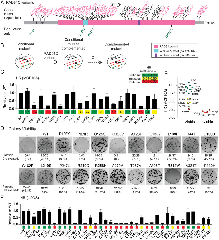
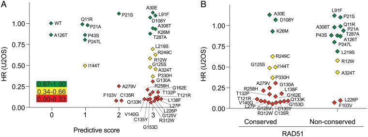
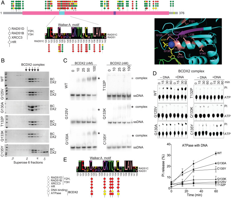
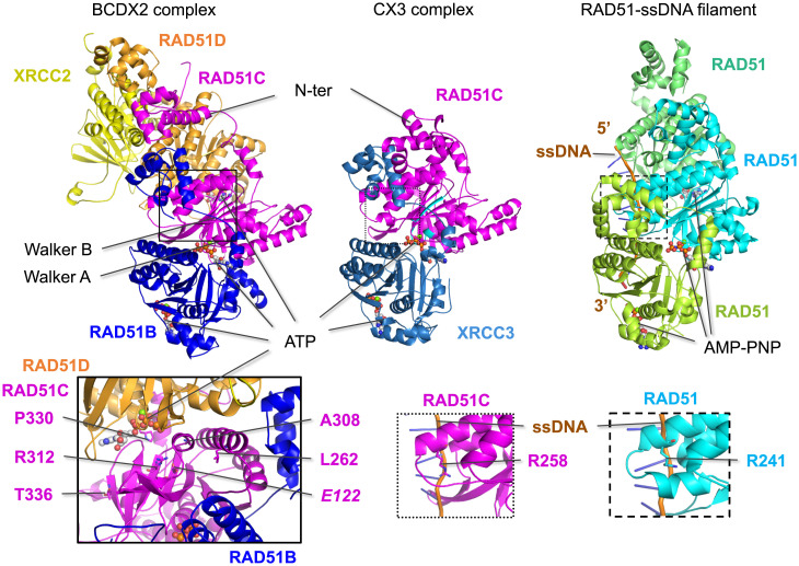
Mutations in homologous recombination (HR) genes, including BRCA1, BRCA2, and the RAD51 paralog RAD51C, predispose to tumorigenesis and sensitize cancers to DNA-damaging agents and poly(ADP ribose) polymerase inhibitors. However, ∼800 missense variants of unknown significance have been identified for RAD51C alone, impairing cancer risk assessment and therapeutic strategies. Here, we interrogated >50 RAD51C missense variants, finding that mutations in residues conserved with RAD51 strongly predicted HR deficiency and disrupted interactions with other RAD51 paralogs. A cluster of mutations was identified in and around the Walker A box that led to impairments in HR, interactions with three other RAD51 paralogs, binding to single-stranded DNA, and ATP hydrolysis. We generated structural models of the two RAD51 paralog complexes containing RAD51C, RAD51B-RAD51C-RAD51D-XRCC2 and RAD51C-XRCC3. Together with our functional and biochemical analyses, the structural models predict ATP binding at the interface of RAD51C interactions with other RAD51 paralogs, similar to interactions between monomers in RAD51 filaments, and explain the failure of RAD51C variants in binding multiple paralogs. Ovarian cancer patients with variants in this cluster showed exceptionally long survival, which may be relevant to the reversion potential of the variants. This comprehensive analysis provides a framework for RAD51C variant classification. Importantly, it also provides insight into the functioning of the RAD51 paralog complexes.

Keywords: DNA repair; Homologous recombination; RAD51 paralog; RAD51C; Variants of unknown significance.


Conflict of interest statement

The authors declare no competing interest.

Figures

Fig. 1.
Fig. 1.
HR-deficient RAD51C variants identified using MCF10A and U2OS cell mutants. (A) RAD51C variants analyzed in this study. Cancer variants are indicated along the top of the schematic. Breast or ovarian cancer (BC/OC) variants are in pink; variants found in other cancer types are in black (see Table 1). Cancer variants that have also been identified as population variants are indicated with an asterisk; four population variants not identified in cancers are indicated under the schematic in green (see Table 2). R258H, annotated with a #, was initially identified as a biallelic mutation in Fanconi-anemia-like syndrome patients. Identified motifs in RAD51C include the Walker A and B motifs that are important for nucleotide binding and hydrolysis. aa, amino acid; N-ter, N terminus. (B) Schematic for testing RAD51C variants in MCF10A cells. Both endogenous alleles of RAD51C are disrupted (asterisks), but cells are viable due to expression from a conditional, ectopically integrated copy of RAD51C flanked by LoxP sites (orange triangles). RAD51C variants are introduced by viral transduction, and their ability to complement is determined after Cre-mediated excision of the conditional RAD51C copy. (C) HR levels in MCF10A cells expressing the indicated variants. A direct repeat HR reporter (DR-GFP) is present in the genome of the MCF10A conditional cell line. Upon introduction of a DSB in the reporter, repair by HR produces a functional GFP gene that is quantified by flow cytometry. HR levels for the variants are expressed relative to the HR level achieved with WT RAD51C. (D) MCF10A colony formation after Cre expression in cells containing the indicated RAD51C variants. The number of colonies that had excised the conditional copy of RAD51C compared to the total that were analyzed is indicated together with the percent excision. After 10 d of growth, three variants (T121R, G125V, and R312W) were unable to give rise to viable colonies, and for a fourth, C135Y, a single clone that had excised the conditional RAD51C copy, did not survive past two passages. (E) Comparison of HR and viability in MCF10A cells. RAD51C variants called as HR proficient or reduced support viability of MCF10A cells, while those called as HR deficient do not, except for R258H, which is on the higher end of HRD. (F) HR levels in RAD51C knockout U2OS cells expressing the indicated RAD51C variants. A distinct HR reporter (SCR-GFP), in which HR is also induced by a DSB, is present in the genome of the U2OS cells. HR was quantified relative to WT RAD51C and categorized as proficient (green), reduced (yellow), and deficient (red), as in C.
Fig. 2.
Fig. 2.
HR-deficient RAD51C variants typically have impaired interactions with other RAD51 paralogs. (A) RAD51 paralog complexes. The BCDX2 complex contains RAD51B, RAD51C, RAD51D, and XRCC2 and the CX3 complex contains RAD51C and XRCC3. RAD51C is the sole member present in subcomplexes. (B) Representative Y2H and Y3H interactions of RAD51C variants with other RAD51 paralogs, as monitored by growth. A Y2H approach was used to test RAD51C interactions with either RAD51B or XRCC3, and a Y3H approach was used for RAD51D because the RAD51C interaction is more robust in the presence of its binding partner RAD51B. RAD51C variants were tested as here as activating domain (AD) fusions as well as reciprocal DNA binding domain (BD) fusions (SI Appendix, Fig. S3A). (C) Heat map indicates averaged protein-protein interactions as determined in the Y2H/Y3H assays. The RAD51C variant interaction with the indicated RAD51 paralog is relative to the WT. Growth was quantified using ImageJ from two or more independent experiments. The last column shows HR quantification from U2OS cells for comparison. Variants R212C and R214C were tested later than the other variants and found to be HR proficient. (D–F) HR levels for the RAD51C variants correlate well with Y2H/Y3H interactions for each of RAD51D (D), RAD51B (E), and XRCC3 (F), with the RAD51D interaction having the highest correlation the best predictor (RAD51D and RAD51B, P < 0.0001; XRCC3, P < 0.001). Linear regression lines are shown in black with 90% confidence intervals shaded in green.
Fig. 3.
Fig. 3.
RAD51C residue conservation with RAD51 is predictive for variant HR function. (A) Comparison of HR activity of the RAD51C variants in U2OS cells with the combined scores from the PolyPhen-2, SIFT, and PROVEAN prediction tools. A predictive score of 0 (benign) or 1 (damaging) is given for each tool, according to the cut off score for each, and then the three scores are added. Thus, a combined score of 0 indicates that all three tools predict that the variant is benign, while a combined score of 3 indicates that all three tools predict that the variant is deleterious. The variants are colored according to whether HR is proficient (green), reduced (yellow), or deficient (red). (B) Comparison of HR activity of the RAD51C variants in U2OS cells with residue conservation with RAD51. Conserved, RAD51C amino acid is identical or strongly similar to the cognate residue in RAD51; nonconserved, RAD51C amino acid is weakly similar or not similar at all to the cognate residue in RAD51.
Fig. 4.
Fig. 4.
RAD51C variants in Walker A region exhibit DNA binding and ATP hydrolysis defects in the context of the BCDX2 complex. (A) A lollipop diagram of RAD51C cancer and population variants summarizes defects observed in Y2H/Y3H and HR analysis (data from Fig. 1 and 2). The three circles represent results from XRCC3 Y2H, RAD51B Y2H, and RAD51D Y3H analyses, and the diamond represents HR activity in U2OS cells. A mutation cluster is observed around the RAD51C Walker A motif, which is expanded below with an alignment to RAD51 for RAD51C amino acids 120 to 142. RAD51C variants at conserved residues show defects in all assays, whereas those in nonconserved residues do not. The Inset shows a detail of a RAD51 structure around the ATPase catalytic site (Protein Data Bank [PDB] 5NWL) with conserved residues highlighted, with numbering from RAD51C. Residues at or around the Walker A motif are colored in salmon and those from the Walker B motif are in purple. ATP and its bound magnesium ion are also shown. (B) WT RAD51B-RAD51C-RAD51D-XRCC2 (BCDX2) complexes or complexes containing the indicated RAD51C mutants were purified in the presence of ATP and MgCl2 and loaded onto a Superpose 6 size exclusion column. Fractions were run on a sodium dodecyl-sulfate polyacrylamide gel electrophoresis gel and visualized by Coomassie staining, and those containing the BCDX2 complexes used for subsequent experiments are indicated (arrows). BC (RAD51B-RAD51C) and DX2 (RAD51D-XRCC2) were run as doublets, with RAD51C and RAD51D as the upper bands in the respective doublets. Complexes with RAD51C C135Y appear less stable with distinct DX2 subcomplexes apparent in the later fractions. Protein markers are as follows: Tg, thyroglobulin; Fe, ferritin; Al, aldolase; Ov, ovalbumin. (C) ssDNA binding. ssDNA binding by the BCDX2 complexes was assessed by EMSAs using an 80mer substrate in the presence of ATP and MgCl2. The ssDNA shifted complex with C135Y runs slightly faster on the gels. (D) ATP hydrolysis. ATP hydrolysis was assessed by incubating BCDX2 complexes with ATP over time in the absence or presence of ssDNA and performing thin-layer chromatography with polyethyleneimine cellulose sheets. Pi indicates released inorganic phosphate after hydrolysis. The graph shows results from three independent experiments in the presence of ssDNA. (E) Summary of defects observed with RAD51C variants in the Walker A mutation cluster that were analyzed biochemically.
Fig. 5.
Fig. 5.
Predicted structures of the BCDX2 and CX3 complexes. Structural predictions of the BCDX2 and CX3 complexes were generated by AlphaFold2. RAD51C and the other RAD51 paralogs are predicted to share structural similarity to RAD51 in the ATPase domain, which contains the Walker A and Walker B motifs (highlighted in cyan for RAD51C) and to bind ATP at the subunit interfaces, similar to RAD51 binding of AMP-PNP in the nucleoprotein filament (PDB 5h1b). RAD51B and XRCC3 are predicted to bind on the same surface of RAD51C, while RAD51D is predicted to bind to the opposite side from RAD51B in the BCDX2 complex. Mutations that interfere with ATP binding are expected to impact subunit interactions and, therefore, complex formation. This is borne out in the biochemical instability of CmutX3 complexes, while BCmutDX2 complexes are more stable, possibly because of the greater number of interactions. Left Inset: Five RAD51C variants that specifically interfere with the RAD51D interaction in Y3H assays are in residues that are predicted to be close to RAD51D, with P330 positioned the closest. Among these is R312, which hydrogen bonds with E122 (italics); notably, both of these residues are conserved among all of the RAD51 paralogs. Middle and Right Insets: A key residue that contacts ssDNA in the RAD51 presynaptic filament is R241, which corresponds to R258 in RAD51C and can be modeled to bind ssDNA.

References

    1. Chen C. C., Feng W., Lim P. X., Kass E. M., Jasin M., Homology-directed repair and the role of BRCA1, BRCA2, and related proteins in genome integrity and cancer. Annu. Rev. Cancer Biol. 2, 313–336 (2018). - PMC - PubMed
    1. Pennington K. P., et al. , Germline and somatic mutations in homologous recombination genes predict platinum response and survival in ovarian, fallopian tube, and peritoneal carcinomas. Clin. Cancer Res. 20, 764–775 (2014). - PMC - PubMed
    1. Tung N., et al. , Frequency of germline mutations in 25 cancer susceptibility genes in a sequential series of patients with breast cancer. J. Clin. Oncol. 34, 1460–1468 (2016). - PMC - PubMed
    1. Pritchard C. C., et al. , Inherited DNA-repair gene mutations in men with metastatic prostate cancer. N. Engl. J. Med. 375, 443–453 (2016). - PMC - PubMed
    1. O’Connor M. J., Targeting the DNA damage response in cancer. Mol. Cell 60, 547–560 (2015). - PubMed