. 2022 Oct:84:104246.
doi: 10.1016/j.ebiom.2022.104246. Epub 2022 Sep 10.

Genetic landscape of a large cohort of Primary Ovarian Insufficiency: New genes and pathways and implications for personalized medicine

Affiliations

Genetic landscape of a large cohort of Primary Ovarian Insufficiency: New genes and pathways and implications for personalized medicine

Abdelkader Heddar et al. EBioMedicine. 2022 Oct.

Abstract

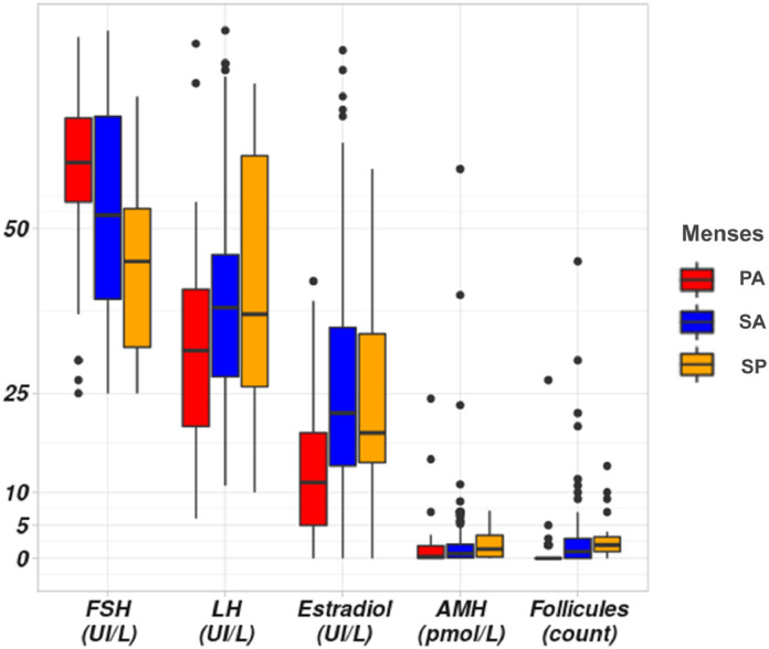
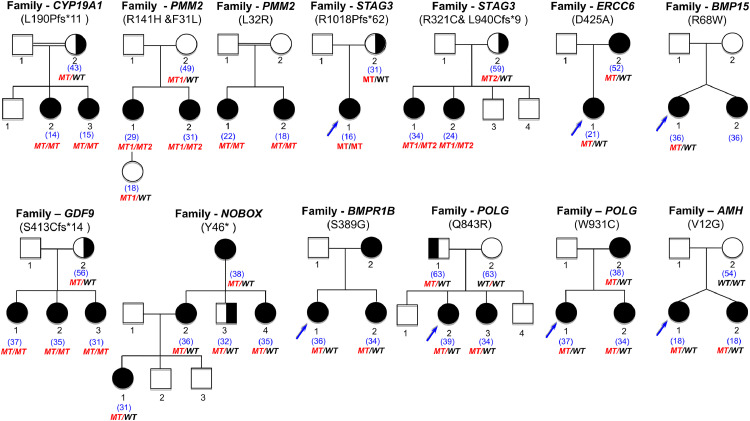
Background: Primary Ovarian Insufficiency (POI), a public health problem, affects 1-3.7% of women under 40 yielding infertility and a shorter lifespan. Most causes are unknown. Recently, genetic causes were identified, mostly in single families. We studied an unprecedented large cohort of POI to unravel its molecular pathophysiology.

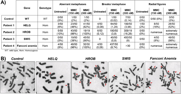
Methods: 375 patients with 70 families were studied using targeted (88 genes) or whole exome sequencing with pathogenic/likely-pathogenic variant selection. Mitomycin-induced chromosome breakages were studied in patients' lymphocytes if necessary.

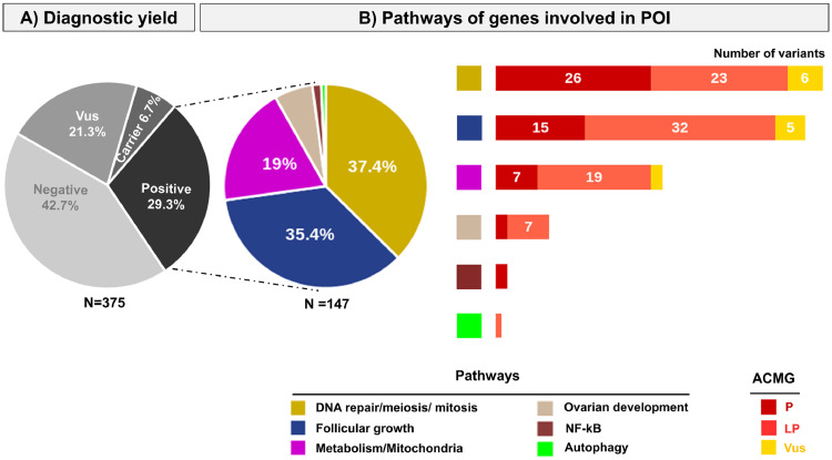
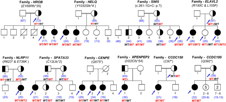
Findings: A high-yield of 29.3% supports a clinical genetic diagnosis of POI. In addition, we found strong evidence of pathogenicity for nine genes not previously related to a Mendelian phenotype or POI: ELAVL2, NLRP11, CENPE, SPATA33, CCDC150, CCDC185, including DNA repair genes: C17orf53(HROB), HELQ, SWI5 yielding high chromosomal fragility. We confirmed the causal role of BRCA2, FANCM, BNC1, ERCC6, MSH4, BMPR1A, BMPR1B, BMPR2, ESR2, CAV1, SPIDR, RCBTB1 and ATG7 previously reported in isolated patients/families. In 8.5% of cases, POI is the only symptom of a multi-organ genetic disease. New pathways were identified: NF-kB, post-translational regulation, and mitophagy (mitochondrial autophagy), providing future therapeutic targets. Three new genes have been shown to affect the age of natural menopause supporting a genetic link.

Interpretation: We have developed high-performance genetic diagnostic of POI, dissecting the molecular pathogenesis of POI and enabling personalized medicine to i) prevent/cure comorbidities for tumour/cancer susceptibility genes that could affect life-expectancy (37.4% of cases), or for genetically-revealed syndromic POI (8.5% of cases), ii) predict residual ovarian reserve (60.5% of cases). Genetic diagnosis could help to identify patients who may benefit from the promising in vitro activation-IVA technique in the near future, greatly improving its success in treating infertility.

Funding: Université Paris Saclay, Agence Nationale de Biomédecine.

Keywords: Meiosis/DNA repair genes; Mitophagy; NF-KB; Personalized medicine; Post - translational regulation; Primary ovarian insufficiency.


Conflict of interest statement

Declaration of interests The authors declare no conflict of interest.

Figures

Figure 1
Figure 1
Hormonal and ultrasonographical datas in our cohort of patients with POI. Hormonal assays (FSH, LH, Estradiol and AMH) in patients according to menses. PA: Primary Amenorrhea, SA: Secondary Amenorrhea, SP: Spaniomenorrhea. The Y scale unit is variable according to each hormonal assay represented in the X-axis. Normal range: AMH (7-20.7 pmol/l); Respectively follicular, ovulatory, luteal phases and menopause: FSH (IU/L): (2.9-12), (6.3-24), (1.5-7), (17-95); LH: (IU/L) (1.5-8), (9.6-80), (0.2-6.5), (8-33); E2 (ng/l): (19.5-144.2),(63.9-356.7),(55.8-214.2),(≤ 32.2).
Figure 2
Figure 2
Genetic studies of the cohort of patients with POI. Patients were studied by a custom-made targeted NGS comprising 88 genes or by whole exome sequencing (see methods). A) Diagnostic yield using ACMG criteria: Variants are classified according to the American College of Medical Genetics (ACMG) guidelines. N= 375: the whole cohort comprises 375 patients with POI. Vus: Variant of unknown significance. Carrier: patients harbouring a heterozygous pathogenic variant in a known autosomal recessive POI gene. Positive: the diagnostic yield corresponds to patients carrying pathogenic (P) or likely pathogenic (LP) variants and is 29.3%. B) Pathways of genes involved in POI. The different pathways are indicated with different colors. The pie chart represents the proportion of patients with P or LP variants in a specific pathway: DNA repair meiosis and mitosis (37.4%), Follicular growth (35.4%), Mitochondria and Metabolism (19%), Ovarian development (6.1%), NF-kB (1.4%), Autophagy (0.7%). The histograms show the number and type of variants detected in each pathway.
Figure 3
Figure 3
Pedigrees of the families with genes not previously involved in Mendelian phenotype or POI. On top of each family, the name of the gene is indicated, with the variant found. Double lines indicate consanguineous unions. The age of the patient at diagnosis is indicated in blue between brackets. Blue arrows indicate the patients and relatives studied with NGS (see text and Methods chapter). All family members available were studied by Sanger Sequencing. The segregation of the variants is shown under each individual sequenced. MT: mutated. MT1: first mutation. MT2: second mutation. WT: wild type. Family CCDC185: the number inside each symbol indicates the number of siblings in the family, either sisters (5) or brothers (2).
Figure 4
Figure 4
Chromosomal instability in patients with molecular defects of DNA repair genes. A). Chromosomal breaks analysis of the patient's lymphocytes, a control (a WT fertile woman), and a patient with Fanconi anemia, in the presence of increasing concentration of mitomycin (MMC). B) Selected figures of metaphases from the patients with molecular defects of SWI5, HELQ, HROB compared to Fanconi anemia and control cells in the presence of 300 nM of MMC. Chromosomal breaks are shown with red arrows and radial figures with asterisks. In the absence of MMC, while no spontaneous breaks are observed in cells of the patient with the SWI5 homozygous splice variant, respectively 6% and 10 % of cells of the patients with homozygous truncated variants of HELQ and HROB presented increased breaks, similarly to cells of the patient with Fanconi anemia (8%). In the presence of 150nM MMC, 86% of cells with the HROB pathogenic variant presented breaks with 3.8 breaks per metaphase very similarly to cells of the patient with Fanconi anemia (96%) and radial figures were observed in numerous cells of both types. Increased breaks were also observed in 46% of cells with the HELQ pathogenic variant (1.28 breaks per metaphase) and 38% of cells with the homozygous SWI5 variant (0.8 breaks per metaphase). Radial figures were observed in 16 % and 8 % of cells respectively. At 300 nM MMC, HROB-cells mimic Fanconi anemia cells, all presenting breaks (16.26 breaks per metaphase) with numerous radial figures, while 90% of SWI5-cells presented breaks with a mean of 9 breaks per metaphase and 82% of HELQ-cells with a mean of 3.98 breaks per metaphase. HROB-cells mimic Fanconi anemia cells with spontaneous chromosomal instability and marked hypersensitivity to MMC while HELQ-cells and SWI5-cells have a milder phenotype, but clearly with increased breaks when compared to control cells, and with radial figures, a pathognomonic feature of Fanconi anemia.
Figure 5
Figure 5
Pedigrees of the family confirming the causal role of genes in POI. Double lines indicate consanguineous union. Blue arrows indicate the patients and relatives studied with NGS, The other family members available are studied by Sanger sequencing. The segregation of the variant is shown under each sequenced individual. MT: Mutated, MT1: first mutation, MT2, second mutation, WT: wild type. The age of patients at diagnosis is indicated in blue between brackets.

References

    1. Huhtaniemi I, Hovatta O, La Marca A, et al. Advances in the molecular pathophysiology, genetics, and treatment of primary ovarian insufficiency. Trends Endocrinol Metab. 2018;29:400–419. doi: 10.1016/j.tem.2018.03.010. - DOI - PubMed
    1. Golezar S, Ramezani Tehrani F, Khazaei S, Ebadi A, Keshavarz Z. The global prevalence of primary ovarian insufficiency and early menopause: a meta-analysis. Climacteric. 2019;22:403–411. doi: 10.1080/13697137.2019.1574738. - DOI - PubMed
    1. Bachelot A, Nicolas C, Bidet M, et al. Long-term outcome of ovarian function in women with intermittent premature ovarian insufficiency. Clin Endocrinol. 2017;86:223–228. doi: 10.1111/cen.13105. - DOI - PubMed
    1. Stentz NC, Koelper N, Barnhart KT, Sammel MD, Senapati S. Infertility and mortality. Am J Obstet Gynecol. 2020;222:251.e1–251.e10. doi: 10.1016/j.ajog.2019.09.007. - DOI - PubMed
    1. Wang Y-X, Farland LV, Wang S, et al. Association of infertility with premature mortality among US women: Prospective cohort study. Lancet Reg Health Am. 2021;7:100122. doi: 10.1016/j.lana.2021.100122. - DOI - PMC - PubMed