. 2022 Sep 12;40(9):1060-1069.e7.
doi: 10.1016/j.ccell.2022.07.005.

A covalent inhibitor of K-Ras(G12C) induces MHC class I presentation of haptenated peptide neoepitopes targetable by immunotherapy

Affiliations

A covalent inhibitor of K-Ras(G12C) induces MHC class I presentation of haptenated peptide neoepitopes targetable by immunotherapy

Ziyang Zhang et al. Cancer Cell. .

Abstract

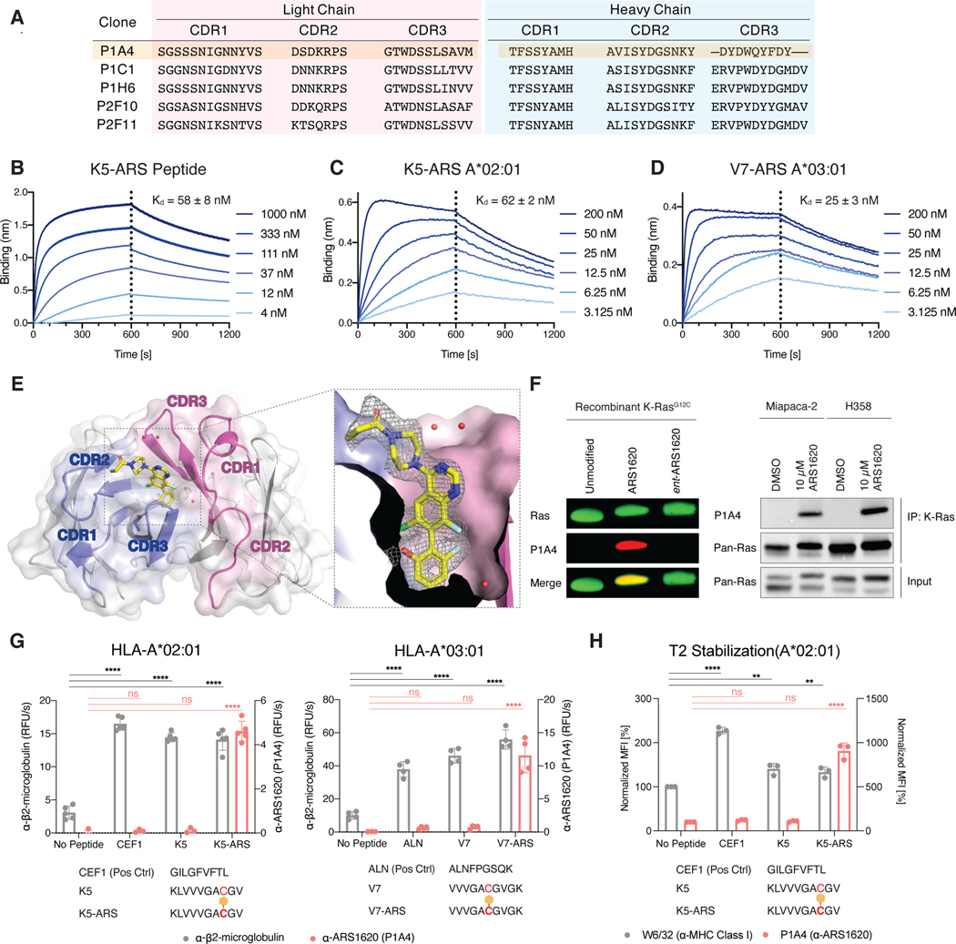
Immunotargeting of tumor-specific antigens is a powerful therapeutic strategy. Immunotherapies directed at MHC-I complexes have expanded the scope of antigens and enabled the direct targeting of intracellular oncoproteins at the cell surface. We asked whether covalent drugs that alkylate mutated residues on oncoproteins could act as haptens to generate unique MHC-I-restricted neoantigens. Here, we report that KRAS G12C mutant cells treated with the covalent inhibitor ARS1620 present ARS1620-modified peptides in MHC-I complexes. Using ARS1620-specific antibodies identified by phage display, we show that these haptenated MHC-I complexes can serve as tumor-specific neoantigens and that a bispecific T cell engager construct based on a hapten-specific antibody elicits a cytotoxic T cell response against KRAS G12C cells, including those resistant to direct KRAS G12C inhibition. With multiple K-RAS G12C inhibitors in clinical use or undergoing clinical trials, our results present a strategy to enhance their efficacy and overcome the rapidly arising tumor resistance.

Keywords: ARS1620; KRas; MHC-I; antibody; cancer; covalent inhibitors; drug resistance; immunotherapy.


Conflict of interest statement

Declaration of interests C.S.C., K.S.M., Z.Z., and P.J.R. are inventors on a provisional patent application covering this work and owned by the University of California, San Francisco (UCSF). K.M.S. is an inventor on patent applications related to this technology owned by UCSF. K.M.S. is an inventor on patents covering covalent inhibitors of K-Ras(G12C) owned by UCSF and licensed to Wellspring Biosciences. K.M.S. is a consultant to and shareholder in the following companies: Revolution Medicines, Black Diamond Therapeutics, BridGene Biosciences, Denali Therapeutics, Dice Molecules, eFFECTOR Therapeutics, Erasca, Genentech/Roche, Janssen Pharmaceuticals, Kumquat Biosciences, Kura Oncology, Mitokinin, Nested, Type6 Therapeutics, Venthera, Wellspring Biosciences (Araxes Pharma), Nextech, Radd, Totus, Vicinitas, Turning Point, Ikena, Initial Therapeutics, Vevo, and BioTheryX.

Figures

Figure 1.
Figure 1.
K-Ras(G12C)-derived peptides covalently modified by the investigational inhibitor ARS1620 form functional complexes with MHC Class I heavy chain and β2-microglobulin. A. Conjugate addition from the acquired cysteine (Cys12) on K-Ras(G12C) to the acrylamide group in ARS1620 yields a covalent ARS1620-K-Ras(G12C) adduct. B. ARS1620-modified peptides form functional complexes with MHC Class I heavy chain and β2-microglobulin. Recombinant MHC-I complexes were prepared by refolding of the indicated heavy chain in the presence of β2-microglobulin and the indicated peptide. For sandwich ELISA detection, the complexes were captured by the conformation-specific MHC Class I heavy chain antibody W6/32 and detected by an β2-microglobulin-specific antibody (BBM.1) (One-way ANOVA with Dunnett’s correction for multiple comparisons, ns, not significant, ****, p<0.0001). Individual data points are shown with mean ±SD indicated. C. ARS1620-modified peptides stabilize MHC Class I on the surface of the TAP-deficient cell line T2 (One-way ANOVA with Dunnett’s correction for multiple comparisons, ns, not significant, ****, p<0.0001). Individual data points are shown with mean ±SD indicated. D. Thermal stability of A*02:01 MHC-I complexes loaded with various K5, K-Ras-derived peptides as determined by differential scanning fluorimetry. Data is represented as mean ± SD of four replicates.
Figure 2.
Figure 2.
P1A4 is a recombinant antibody that specifically recognizes the K-Ras(G12C) inhibitor ARS1620. A. Amino acid sequences of the CDRs of five unique Fabs identified in the phage display selection. B. Biolayer interferometry sensograms of P1A4 Fab binding to the peptide Biotin-KLVVVGAC*GV, where the cysteine residue is modified by ARS1620. Dissociation constant (Kd) was determined by fitting the steady-state response to a 1:1 equilibrium binding model. C. Biolayer interferometry sensograms of P1A4 Fab binding to the K5-ARS A*02:01 MHC-I complex. Fit as described in A. D. Biolayer interferometry sensograms of P1A4 Fab binding to the V7-ARS A*03:01 MHC-I complex. Fit as described in A. E. X-ray crystal structure of ARS1620 bound to Fab P1A4 (PDB: 7KKH). The heavy chain CDRs are shown in blue and the light chain CDRs in purple. Ordered water molecules in the pocket are shown as red spheres. Fo-Fc omit map for ARS1620 is shown in mesh, contoured at 1.0 σ. F. P1A4 IgG detects ARS1620-modified K-Ras(G12C) as a recombinant protein or from ARS1620-treated cell lysates. G. Sandwich ELISA of recombinant MHC-I complexes prepared by refolding of the indicated heavy chain in the presence of β2-microglobulin and the indicated peptide. The complexes were captured by the conformation-specific antibody W6/32 and detected by an β2-microglobulin-specific antibody (BBM.1) or an ARS1620-specific antibody (P1A4) (One-way ANOVA with Dunnett’s correction for multiple comparisons, ns, not significant, ****, p<0.0001). Individual data points are shown with mean ±SD indicated. H. ARS1620-modified peptide-stabilized MHC complexes on the surface of the TAP-deficient cell line T2 are detectable by the conformational specific antibody W6/32 (left y axis) as well as by P1A4 (right y axis) (One-way ANOVA with Dunnett’s correction for multiple comparisons, ns, not significant, **, p<0.01, ***, p<0.001, ****, p<0.0001). Individual data points are shown with mean ±SD indicated.
Figure 3.
Figure 3.
ARS1620-modified peptides are presented by MHC Class I on K-Ras(G12C)-mutant cells. A. H358 cells treated with 10 μM ARS1620 show increased surface staining by P1A4. B. ARS1620 treatment leads to increase surface staining by P1A4 for three K-Ras(G12C) mutant cell lines (unpaired two-tailed t-test). Individual data points are shown with mean ±SD indicated. C. MHC Class I heavy chain and β2-microglobulin coimmunoprecipitate with ARS1620 in drug-treated cells. D. Proximity ligation assay reveals colocalization of ARS1620 and MHC Class I on the surface of K-Ras(G12C) mutant cells lines.
Figure 4.
Figure 4.
Bispecific antibodies induce ARS1620 dependent killing of K-Ras(G12C)-mutant cells. A. K-Ras(G12C) mutant cell lines were treated with ARS1620 and cell viability was assessed after 72 h. Data is represented as mean ± SD of three replicates. B. SW1573 cells stably expressing nucleus-restricted mKate fluorescent protein were pulse-treated with ARS1620 and co-incubated with unstimulated PBMCs at 10:1 effector:target ratio in the presence or absence of P1A4xCD3, and cell viability was monitored by live fluorescence imaging for 72 h (One-way ANOVA with Dunnett’s correction for multiple comparisons, ns, not significant, *, p<0.05, **, p<0.01, ***, p<0.001, ****, p<0.0001). Data is presented as viable cell count relative to time 0. Individual data points are shown with mean ±SD indicated. C. At the end of the experiment in panel B, PBMCs were analyzed by flow cytometry. D. P1A4xCD3 induces ARS1620-dependent killing of K-Ras(G12C) mutant cell lines in a dose-dependent fashion (unpaired two-tailed t-test with Holm-Šídák correction for multiple comparisons, ns, not significant, *, p<0.05, **, p<0.01, ***, p<0.001). Individual data points are shown with mean ±SD indicated. E. SW1573 cells stably expressing nucleus-restricted mKate fluorescent protein were grown in media containing DMSO or 10 μM ARS1620 for 14 days, co-incubated with unstimulated PBMCs at 10:1 effector:target ratio in the presence or absence of P1A4xCD3, and cell viability was monitored by live fluorescence imaging for 72 h (unpaired two-tailed t-test, ns, not significant, *, p<0.05, **, p<0.01, ***, p<0.001). Data is presented as viable cell count relative to time 0. Individual data points are shown with mean ±SD indicated. F. H358 cells (H358 Parent) or H358 cells stably expressing K-Ras(G12V) (H358-G12V), each stably expressing nucleus-restricted mKate fluorescent protein were pulse-treated with ARS1620 and co-incubated with unstimulated PBMCs at 10:1 effector:target ratio in the presence or absence of P1A4xCD3, and cell viability was monitored by live fluorescence imaging for 72 h (One-way ANOVA with Dunnett’s correction for multiple comparisons, ns, not significant, *, p<0.05, **, p<0.01, ***, p<0.001, ****, p<0.0001). Data is presented as viable cell count relative to time 0. Individual data points are shown with mean ±SD indicated. G. mice bearing H358 xenografts were treated with covalent K-Ras(G12C) inhibitors, and tumors were dissected and analyzed by flow cytometry.

Comment in

References

    1. Adams PD, Afonine PV, Bunkóczi G, Chen VB, Davis IW, Echols N, Headd JJ, Hung L-W, Kapral GJ, Grosse-Kunstleve RW, et al. (2010). {\it PHENIX}: a comprehensive Python-based system for macromolecular structure solution. Acta Crystallographica Section D 66, 213–221. 10.1107/S0907444909052925. - DOI - PMC - PubMed
    1. Awad MM, Liu S, Rybkin II, Arbour KC, Dilly J, Zhu VW, Johnson ML, Heist RS, Patil T, Riely GJ, et al. (2021). Acquired Resistance to KRASG12C Inhibition in Cancer. New England Journal of Medicine 384, 2382–2393. 10.1056/NEJMoa2105281. - DOI - PMC - PubMed
    1. Bargou R, Leo E, Zugmaier G, Klinger M, Goebeler M, Knop S, Noppeney R, Viardot A, Hess G, Schuler M, et al. (2008). Tumor regression in cancer patients by very low doses of a T cell-engaging antibody. Science 321, 974–977. 10.1126/science.1158545. - DOI - PubMed
    1. Canon J, Rex K, Saiki AY, Mohr C, Cooke K, Bagal D, Gaida K, Holt T, Knutson CG, Koppada N, et al. (2019). The clinical KRAS(G12C) inhibitor AMG 510 drives anti-tumour immunity. Nature 575, 217–223. 10.1038/s41586-019-1694-1. - DOI - PubMed
    1. Chang AY., Dao T., Gejman RS., Jarvis CA., Scott A., Dubrovsky L., Mathias MD., Korontsvit T., Zakhaleva V., Curcio M., et al.. (2017). A therapeutic T cell receptor mimic antibody targets tumor-associated PRAME peptide/HLA-I antigens. J Clin Invest 127, 2705–2718. 10.1172/JCI92335. - DOI - PMC - PubMed

MeSH terms