. 2022 Sep 20;3(9):100740.
doi: 10.1016/j.xcrm.2022.100740. Epub 2022 Sep 12.

A genetically modified minipig model for Alzheimer's disease with SORL1 haploinsufficiency

Affiliations

A genetically modified minipig model for Alzheimer's disease with SORL1 haploinsufficiency

Olav M Andersen et al. Cell Rep Med. .

Abstract

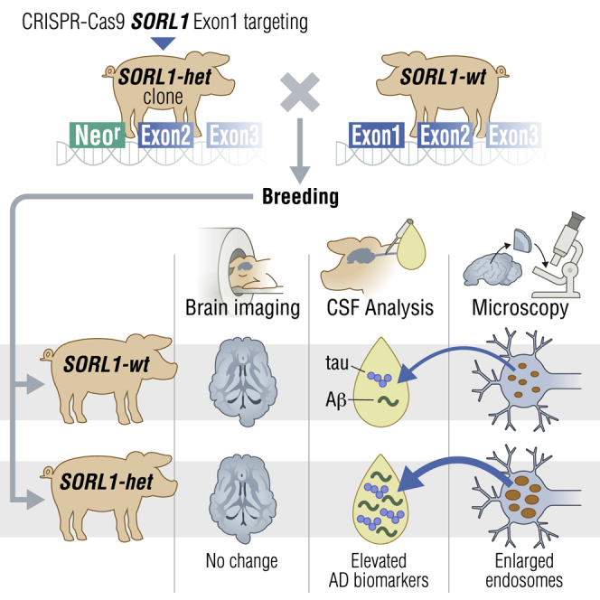
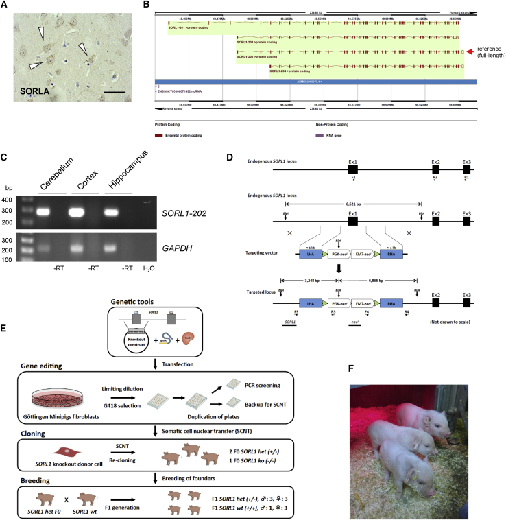
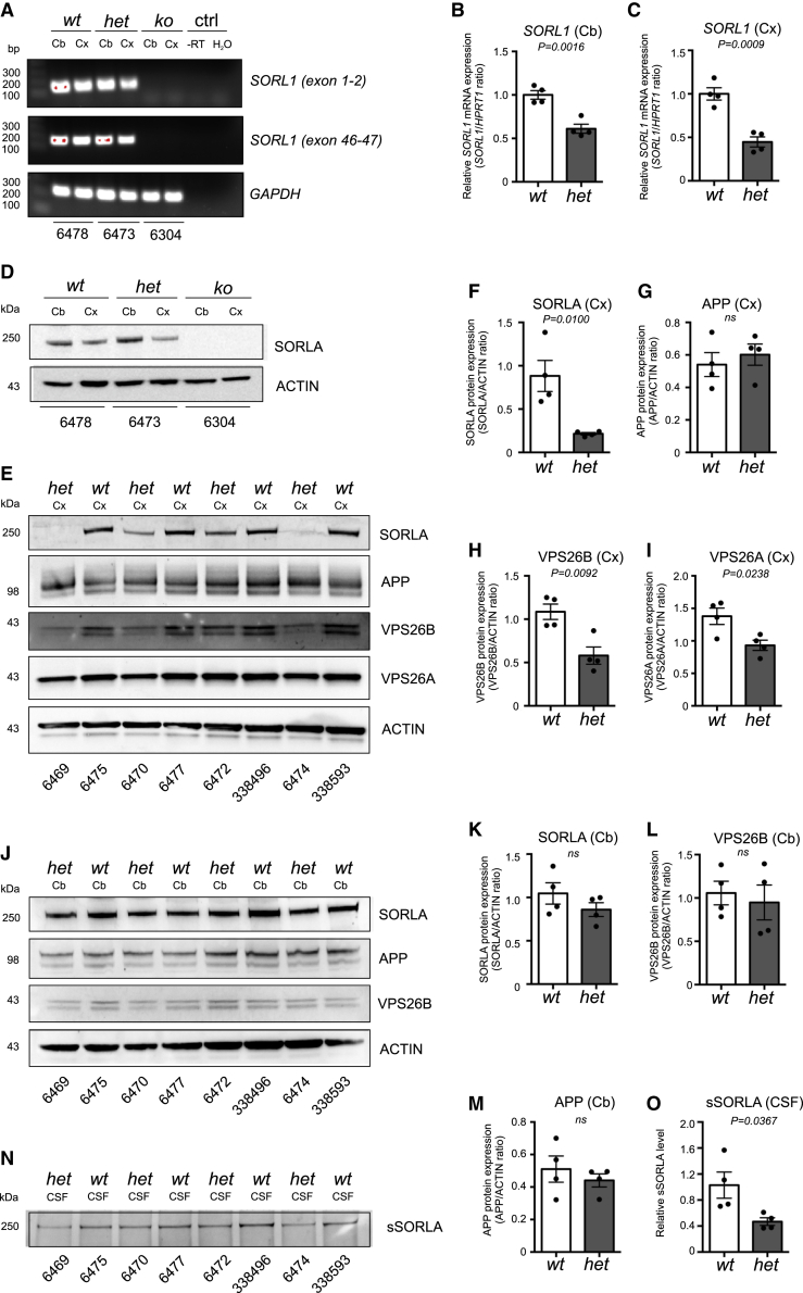
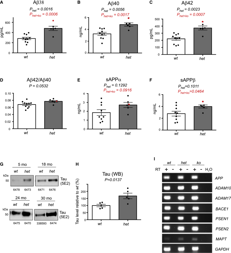
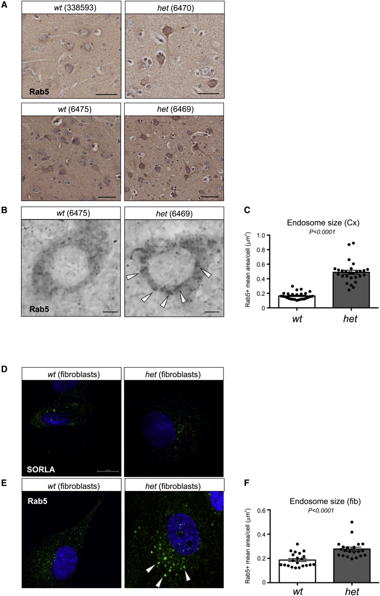
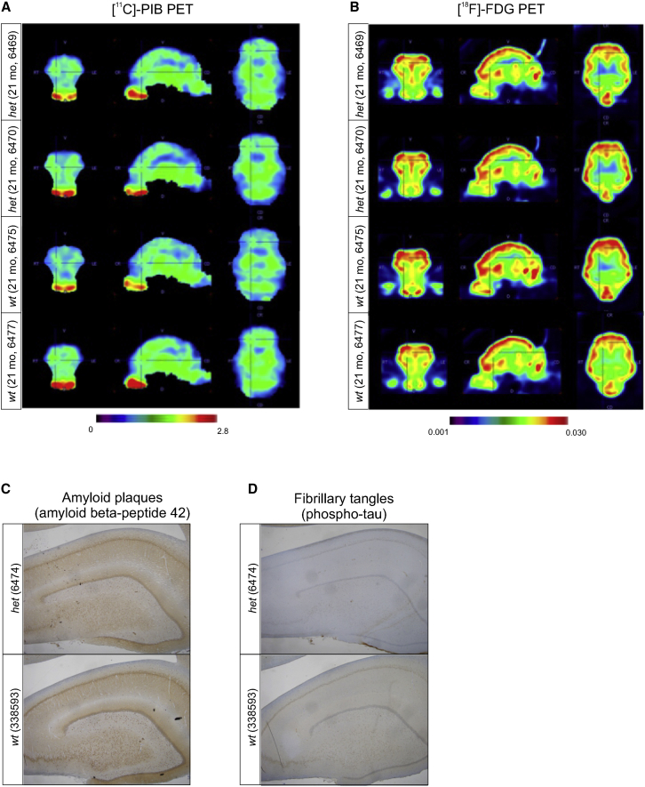
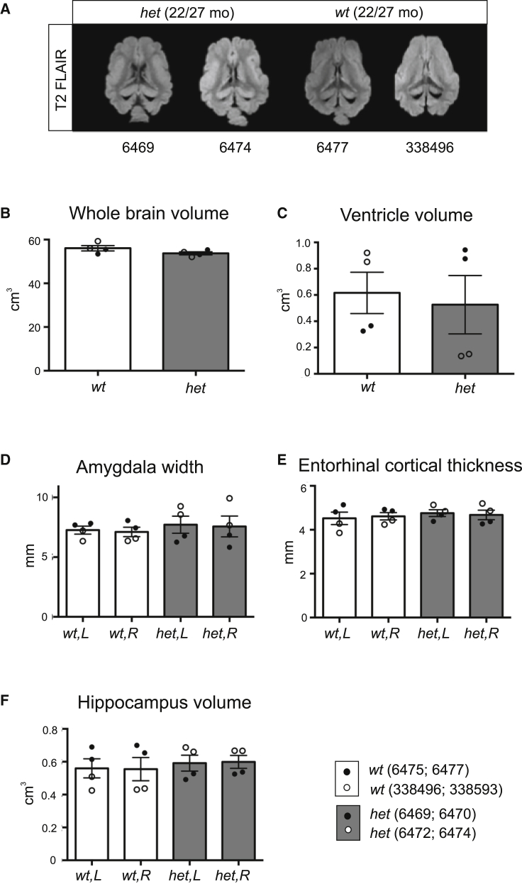
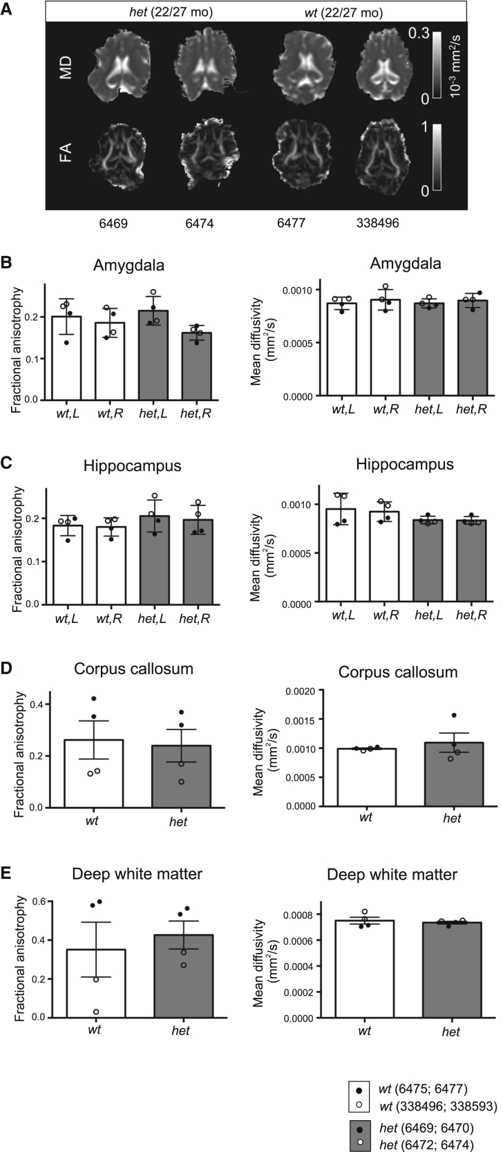
The established causal genes in Alzheimer's disease (AD), APP, PSEN1, and PSEN2, are functionally characterized using biomarkers, capturing an in vivo profile reflecting the disease's initial preclinical phase. Mutations in SORL1, encoding the endosome recycling receptor SORLA, are found in 2%-3% of individuals with early-onset AD, and SORL1 haploinsufficiency appears to be causal for AD. To test whether SORL1 can function as an AD causal gene, we use CRISPR-Cas9-based gene editing to develop a model of SORL1 haploinsufficiency in Göttingen minipigs, taking advantage of porcine models for biomarker investigations. SORL1 haploinsufficiency in young adult minipigs is found to phenocopy the preclinical in vivo profile of AD observed with APP, PSEN1, and PSEN2, resulting in elevated levels of β-amyloid (Aβ) and tau preceding amyloid plaque formation and neurodegeneration, as observed in humans. Our study provides functional support for the theory that SORL1 haploinsufficiency leads to endosome cytopathology with biofluid hallmarks of autosomal dominant AD.

Keywords: Alzheimer’s disease; CRISPR-Cas9; SORL1; SORLA; genome editing; large animal model; retromer-dependent endosomal recycling.


Conflict of interest statement

Declaration of interests L.B. and M.D. are employees of AbbVie and own AbbVie stock. AbbVie participated in the design and study conduct for this research as well as in the interpretation of data, review, and approval of the publication. Ellegaard Göttingen Minipigs A/S has the commercialization rights to the genetically altered Göttingen minipig SORL1 model. S.A.S. is a co-founder of Retromer Therapeutics, has equity in the company, and is a paid consultant to the company. O.M.A. also has commercial interests in Retromer Therapeutics, but the company was not involved in any aspects of the study.

Figures

None
Graphical abstract
Figure 1
Figure 1
Generation of SORL1-deficient Göttingen minipigs (A) Immunohistochemical detection of endogenous SORLA predominantly in neuronal cell somata (indicated by arrowheads) in the frontal Cx from an adult wt Göttingen minipig. Scale bar, 20 μm. (B) Schematic of porcine SORL1 transcripts from Ensembl Sscrofa 11.1. Four SORL1 isoforms are listed, of which SORL1 -202, comprising 48 exons, is considered the reference transcript. (C) RT-PCR validation of the porcine reference SORL1 -202 transcript. cDNA obtained from RT of total RNA, isolated from the Cx, hippocampus, and Cb from a wt Göttingen minipig, was used as a template in RT-PCR using primers specific for the 5′ end of the reference SORL1 -202 transcript (exons 1–3) or for porcine GAPDH. Reactions carried out in parallel using RNA without RT (−RT) or with water (H2O) as template served as negative controls. (D) Schematic of the endogenous SORL1 locus (top panel). The three first exons of the endogenous porcine SORL1 gene are shown as black boxes. Primers used for RT-PCR (F1+R1 and F1+R2, respectively) to validate the presence of the reference SORL1-202 transcript in the Göttingen minipig breed and in SORL1 wt, het, and ko animals, are illustrated as horizontal arrows. Also shown is a schematic of the endogenous SORL1 locus, the gene-targeting vector, and the targeted SORL1 locus (bottom panel). The targeting vector comprises a left and right homology arm (shown as blue boxes; LHA and RHA, respectively) flanked by loxP sites (green triangles). The LHA and RHA are separated by a PGK-neor/EM7-zeor expression cassette for mammalian and bacterial selection, respectively. A region of 609 bp, comprising the entire SORL1 exon 1 and its flanking regions, is replaced by the PGK-neor/EM7-zeor cassette upon successful gene editing, resulting in DNA fragments of 3,248 bp and 6,865 bp when BlpI-digested genomic DNA is hybridized with the SORL1 and neor probe, respectively. Positions of the PCR screening primer pairs F3/R3 and F4/R4 are illustrated as horizontal arrows. BlpI restriction sites are indicated as vertical arrows, and Southern blot probes (SORL1probe and neorprobe) are shown as black horizontal bars. (E) Generation of SORL1-deficient Göttingen minipigs. Gene editing was performed by co-transfecting primary porcine fibroblasts, isolated from newborn female Göttingen minipigs, with the gene-targeting vector, the hCas9 plasmid, and the gRNA vector. Two days after transfection, the transfected cells were trypsinized, and half of the cell suspension was subjected to limiting dilution by reseeding the cells into 96-well plates. After selection for 2 weeks, G418-resistant cell clones were trypsinized and, one third of the resulting cell suspension was transferred to 96-well PCR plates for PCR screening, one third was cultured for Southern blot analysis, and one third was cultured in 96-well plates for freezing at early passages and subsequent usage as nuclear donor cells for cloning of minipigs by SCNT. Two surviving piglets with heterozygous KO of SORL1 (SORL1 het) and one surviving piglet with homozygous KO of SORL1 (SORL1 ko) were obtained after SCNT and one re-cloning. The cloned SORL1 het founders (F0 generation) were used for conventional breeding to obtain SORL1 het and SORL1 wt offspring (F1 generation). (F) Eight-day-old cloned Göttingen minipigs. All piglets were born with no apparent gross abnormalities, but apart from one SORL1 ko piglet, all SORL1 het piglets died neonatally. Fibroblasts isolated from an ear biopsy from the SORL1 het piglet shown at the front in the photo were used for re-cloning to obtain viable SORL1 het pigs.
Figure 2
Figure 2
Decreased SORLA expression in the brain of SORL1-deficient Göttingen minipigs (A) RT-PCR validation of SORL1-depleted Göttingen minipigs. cDNA obtained from reverse transcription of total RNA isolated from the cerebellum (Cb) or frontal cortex (Cx) from wt, het, and ko SORL1 Göttingen minipigs was used as a template in RT-PCR using primers specific for SORL1 exons 1 and 2 (see primers F1+R2 in Figure 1D), SORL1 exons 46 and 47 of the SORL1 -202 transcript, or for porcine GAPDH (reference gene). Negative controls (ctrl) correspond to reactions carried out in parallel using −RT) or with H2O as templates. (B and C) Relative SORL1 mRNA expression in SORL1 het (n = 4) minipigs was measured by qPCR, calculated relative to the reference gene HPRT1, and normalized to SORL1 expression in SORL1 wt minipigs (n = 4) for samples from the Cb (B) and Cx (C). (D) WB analysis confirming complete absence of SORLA expression in Cb or frontal Cx homogenates from the SORL1 ko compared with a wt and a het SORL1 Göttingen minipig (D). (E) Homogenates of Cx from het and wt SORL1 minipigs were analyzed by WB to determine expression levels of SORLA, APP, VPS26B, and VPS26A, using the level of ACTIN as a loading ctrl. (F–I) The expression levels of SORLA (F), APP (G), VPS26B (H), and VPS26A (I) were quantified relative to the level of ACTIN. (J) Homogenates of Cb from het and wt SORL1 minipigs were analyzed by WB to determine expression levels of SORLA, APP, and VPS26B, using the level of ACTIN as a loading ctrl. (K–M) The expression levels of SORLA (K), APP (L), and VPS26B (M) were quantified relative to the level of ACTIN. (N and O) Equal amounts of CSF from het and wt SORL1 minipigs (n = 4 in both groups) were immunoprecipitated and analyzed by WB using an antibody for SORLA detection (N), and the relative levels of sSORLA were quantified (O). Identification numbers of individual minipigs are provided below every lane for all WB analyses. Two-tailed unpaired Student’s t test was used for all statistical analyses, with p values below 0.05 considered significantly changed. Data are expressed as mean ± SEM.
Figure 3
Figure 3
Increased Aβ and tau levels in CSF from SORL1-deficient Göttingen minipigs (A–F) Quantification of the APP processing products Aβ38 (A), Aβ40 (B), Aβ42 (C), Aβ42/Aβ40 ratio (D), and soluble APPα (E) and APPβ (F) in CSF from SORL1-deficient (n = 6) and age-matched wt (n = 9/10) Göttingen minipigs. The average ages of the two groups of pigs were similar. The group of SORL1 het minipigs is depicted, including data obtained from the ko pig (6304) shown in red and statistical analysis shown for data excluding (black) or including (red) the data point for the SORL1 ko pig. Quantifications were performed using MSD assays for human APP fragments because of 100% conservation of the 42 amino acids comprising the Aβ sequence. (G) WB analysis of tau in CSF (first-time isolation) from wt and het SORL1 minipigs at 5, 18, 24, or 30 months of age. Identification numbers of individual minipigs are provided below every lane. Detection was performed with the 5E2 anti-tau antibody that binds to a region of tau that is 100% conserved between the human and pig protein (Figure S5A). (H) Quantification of CSF tau WB analysis for the 18-month-old (1 wt/het pair), 24-month-old (2 wt/het pairs), and 30-month-old (2 wt/het pairs) SORL1 minipigs. The signal for SORL1 wt pigs was set to 100% for each age, and the signal for the paired SORL1 het was expressed relative to this. (A–F and H) Two-tailed unpaired Student’s t test was used for all statistical analyses, with p values below 0.05 considered significantly changed. Data are expressed as mean ± SEM. (I) RT-PCR analysis of genes involved in generation of amyloid and tau. Gene expression of APP, α-secretases (ADAM10 and ADAM17), β-secretase (BACE1), subunits of the γ-secretase (PSEN1 and PSEN2), and MAPT (encoding tau) were analyzed in Cx tissue from a wt (6475), a het (6469), and the ko (6304) SORL1 minipig, validating that CRISPR-Cas9 had no detrimental effect on these genes. GAPDH served as a ctrl for successful cDNA synthesis, whereas −RT samples and water were used as a negative ctrl.
Figure 4
Figure 4
Enlarged early endosomes in SORL1 het Göttingen minipigs (A) Paraffin sections of cortical brain regions from SORL1 wt (338593 and 6475) and SORL1 het (6470 and 6469) minipigs immunolabeled with Rab5 antibody to identify early endosomes, occurring as a brown signal from 3,3′-diaminobenzidine (DAB) detection of the horseradish peroxidase (HRP)-conjugated secondary antibody. (B) High-magnification images from bright-field microscopy of early endosome compartments (arrowheads), showing an increase in Rab5-positive structures in neurons from the het compared with the wt SORL1 minipig. (C) The mean area of Rab5-positive (early endosome) structures was measured across neurons from wt (mean area = 0.1640 μm2 ± 0.009565, n = 27 cells) and het (mean area = 0.4887 μm2 ± 0.002815, n = 27 cells) minipig Cx areas as depicted in (B). (D) Immunofluorescence confocal images showing SORLA expression in cultured fibroblasts from wt (6475) and het (6469) SORL1 minipigs, demonstrating lower expression levels in cells from SORL1 het minipigs, as seen by fewer positive signals. (E) Endosome abnormalities were also present in fibroblasts from SORL1 het minipigs, as visualized by Rab5-positive structures with increased size. (F) The mean area of Rab5-positive (early endosome) structures was measured for cultured primary fibroblasts from wt (mean area = 0.1862 ± 0.01223 μm2, n = 21 cells) and het (mean area = 0.2771 ± 0.01566 μm2, n = 21 cells) SORL1 minipigs, as depicted in (E). Scale bars equal 50 μm (A) and 10 μm (B and D). Two-tailed unpaired Student’s t test was used for all statistical analyses, with p values below 0.05 considered significantly changed. Data are expressed as mean ± SEM.
Figure 5
Figure 5
[11C]-PIB and [18F]-FDG PET analysis of young adult SORL1 het and SORL1 wt Göttingen minipigs Shown is [11C]-PIB- and [18F]-FDG PET analyses of four female 21-month-old SORL1 minipigs (het, 6469 and 6470; wt, 6475 and 6477). (A) [11C]-PIB-PET summed images from the 30- to 90-min portion of the dynamic scan, divided by averaged whole brain activity. (B) [18F]-FDG PET standard uptake volume (SUV) images, corrected for full brain activity. (C) Absence of amyloid plaque pathology in a wt and a het SORL1 minipig (29 and 30 months old, respectively), as evidenced by lack of signals in immunostaining for deposits using an antibody for Aβ42 that is routinely applied in clinical settings for validation of AD pathology. (D) Absence of fibrillar tangles in the wt and het SORL1 minipigs shown in (C), visualized by lack of signals in immunostaining with anti-tau phospho-Thr231 antibody, which is routinely applied in clinical settings for validation of AD pathology.
Figure 6
Figure 6
Anatomical MRI analysis of young adult SORL1 het and SORL1 wt Göttingen minipigs (A–F) Examples of 3D T2 FLAIR magnetic resonance images used for volume measurements (extracerebral tissue stripped for visualization) in female (6469) and male (6474) SORL1 het minipigs and female (6477) and male (338496) SORL1 wt minipigs (A). Also shown are quantification of whole brain volume (B), ventricle volume (C), amygdala width (D), entorhinal cortical thickness (E), and hippocampus volume (F) from anatomical MRI scanning. The four female minipigs (wt, 6475 and 6477; het, 6469 and 6470) and the four male minipigs (wt, 338496 and 338593; het, 6472 and 6474) were 22 and 27 months of age, respectively, when scanned. Data are expressed as mean ± SEM.
Figure 7
Figure 7
Normal brain microstructure in SORL1 het and SORL1 wt Göttingen minipigs (A) Examples of mean diffusivity (MD) and fractional anisotropy (FA) maps, representing tissue microstructure, from diffusion tensor MRI in female (6469) and male (6474) SORL1 het minipigs and female (6477) and male (338,496) SORL1 wt minipigs. (B–E) FA and MD of the amygdala (B), hippocampus (C), corpus callosum (D), and deep white matter (E) were determined with DTI. The four female minipigs (wt, 6475 and 6477; het, 6469 and 6470) and the four male minipigs (wt, 338,496 and 338,593; het, 6472 and 6474) were 22 and 27 months of age, respectively, when scanned. Data are expressed as mean ± SEM.

References

    1. Cacace R., Sleegers K., Van Broeckhoven C. Molecular genetics of early-onset Alzheimer's disease revisited. Alzheimers Dement. 2016;12:733–748. doi: 10.1016/j.jalz.2016.01.012. - DOI - PubMed
    1. Scheltens P., De Strooper B., Kivipelto M., Holstege H., Chételat G., Teunissen C.E., Cummings J., van der Flier W.M. Alzheimer's disease. Lancet. 2021;397:1577–1590. doi: 10.1016/S0140-6736(20)32205-4. - DOI - PMC - PubMed
    1. Jensen M., Schröder J., Blomberg M., Engvall B., Pantel J., Ida N., Basun H., Wahlund L.O., Werle E., Jauss M., et al. Cerebrospinal fluid A beta42 is increased early in sporadic Alzheimer's disease and declines with disease progression. Ann. Neurol. 1999;45:504–511. doi: 10.1002/1531-8249(199904)45:4<504::aid-ana12>3.0.co;2-9. - DOI - PubMed
    1. Bateman R.J., Xiong C., Benzinger T.L.S., Fagan A.M., Goate A., Fox N.C., Marcus D.S., Cairns N.J., Xie X., Blazey T.M., et al. Clinical and biomarker changes in dominantly inherited Alzheimer's disease. N. Engl. J. Med. 2012;367:795–804. doi: 10.1056/NEJMoa1202753. - DOI - PMC - PubMed
    1. Maia L.F., Kaeser S.A., Reichwald J., Lambert M., Obermüller U., Schelle J., Odenthal J., Martus P., Staufenbiel M., Jucker M. Increased CSF Abeta during the very early phase of cerebral Abeta deposition in mouse models. EMBO Mol. Med. 2015;7:895–903. doi: 10.15252/emmm.201505026. - DOI - PMC - PubMed

Publication types